- Download PDF
- Share X Facebook Email LinkedIn
- Permissions

Improving Traumatic Brain Injury Care and Research : A Report From the National Academies of Sciences, Engineering, and Medicine
- 1 National Academies of Sciences, Engineering, and Medicine, Washington, DC
- 2 Institute for Healthcare Improvement, Boston, Massachusetts
- JAMA Insights Mild Traumatic Brain Injury in 2019-2020 Noah D. Silverberg, PhD; Ann-Christine Duhaime, MD; Mary Alexis Iaccarino, MD JAMA
- Special Communication CDC Guideline on the Diagnosis and Management of Mild Traumatic Brain Injury Among Children Angela Lumba-Brown, MD; Keith Owen Yeates, PhD; Kelly Sarmiento, MPH; Matthew J. Breiding, PhD; Tamara M. Haegerich, PhD; Gerard A. Gioia, PhD; Michael Turner, MD; Edward C. Benzel, MD; Stacy J. Suskauer, MD; Christopher C. Giza, MD; Madeline Joseph, MD; Catherine Broomand, PhD; Barbara Weissman, MD; Wayne Gordon, PhD; David W. Wright, MD; Rosemarie Scolaro Moser, PhD; Karen McAvoy, PhD; Linda Ewing-Cobbs, PhD; Ann-Christine Duhaime, MD; Margot Putukian, MD; Barbara Holshouser, PhD; David Paulk, EdD; Shari L. Wade, PhD; Stanley A. Herring, MD; Mark Halstead, MD; Heather T. Keenan, MD, PhD; Meeryo Choe, MD; Cindy W. Christian, MD; Kevin Guskiewicz, PhD, ATC; P. B. Raksin, MD; Andrew Gregory, MD; Anne Mucha, PT, DPT; H. Gerry Taylor, PhD; James M. Callahan, MD; John DeWitt, PT, DPT, ATC; Michael W. Collins, PhD; Michael W. Kirkwood, PhD; John Ragheb, MD; Richard G. Ellenbogen, MD; Theodore J. Spinks, MD; Theodore G. Ganiats, MD; Linda J. Sabelhaus, MLS; Katrina Altenhofen, MPH; Rosanne Hoffman, MPH; Tom Getchius, BA; Gary Gronseth, MD; Zoe Donnell, MA; Robert E. O’Connor, MD, MPH; Shelly D. Timmons, MD, PhD JAMA Pediatrics
- Review Pharmacological Interventions and Symptom Reduction for Mild Traumatic Brain Injury Charles Feinberg, BA; Catherine Carr, MLIS; Roger Zemek, MD; Keith Owen Yeates, PhD; Christina Master, PhD; Kathryn Schneider, PT, PhD; Michael J. Bell, MD; Stephen Wisniewski, PhD; Rebekah Mannix, MD, MPH JAMA Neurology
Traumatic brain injury (TBI) takes a substantial toll on health and health care costs in the US. Yet TBI is often unrecognized, misclassified, undertreated (especially in its longer-term manifestations), and, in proportion to its public health consequences, underresearched. Despite the dedication of an increasing number of professionals, disciplines, and organizations devoted to TBI care and research, including innovative programs for military service members and veterans, care often fails to meet the needs of affected individuals, families, and communities. The US lacks consolidated leadership for achieving improvements in TBI care and outcomes, and, partly as a result, it lacks a strategic plan for fostering change and overseeing progress. With stronger leadership and proper redesign, the health care system could reduce the morbidity and disability associated with TBI, while enhancing the effectiveness of TBI care.
Read More About
Bowman K , Matney C , Berwick DM. Improving Traumatic Brain Injury Care and Research : A Report From the National Academies of Sciences, Engineering, and Medicine . JAMA. 2022;327(5):419–420. doi:10.1001/jama.2022.0089
Manage citations:
© 2024
Artificial Intelligence Resource Center
Cardiology in JAMA : Read the Latest
Browse and subscribe to JAMA Network podcasts!
Others Also Liked
Select your interests.
Customize your JAMA Network experience by selecting one or more topics from the list below.
- Academic Medicine
- Acid Base, Electrolytes, Fluids
- Allergy and Clinical Immunology
- American Indian or Alaska Natives
- Anesthesiology
- Anticoagulation
- Art and Images in Psychiatry
- Artificial Intelligence
- Assisted Reproduction
- Bleeding and Transfusion
- Caring for the Critically Ill Patient
- Challenges in Clinical Electrocardiography
- Climate and Health
- Climate Change
- Clinical Challenge
- Clinical Decision Support
- Clinical Implications of Basic Neuroscience
- Clinical Pharmacy and Pharmacology
- Complementary and Alternative Medicine
- Consensus Statements
- Coronavirus (COVID-19)
- Critical Care Medicine
- Cultural Competency
- Dental Medicine
- Dermatology
- Diabetes and Endocrinology
- Diagnostic Test Interpretation
- Drug Development
- Electronic Health Records
- Emergency Medicine
- End of Life, Hospice, Palliative Care
- Environmental Health
- Equity, Diversity, and Inclusion
- Facial Plastic Surgery
- Gastroenterology and Hepatology
- Genetics and Genomics
- Genomics and Precision Health
- Global Health
- Guide to Statistics and Methods
- Hair Disorders
- Health Care Delivery Models
- Health Care Economics, Insurance, Payment
- Health Care Quality
- Health Care Reform
- Health Care Safety
- Health Care Workforce
- Health Disparities
- Health Inequities
- Health Policy
- Health Systems Science
- History of Medicine
- Hypertension
- Images in Neurology
- Implementation Science
- Infectious Diseases
- Innovations in Health Care Delivery
- JAMA Infographic
- Law and Medicine
- Leading Change
- Less is More
- LGBTQIA Medicine
- Lifestyle Behaviors
- Medical Coding
- Medical Devices and Equipment
- Medical Education
- Medical Education and Training
- Medical Journals and Publishing
- Mobile Health and Telemedicine
- Narrative Medicine
- Neuroscience and Psychiatry
- Notable Notes
- Nutrition, Obesity, Exercise
- Obstetrics and Gynecology
- Occupational Health
- Ophthalmology
- Orthopedics
- Otolaryngology
- Pain Medicine
- Palliative Care
- Pathology and Laboratory Medicine
- Patient Care
- Patient Information
- Performance Improvement
- Performance Measures
- Perioperative Care and Consultation
- Pharmacoeconomics
- Pharmacoepidemiology
- Pharmacogenetics
- Pharmacy and Clinical Pharmacology
- Physical Medicine and Rehabilitation
- Physical Therapy
- Physician Leadership
- Population Health
- Primary Care
- Professional Well-being
- Professionalism
- Psychiatry and Behavioral Health
- Public Health
- Pulmonary Medicine
- Regulatory Agencies
- Reproductive Health
- Research, Methods, Statistics
- Resuscitation
- Rheumatology
- Risk Management
- Scientific Discovery and the Future of Medicine
- Shared Decision Making and Communication
- Sleep Medicine
- Sports Medicine
- Stem Cell Transplantation
- Substance Use and Addiction Medicine
- Surgical Innovation
- Surgical Pearls
- Teachable Moment
- Technology and Finance
- The Art of JAMA
- The Arts and Medicine
- The Rational Clinical Examination
- Tobacco and e-Cigarettes
- Translational Medicine
- Trauma and Injury
- Treatment Adherence
- Ultrasonography
- Users' Guide to the Medical Literature
- Vaccination
- Venous Thromboembolism
- Veterans Health
- Women's Health
- Workflow and Process
- Wound Care, Infection, Healing
- Register for email alerts with links to free full-text articles
- Access PDFs of free articles
- Manage your interests
- Save searches and receive search alerts
An official website of the United States government
The .gov means it’s official. Federal government websites often end in .gov or .mil. Before sharing sensitive information, make sure you’re on a federal government site.
The site is secure. The https:// ensures that you are connecting to the official website and that any information you provide is encrypted and transmitted securely.
- Publications
- Account settings
- My Bibliography
- Collections
- Citation manager
Save citation to file
Email citation, add to collections.
- Create a new collection
- Add to an existing collection
Add to My Bibliography
Your saved search, create a file for external citation management software, your rss feed.
- Search in PubMed
- Search in NLM Catalog
- Add to Search
Traumatic brain injury: progress and challenges in prevention, clinical care, and research
- PMID: 36183712
- PMCID: PMC10427240
- DOI: 10.1016/S1474-4422(22)00309-X
- Correction to Lancet Neurol 2022; published online Sept 29. https://doi.org/10.1016/ S1474-4422(22)00309-X. [No authors listed] [No authors listed] Lancet Neurol. 2022 Dec;21(12):e10. doi: 10.1016/S1474-4422(22)00411-2. Epub 2022 Oct 7. Lancet Neurol. 2022. PMID: 36216016 No abstract available.
Traumatic brain injury (TBI) has the highest incidence of all common neurological disorders, and poses a substantial public health burden. TBI is increasingly documented not only as an acute condition but also as a chronic disease with long-term consequences, including an increased risk of late-onset neurodegeneration. The first Lancet Neurology Commission on TBI, published in 2017, called for a concerted effort to tackle the global health problem posed by TBI. Since then, funding agencies have supported research both in high-income countries (HICs) and in low-income and middle-income countries (LMICs). In November 2020, the World Health Assembly, the decision-making body of WHO, passed resolution WHA73.10 for global actions on epilepsy and other neurological disorders, and WHO launched the Decade for Action on Road Safety plan in 2021. New knowledge has been generated by large observational studies, including those conducted under the umbrella of the International Traumatic Brain Injury Research (InTBIR) initiative, established as a collaboration of funding agencies in 2011. InTBIR has also provided a huge stimulus to collaborative research in TBI and has facilitated participation of global partners. The return on investment has been high, but many needs of patients with TBI remain unaddressed. This update to the 2017 Commission presents advances and discusses persisting and new challenges in prevention, clinical care, and research.
In LMICs, the occurrence of TBI is driven by road traffic incidents, often involving vulnerable road users such as motorcyclists and pedestrians. In HICs, most TBI is caused by falls, particularly in older people (aged ≥65 years), who often have comorbidities. Risk factors such as frailty and alcohol misuse provide opportunities for targeted prevention actions. Little evidence exists to inform treatment of older patients, who have been commonly excluded from past clinical trials—consequently, appropriate evidence is urgently required. Although increasing age is associated with worse outcomes from TBI, age should not dictate limitations in therapy. However, patients injured by low-energy falls (who are mostly older people) are about 50% less likely to receive critical care or emergency interventions, compared with those injured by high-energy mechanisms, such as road traffic incidents.
Mild TBI, defined as a Glasgow Coma sum score of 13–15, comprises most of the TBI cases (over 90%) presenting to hospital. Around 50% of adult patients with mild TBI presenting to hospital do not recover to pre-TBI levels of health by 6 months after their injury. Fewer than 10% of patients discharged after presenting to an emergency department for TBI in Europe currently receive follow-up. Structured follow-up after mild TBI should be considered good practice, and urgent research is needed to identify which patients with mild TBI are at risk for incomplete recovery.
The selection of patients for CT is an important triage decision in mild TBI since it allows early identification of lesions that can trigger hospital admission or life-saving surgery. Current decision making for deciding on CT is inefficient, with 90–95% of scanned patients showing no intracranial injury but being subjected to radiation risks. InTBIR studies have shown that measurement of blood-based biomarkers adds value to previously proposed clinical decision rules, holding the potential to improve efficiency while reducing radiation exposure. Increased concentrations of biomarkers in the blood of patients with a normal presentation CT scan suggest structural brain damage, which is seen on MR scanning in up to 30% of patients with mild TBI. Advanced MRI, including diffusion tensor imaging and volumetric analyses, can identify additional injuries not detectable by visual inspection of standard clinical MR images. Thus, the absence of CT abnormalities does not exclude structural damage—an observation relevant to litigation procedures, to management of mild TBI, and when CT scans are insufficient to explain the severity of the clinical condition.
Although blood-based protein biomarkers have been shown to have important roles in the evaluation of TBI, most available assays are for research use only. To date, there is only one vendor of such assays with regulatory clearance in Europe and the USA with an indication to rule out the need for CT imaging for patients with suspected TBI. Regulatory clearance is provided for a combination of biomarkers, although evidence is accumulating that a single biomarker can perform as well as a combination. Additional biomarkers and more clinical-use platforms are on the horizon, but cross-platform harmonisation of results is needed. Health-care efficiency would benefit from diversity in providers.
In the intensive care setting, automated analysis of blood pressure and intracranial pressure with calculation of derived parameters can help individualise management of TBI. Interest in the identification of subgroups of patients who might benefit more from some specific therapeutic approaches than others represents a welcome shift towards precision medicine. Comparative-effectiveness research to identify best practice has delivered on expectations for providing evidence in support of best practices, both in adult and paediatric patients with TBI.
Progress has also been made in improving outcome assessment after TBI. Key instruments have been translated into up to 20 languages and linguistically validated, and are now internationally available for clinical and research use. TBI affects multiple domains of functioning, and outcomes are affected by personal characteristics and life-course events, consistent with a multifactorial bio-psycho-socio-ecological model of TBI, as presented in the US National Academies of Sciences, Engineering, and Medicine (NASEM) 2022 report. Multidimensional assessment is desirable and might be best based on measurement of global functional impairment. More work is required to develop and implement recommendations for multidimensional assessment. Prediction of outcome is relevant to patients and their families, and can facilitate the benchmarking of quality of care. InTBIR studies have identified new building blocks (eg, blood biomarkers and quantitative CT analysis) to refine existing prognostic models. Further improvement in prognostication could come from MRI, genetics, and the integration of dynamic changes in patient status after presentation.
Neurotrauma researchers traditionally seek translation of their research findings through publications, clinical guidelines, and industry collaborations. However, to effectively impact clinical care and outcome, interactions are also needed with research funders, regulators, and policy makers, and partnership with patient organisations. Such interactions are increasingly taking place, with exemplars including interactions with the All Party Parliamentary Group on Acquired Brain Injury in the UK, the production of the NASEM report in the USA, and interactions with the US Food and Drug Administration. More interactions should be encouraged, and future discussions with regulators should include debates around consent from patients with acute mental incapacity and data sharing. Data sharing is strongly advocated by funding agencies. From January 2023, the US National Institutes of Health will require upload of research data into public repositories, but the EU requires data controllers to safeguard data security and privacy regulation. The tension between open data-sharing and adherence to privacy regulation could be resolved by cross-dataset analyses on federated platforms, with the data remaining at their original safe location. Tools already exist for conventional statistical analyses on federated platforms, however federated machine learning requires further development. Support for further development of federated platforms, and neuroinformatics more generally, should be a priority.
This update to the 2017 Commission presents new insights and challenges across a range of topics around TBI: epidemiology and prevention (section 1); system of care (section 2); clinical management (section 3); characterisation of TBI (section 4); outcome assessment (section 5); prognosis (Section 6); and new directions for acquiring and implementing evidence (section 7). Table 1 summarises key messages from this Commission and proposes recommendations for the way forward to advance research and clinical management of TBI.
PubMed Disclaimer
Conflict of interest statement
Declaration of interests No funding was provided specifically for this Commission paper; however, most authors are involved in the International Initiative for Traumatic Brain Injury Research (InTBIR) as a scientific participant or an investigator. This Commission would not have been possible without the indirect facilitation provided by the InTBIR network. AIRM declares consulting fees from PresSura Neuro, Integra Life Sciences, and NeuroTrauma Sciences. DKM reports research support, and educational and consulting fees from Lantmannen AB, GlaxoSmithKline, Calico, PresSura Neuro, NeuroTrauma Sciences, and Integra Neurosciences. GTM declares grants from the US National Institutes of Health-National Institute of Neurological Disorders and Stroke (grant U01NS086090), the US Department of Defense (grant W81XWH-14-2-0176, grant W81XWH-18-2-0042, and contract W81XWH-15-9-0001). MC reports licensing fees for ICM+ software from Cambridge Enterprise and was an honorary (unpaid) director for Medicam. PS reports licensing fees for ICM+ software from Cambridge Enterprise. MBS has in the past 3 years received consulting income from Acadia Pharmaceuticals, Aptinyx, atai Life Sciences, Boehringer Ingelheim, Bionomics, BioXcel Therapeutics, Clexio, Eisai, EmpowerPharm, Engrail Therapeutics, Janssen, Jazz Pharmaceuticals, and Roche/Genentech. MBS also has stock options in Oxeia Biopharmaceuticals and EpiVario and is paid for editorial work on Depression and Anxiety (Editor-in-Chief), Biological Psychiatry (Deputy Editor), and UpToDate (Co-Editor-in-Chief for Psychiatry). KKWW holds stock options in Gryphon Bio. All other authors declare no competing interests.
Figure 1:. Global incidence and prevalence of…
Figure 1:. Global incidence and prevalence of traumatic brain injury compared with other common neurological…
Figure 2:. Between-country variations in mechanism of…
Figure 2:. Between-country variations in mechanism of traumatic brain injury according to the Human Development…
Figure 3:. Estimated frequency of hospital discharges…
Figure 3:. Estimated frequency of hospital discharges and deaths in cases of traumatic brain injury…
Figure 4:. Advances and remaining challenges in…
Figure 4:. Advances and remaining challenges in the provision of health care for people with…
Figure 5:. Consensus-derived matrix for de-escalation of…
Figure 5:. Consensus-derived matrix for de-escalation of therapy in suspected intracranial hypertension
This decision-support heatmap…
Figure 6:. Between-centre differences in surgery in…
Figure 6:. Between-centre differences in surgery in acute traumatic brain injury
(A) Acute surgery in…
Figure 7:. UpSet plot of pathoanatomic common…
Figure 7:. UpSet plot of pathoanatomic common data elements reported on early CT, by traumatic…
Figure 8:. Outcomes after a traumatic brain…
Figure 8:. Outcomes after a traumatic brain injury: the bio-psycho-socio-ecological model
The pyramid represents how…
Figure 9:. UpSet plots of impaired scores…
Figure 9:. UpSet plots of impaired scores on outcomes from (A) the CENTER-TBI study and…
Figure 10:. Calibration plots for external validation…
Figure 10:. Calibration plots for external validation of the IMPACT lab models for mortality and…
Figure 11:. Absolute incremental value (delta R…
Figure 11:. Absolute incremental value (delta R 2 ) of biomarkers when added to the…
Figure 12:. Caterpillar plots of between-centre differences…
Figure 12:. Caterpillar plots of between-centre differences in interventions and outcomes in the CENTER-TBI study
- Towards further progress in traumatic brain injury. Wienhoven M, Piercy J. Wienhoven M, et al. Lancet Neurol. 2023 Feb;22(2):109. doi: 10.1016/S1474-4422(22)00528-2. Lancet Neurol. 2023. PMID: 36681439 No abstract available.
Similar articles
- Head and Spinal Injuries in Equestrian Sports: Update on Epidemiology, Clinical Outcomes, and Injury Prevention. Gates JK, Lin CY. Gates JK, et al. Curr Sports Med Rep. 2020 Jan;19(1):17-23. doi: 10.1249/JSR.0000000000000674. Curr Sports Med Rep. 2020. PMID: 31913919 Review.
- Adult sports-related traumatic brain injury in United States trauma centers. Winkler EA, Yue JK, Burke JF, Chan AK, Dhall SS, Berger MS, Manley GT, Tarapore PE. Winkler EA, et al. Neurosurg Focus. 2016 Apr;40(4):E4. doi: 10.3171/2016.1.FOCUS15613. Neurosurg Focus. 2016. PMID: 27032921
- Traumatic brain injury: integrated approaches to improve prevention, clinical care, and research. Maas AIR, Menon DK, Adelson PD, Andelic N, Bell MJ, Belli A, Bragge P, Brazinova A, Büki A, Chesnut RM, Citerio G, Coburn M, Cooper DJ, Crowder AT, Czeiter E, Czosnyka M, Diaz-Arrastia R, Dreier JP, Duhaime AC, Ercole A, van Essen TA, Feigin VL, Gao G, Giacino J, Gonzalez-Lara LE, Gruen RL, Gupta D, Hartings JA, Hill S, Jiang JY, Ketharanathan N, Kompanje EJO, Lanyon L, Laureys S, Lecky F, Levin H, Lingsma HF, Maegele M, Majdan M, Manley G, Marsteller J, Mascia L, McFadyen C, Mondello S, Newcombe V, Palotie A, Parizel PM, Peul W, Piercy J, Polinder S, Puybasset L, Rasmussen TE, Rossaint R, Smielewski P, Söderberg J, Stanworth SJ, Stein MB, von Steinbüchel N, Stewart W, Steyerberg EW, Stocchetti N, Synnot A, Te Ao B, Tenovuo O, Theadom A, Tibboel D, Videtta W, Wang KKW, Williams WH, Wilson L, Yaffe K; InTBIR Participants and Investigators. Maas AIR, et al. Lancet Neurol. 2017 Dec;16(12):987-1048. doi: 10.1016/S1474-4422(17)30371-X. Epub 2017 Nov 6. Lancet Neurol. 2017. PMID: 29122524 Review. No abstract available.
- How the 1950s changed our understanding of traumatic encephalopathy and its sequelae. Casper ST. Casper ST. CMAJ. 2018 Feb 5;190(5):E140-E142. doi: 10.1503/cmaj.171204. CMAJ. 2018. PMID: 30986191 Free PMC article. No abstract available.
- The four stages of youth sports TBI Policymaking: engagement, enactment, research, and reform. Harvey HH, Koller DL, Lowrey KM. Harvey HH, et al. J Law Med Ethics. 2015 Spring;43 Suppl 1:87-90. doi: 10.1111/jlme.12225. J Law Med Ethics. 2015. PMID: 25846174 No abstract available.
- Incidence of anxiety after traumatic brain injury: a systematic review and meta-analysis. Dehbozorgi M, Maghsoudi MR, Mohammadi I, Firouzabadi SR, Mohammaditabar M, Oraee S, Aarabi A, Goodarzi M, Shafiee A, Bakhtiyari M. Dehbozorgi M, et al. BMC Neurol. 2024 Aug 22;24(1):293. doi: 10.1186/s12883-024-03791-0. BMC Neurol. 2024. PMID: 39174923 Free PMC article.
- Bridging the gap: enhancing TBI care in Pakistan's primary and secondary healthcare settings. Sahitia N, Yaqoob E, Javed S. Sahitia N, et al. Neurosurg Rev. 2024 Aug 23;47(1):461. doi: 10.1007/s10143-024-02705-5. Neurosurg Rev. 2024. PMID: 39174684 Review.
- Wrapping stem cells with wireless electrical nanopatches for traumatic brain injury therapy. Wang L, Du J, Liu Q, Wang D, Wang W, Lei M, Li K, Li Y, Hao A, Sang Y, Yi F, Zhou W, Liu H, Mao C, Qiu J. Wang L, et al. Nat Commun. 2024 Aug 22;15(1):7223. doi: 10.1038/s41467-024-51098-y. Nat Commun. 2024. PMID: 39174514 Free PMC article.
- Exploring the biological basis of acupuncture treatment for traumatic brain injury: a review of evidence from animal models. Wu M, Song W, Teng L, Li J, Liu J, Ma H, Zhang G, Zhang J, Chen Q. Wu M, et al. Front Cell Neurosci. 2024 Aug 7;18:1405782. doi: 10.3389/fncel.2024.1405782. eCollection 2024. Front Cell Neurosci. 2024. PMID: 39171199 Free PMC article.
- Longitudinal assessment of glymphatic changes following mild traumatic brain injury: Insights from perivascular space burden and DTI-ALPS imaging. Zhuo J, Raghavan P, Li J, Roys S, Njonkou Tchoquessi RL, Chen H, Wickwire EM, Parikh GY, Schwartzbauer GT, Grattan LM, Wang Z, Gullapalli RP, Badjatia N. Zhuo J, et al. Front Neurol. 2024 Aug 7;15:1443496. doi: 10.3389/fneur.2024.1443496. eCollection 2024. Front Neurol. 2024. PMID: 39170078 Free PMC article.
- Maas AIR, Menon DK, Adelson PD, et al. Traumatic brain injury: integrated approaches to improve prevention, clinical care, and research. Lancet Neurol 2017; 16: 987–1048. - PubMed
- Menon DK, Bryant C. Time for change in acquired brain injury. Lancet Neurol 2019; 18: 28. - PubMed
- Majdan M, Plancikova D, Brazinova A, et al. Epidemiology of traumatic brain injuries in Europe: a cross-sectional analysis. Lancet Public Health 2016; 1: e76–83. - PubMed
- Bell MJ, Kochanek PM. International traumatic brain injury research: an annus mirabilis? Lancet Neurol 2019; 18: 904–05. - PubMed
- Tosetti P, Hicks RR, Theriault E, Phillips A, Koroshetz W, Draghia-Akli R. Toward an international initiative for traumatic brain injury research. J Neurotrauma 2013; 30: 1211–22. - PMC - PubMed
Publication types
- Search in MeSH
Related information
Grants and funding.
- 90DPTB0011/ACL/ACL HHS/United States
- U01 NS086090/NS/NINDS NIH HHS/United States
LinkOut - more resources
Full text sources.
- Archivio Istituzionale della Ricerca Unimi
- Elsevier Science
- Europe PubMed Central
- PubMed Central
- MedlinePlus Health Information
Miscellaneous
- NCI CPTAC Assay Portal
- Citation Manager
NCBI Literature Resources
MeSH PMC Bookshelf Disclaimer
The PubMed wordmark and PubMed logo are registered trademarks of the U.S. Department of Health and Human Services (HHS). Unauthorized use of these marks is strictly prohibited.
REVIEW article
Thirty years of research on traumatic brain injury rehabilitation: a bibliometric study.

- 1 School of Sports Medicine and Rehabilitation, Beijing Sport University, Beijing, China
- 2 Viterbi School of Engineering, University of Southern California, Los Angeles, CA, United States
Background: Traumatic brain injury (TBI) is a major public health concern with far-reaching consequences on individuals’ lives. Despite the abundance of works published on TBI rehabilitation, few studies have bibliometrically analyzed the published TBI rehabilitation research. This study aims to characterize current international trends and global productivity by analyzing articles on TBI rehabilitation using bibliometric approaches and visualization methods.
Methods: We conducted a bibliometric analysis of data retrieved and extracted from the Web of Science Core Collection database to examine the evolution and thematic trends in TBI rehabilitation research up until December 31, 2022. The specific characteristics of the research articles on TBI rehabilitation were evaluated, such as publication year, countries/regions, institutions, authors, journals, research fields, references, and keywords.
Results: Our analysis identified 5,541 research articles on TBI rehabilitation and observed a progressive increase in publications and citations over the years. The United States (US, 2,833, 51.13%), Australia (727, 13.12%), and Canada (525, 9.47%) were the most prolific countries/regions. The University of Washington (226, 4.08%) and Hammond FM (114, 2.06%) were the most productive institution and author, respectively. The top three productive journals were Brain Injury (862; 15.56%), Archives of Physical Medicine and Rehabilitation (630; 11.37%), and Journal of Head Trauma Rehabilitation (405, 7.31%). The most frequent research fields were Rehabilitation, Neurosciences, and Clinical Neurology. Co-citation references primarily addressed “outcome assessment,” “community integration” and “TBI management,” and “injury chronicity” and “sequelae” have gained more attention in recent years. “Mild TBI,” “outcome,” “stroke” and “children” were the commonly used keywords. Additionally, the analysis unveiled emerging research frontiers, including “return to work,” “disorder of consciousness,” “veterans,” “mild TBI,” “pediatric,” “executive function” and “acquired brain injury.”
Conclusion: This study provides valuable insights into the current state of TBI rehabilitation research, which has experienced a rapid increase in attention and exponential growth in publications and citations in the last three decades. TBI rehabilitation research is characterized by its multi-disciplinary approach, involving fields such as Rehabilitation, Neurosciences, and Clinical Neurology. The analysis revealed emerging research subjects that could inform future research directions.
1. Introduction
Traumatic brain injury (TBI) constitutes a significant global public health and socioeconomic issue, with far-reaching consequences on patients’ physical, cognitive, psychological, social, emotional, and behavioral well-being. TBI is a leading cause of death and disability in young adults ( 1 ), with an annual incidence proportion of 295 per 100,000 of all ages ( 2 ). Among more than 69 million new cases of TBI diagnosed globally each year ( 3 ), over 55.5 million cases result in disability ( 2 ). Common injury mechanisms of TBI include unintentional falls ( 4 ), automobile accidents, firearm-related injury ( 5 , 6 ), sports, and assault. With the increase in the elderly population in developed countries, TBI due to unintentional falls (secondary) could become an increasing public health and socioeconomic issue. Between 2006 and 2014, the age-adjusted rates of TBI-related emergency department (ED) visits rose by almost 54%, from 521.6 to 801.9 per 100,000 population, while the death rate decreased by 6% ( 7 ). Faster transport to trauma centers and more effective treatment may account for the decline in mortality, which has led to an increase in the number of TBI survivors who require rehabilitation and related care ( 8 ). The overall goal of rehabilitation following TBI is to assist the person in achieving the highest degree of cognitive, functional, and physical capacity to maximize an independent post-injury life ( 9 ). Given the significant impact of TBI on individuals and society, understanding the current state of TBI rehabilitation research is crucial.
Bibliometrics refers to the statistical analysis of bibliographic data from scientific publications, such as articles and books, which is utilized to evaluate and reveal the structure of research and its productivity and trends ( 10 ). This analysis facilitates the examination of thousands of publications within a specific subject or research field, enabling the identification of the most influential publications, as well as collaboration among countries, authors, institutions, and active journals ( 11 ). To our knowledge, only one bibliometric study has been published explicitly concentrating on TBI rehabilitation. However, this study had limitations in terms of bibliometric methods and the time frame examined. Mojgani et al. ( 8 ) developed a Python script to analyze relevant articles on TBI rehabilitation as of 2017 and found that mild TBI and concussion were highly discussed hot topics. While the earlier publication offered insights into facets of TBI rehabilitation, a more comprehensive bibliometrics analysis could potentially enrich the field by elucidating research themes that have garnered attention in the past decades. Recently, the fusion of visualization and data mining techniques has bolstered bibliometric approaches, but these novel methods have not yet been applied to studies examining TBI rehabilitation.
This study aims to provide a comprehensive assessment and a clear understanding of the scientific articles on TBI rehabilitation up until December 31, 2022, through state-of-the-art bibliometric approaches. The primary objective is to understand the patterns of TBI rehabilitation studies from multiple perspectives, including the temporal evolution of scientific publications, geographic distribution, lead authors and journals, research fields, and current trends.
2.1. Search strategy
We conducted a systematic search for English articles from the Web of Science Core Collection (WoSCC) database using the medical subject headings and topic terms: “Traumatic Brain Injury” and “Rehabilitation.” Our search strategy was as follows: TS = (Traumatic Brain Injury) AND TS = (Rehabilitation) AND Document Type = (Article) AND Language = (English), with a Publication Date set from 1970 to 2022. To minimize potential bias from frequent database updates, we retrieved literature up to December 31, 2022.
2.2. Data collection
Two researchers (YL and XY) independently retrieved the literature, extracted the data, and cross-checked their results. Any discrepancies were resolved through discussion or consultation with the senior author (JQ). The data of the included articles from WoSCC were downloaded using the “Custom selection” option, which selected all 29 fields for custom export in “plain text file” format ( 12 ). Missing data was manually completed based on the original literature data.
2.3. Statistical analysis
Co-Occurrence (COOC, version 13.4) ( 13 ) is a bibliometrics and knowledge graph visualization software. To prepare the data for analysis, COOC was used to merge the downloaded txt format files, eliminate duplicates, convert files to an xlsx format file, merge synonyms of keywords, and clean the data. VOSviewer (1.6.19), a visualizing bibliometric networks software ( 14 ), was used to analyze cooperation among countries, institutions, and authors, as well as author co-citation and keyword co-occurrence. CiteSpace (6.1.R6 64-bit Advanced), a visual analytic computer program developed by Dr. Chen Chaomei in Java, was used for the reference co-citation analysis and mapping of visualization knowledge domains ( 15 , 16 ). We extracted the academic journal information, including impact factors and category rank, from the Journal Citation Reports ™ (JCR) of 2021. Statistical analysis was conducted using Microsoft Excel 2022 (Microsoft, Redmond, Washington, United States). Datawrapper, a web-based tool ( 17 ), was used to draw the world choropleth map and institution symbol map. The threshold for entering the next stage of publication was set at publishing more than 100 articles per year.
We used the country/region, author, reference, and keyword as the node to draw the corresponding network maps for visual analysis, and the cluster labels for reference analysis that reveal the main topics were extracted from the title word lists using log-likelihood ratio ( 18 ). The modularity value and silhouette score are usually used to assess the clusters. If the modularity value is over 0.3 and silhouette score is over 0.7, it means the cluster community structure is significant and the members have high homogeneity ( 15 , 19 ), indicating the clustering result is meaningful and efficient ( 20 ). In these network maps, node size is positively correlated with the frequency of occurrence, while the connecting lines indicate co-occurrence between two nodes, and the thickness of the lines indicates the strength of the co-occurrence. The cluster timeline map permits the clearly identification of different research trends and their evolution ( 18 ).
3.1. Study selection and data processing
According to our search strategy, the earliest retrievable record from WOSCC was published in 1988. Thus, this study period spanned from 1988 to the end of 2022. Our search strategy yielded a total of 5,760 articles after three duplicates were excluded. We also eliminated 214 proceeding papers and two book chapters, leaving us with a total of 5,541 studies for analysis ( Figure 1 ).
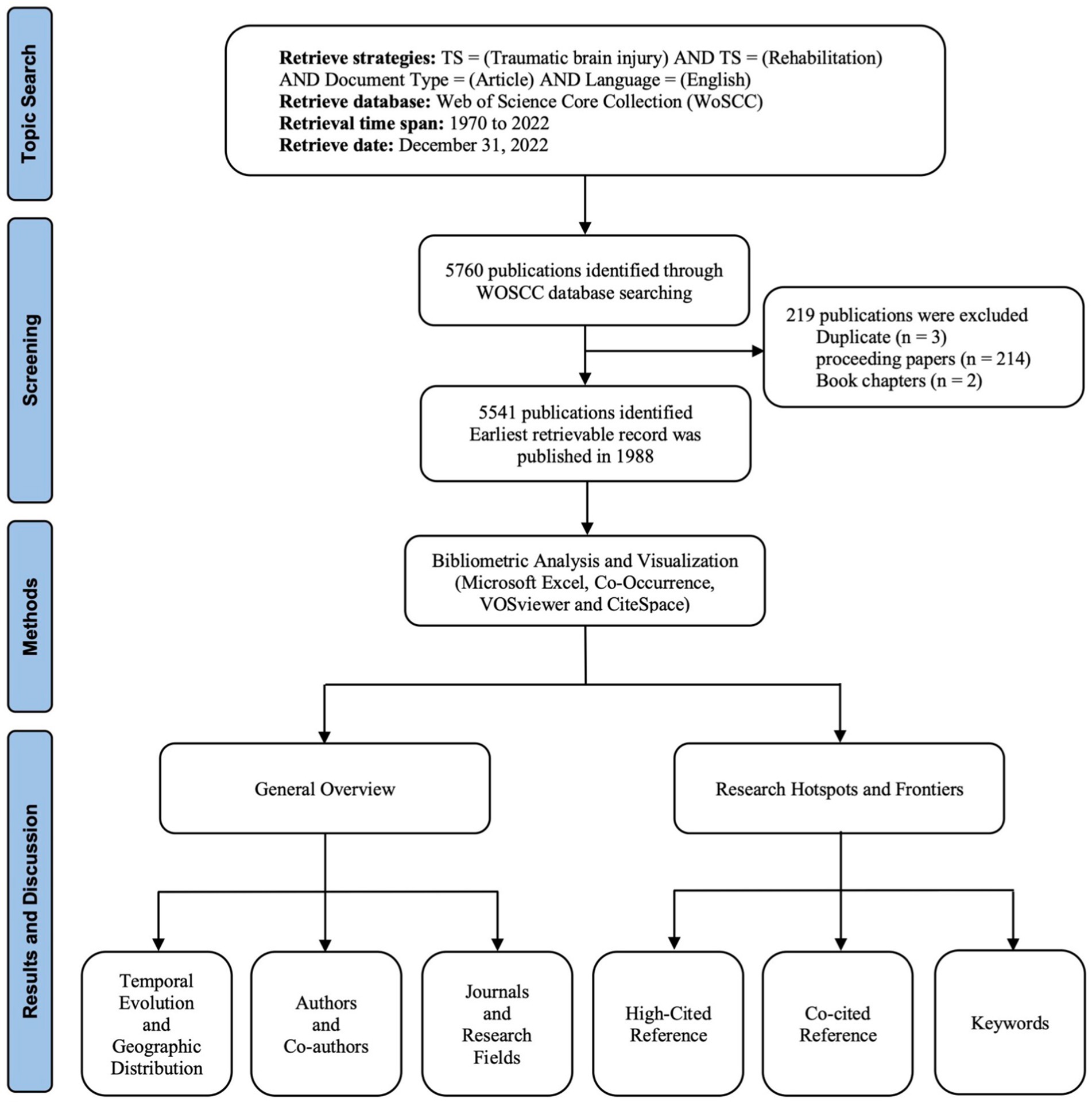
Figure 1 . Flowchart of literature screening included in this study.
There were 1,017 missing fields of the author keywords, and 65 keywords were still missing after copied the supplementary keywords to the author keywords. Consequently, the 65 pieces of literature were excluded from the keyword analysis. We utilized COOC to merge 1,147 synonyms for the identified keywords. After we merged synonyms and removed meaningless words, 6,953 keywords appeared in 5,541 studies. Additionally, we observed 49 missing fields for institutions and countries/regions, which we manually completed based on the original literature.
3.2. Analysis of annual global publications output
We conducted an analysis of the temporal evolution of publications in TBI rehabilitation research from 1988 to 2022. The output of annual publications on TBI rehabilitation is shown in Figure 2A , which reveals two stages: stage 1 (1988–2002) and stage 2 (2003–2022). During the 15-year period of stage 1, the number of publications grew slowly, with an average of 35.87 publications per year. In stage 2, which spanned 20 years after 2002, there was an average of 250.15 articles published per year. The number of annual global publications increased from 56 in 2002 to 424 in 2022, reflecting a growth of 657.1%. The output exceeded 300 between 2017 and 2022 and peaked at 424 in 2022. The fitted curve ( R 2 = 0.9853) shows that the number of publications increases exponentially, with the regression analysis indicating that 461 articles will be published in 2023.

Figure 2 . The publications counts (A) and the citations number (B) per year on TBI rehabilitation research between 1988 and 2022.
Figure 2B displays the number of citations obtained from WOSCC’s citation report, which also fluctuates and increases year by year. A total of 147,831 citations have been cited, with an average of 26.67 citations per article. This increase has been particularly heightened after 2002 and is reflected in the fitted curve ( R 2 = 0.9846), indicating an exponentially increasing trend that coincides with the analysis of published articles.
3.3. Analysis of geographic distribution
TBI rehabilitation had 101 countries/regions and 5,147 institutions publishing at least one article in this field from 1988 to 2022. Figure 3 illustrates the top 15 countries/regions and institutions with the highest number of published articles. The US was a dominant contributor to collaborative TBI rehabilitation research, with 2,833 articles published, representing 51.13% of the total articles. Australia and Canada also made significant contributions to TBI rehabilitation research, with a total of 1,252 articles (accounting for 22.59% of all publications). This finding underscores the potential for co-research collaborations to support other developing countries/regions. Additionally, an analysis of the collaborations between countries/regions revealed that regional clusters were generally formed based on geographical location.
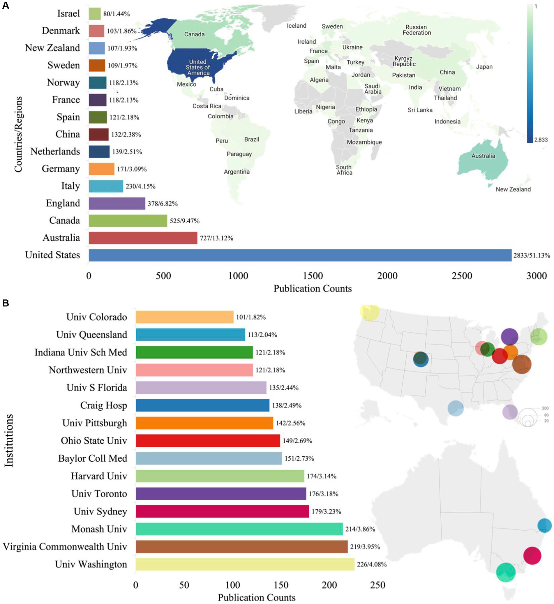
Figure 3 . The distribution of the top productive world countries/regions (A) and institutions (B) on TBI rehabilitation (Univ: University; Sch: School; S: South; Hosp: Hospital; Coll: College; MED: Medicine).
The University of Washington made the greatest contribution to TBI rehabilitation research and participated in publishing 226 articles, accounting for 4.08% of the total. The top 15 institutions are all located in the US, Canada, and Australia. Collaboration network maps of the countries/regions and institutions were plotted to provide a visual representation of the cooperative relationships involved in TBI rehabilitation research ( Supplementary Figure S1 ). The maps show the 30 countries/regions ( Supplementary Figure S1A ) and 149 institutions ( Supplementary Figure S1B ) that published 20 or more articles during the same period. Notably, the US is the leading country in this field, with close collaborative relationships observed between the US, Canada, and Australia. Moreover, the dense lines in the collaboration network maps suggest a high level of cooperation among the institutions involved in this field.
3.4. Analysis of authorship
During the period from 1988 to 2022, a total of 17,410 authors participated in TBI rehabilitation studies. The top 10 most productive authors are displayed in Table 1 , with Hammond FM from Indiana University School of Medicine ranking first with 114 articles and having the highest total link strength. To further examine the collaborations between authors in this field, we constructed a co-occurrence network map ( Supplementary Figure S2A ) of the 78 authors who published at least 20 articles. The abundance of interconnections between researchers indicates prevalent cooperation within the field.
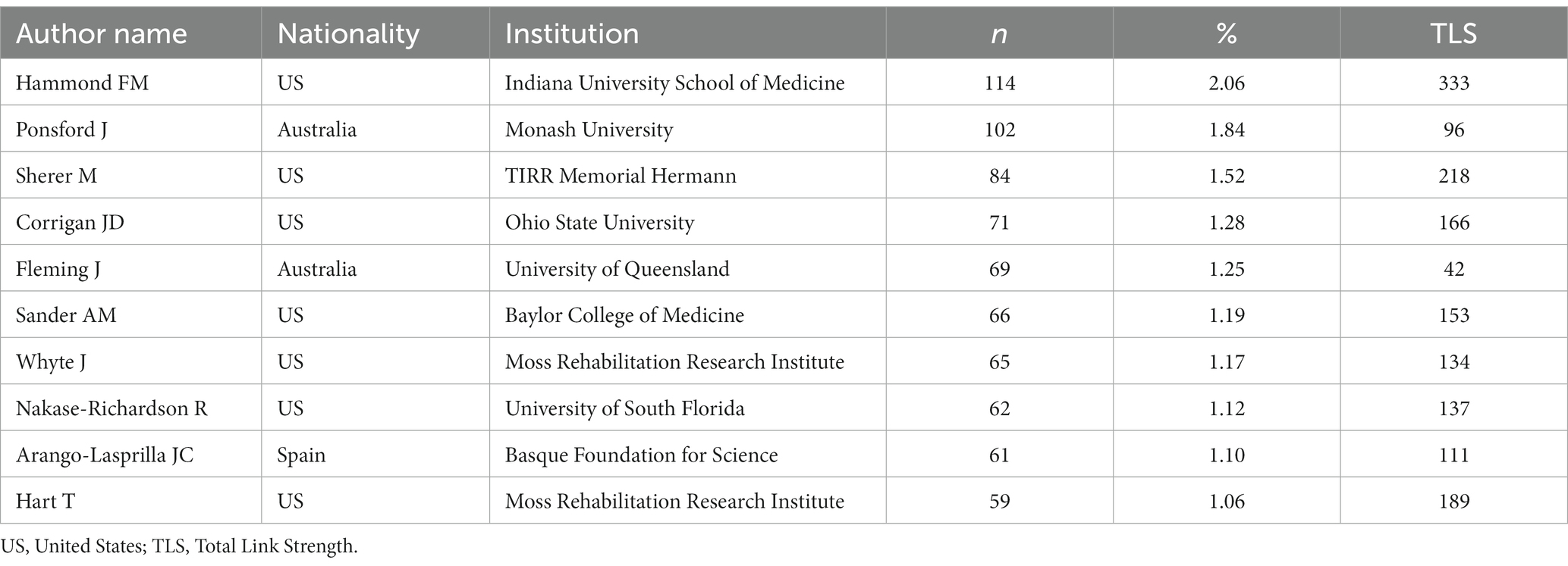
Table 1 . Top 10 most productive authors in TBI rehabilitation research between 1988 and 2022.
Author co-citation is a significant indicator of an author’s influence within a specific field. Supplementary Figure S2B depicted a co-citation network map of corresponding authors, which includes 73 authors who were co-cited at least 200 times. Node size corresponds to their co-citation counts, while lines indicate their co-citation relationships. The top ten most co-cited authors include Corrigan JD (co-citations n = 1,121), Cicerone KD (875), Ponsford J (850), Levin HS (834), Prigatano GP (749), Teasdale G (705), Kreutzer JS (684), Sherer M (678), Wechsler D (616), and Malec JF (609). Five of these authors are also among the top 10 most productive authors.
3.5. Analysis of journals and research fields
From 1988 to 2022, 697 journals published at least one article on TBI rehabilitation. The top 10 most productive journals have been identified, accounting for about 51.87% of the total publication. As shown in Table 2 , Brain Injury published the largest number of articles, accounting for 15.56% of the total. Archives Of Physical Medicine And Rehabilitation (Ranked second, published 630 articles, accounting for 11.37%) has maintained a leading position in the research field of rehabilitation and sports science for many years in a row (Q1, Q2), with the highest total citations. We also obtained the top 10 journals quality updates according to the JCR of 2021 ( Table 3 ). Among the top 10 productive journals, six journals ranked in the second quarter or higher in the relevant research category.
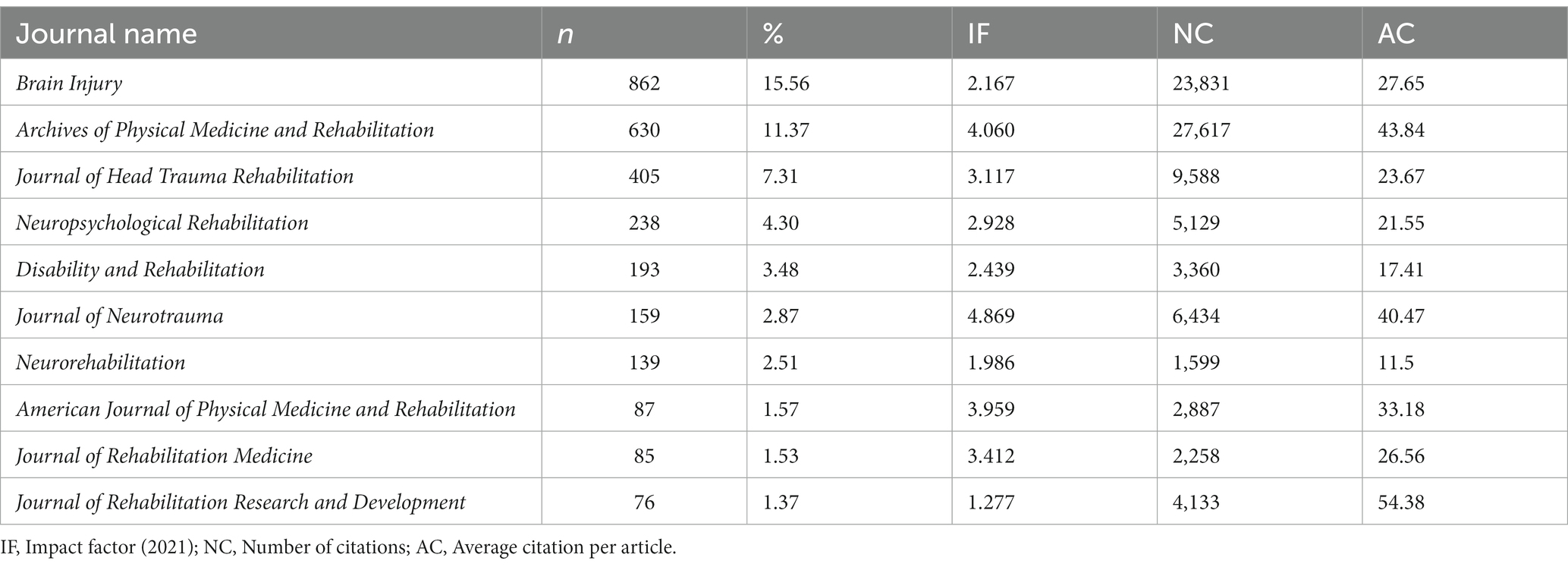
Table 2 . Top 10 most productive journals and their newest impact fact.
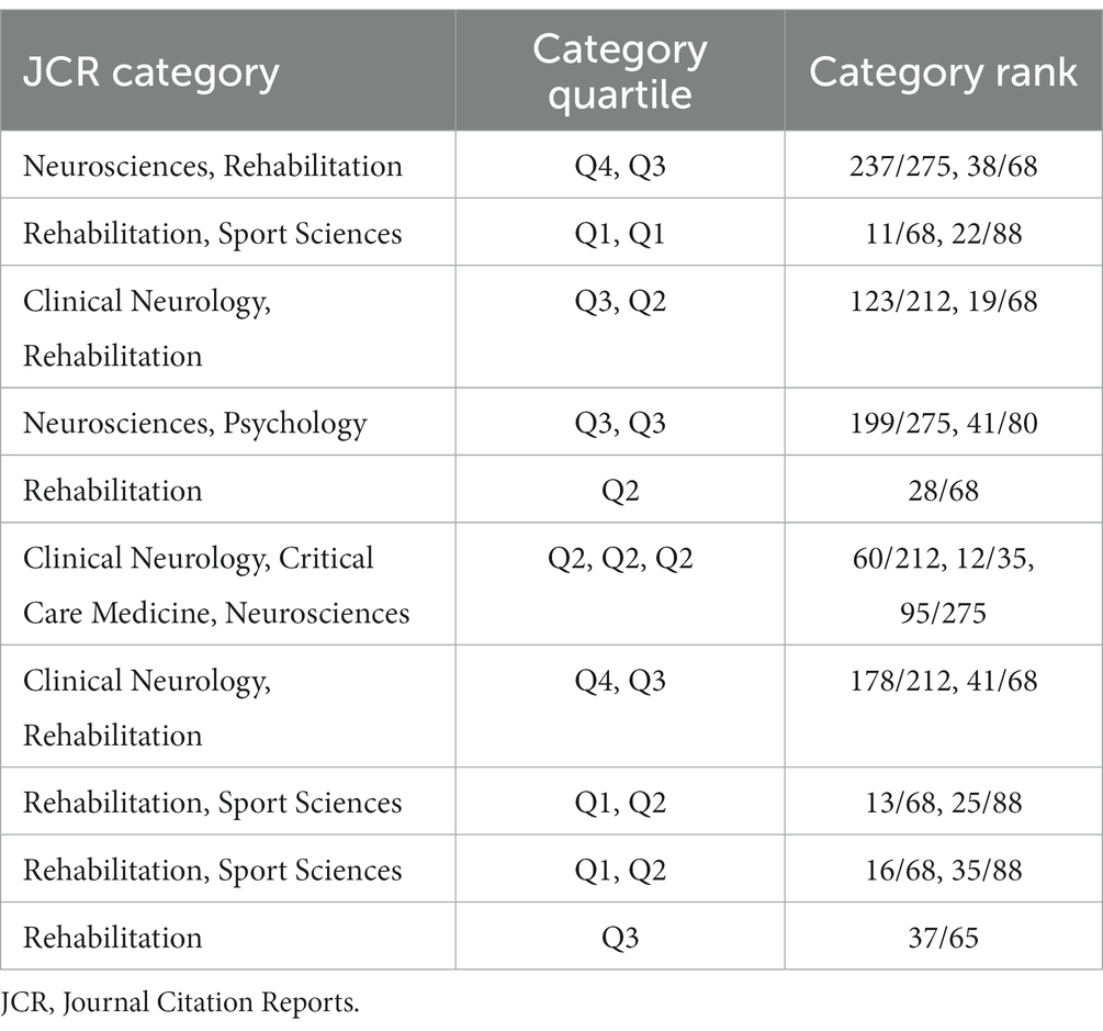
Table 3 . JCR categories and journal quality information of the top 10 most productive journal.
Furthermore, we conducted an analysis of the research fields involved in TBI rehabilitation and identified 117 research fields. Among these, we determined the top 10 research fields, which collectively accounted for 85.67% of total publications ( Figure 4 ). Rehabilitation was found to be the most prevalent research field with 3,118 publication counts, representing 29.58% of all publications. Following closely behind were Neurosciences ( n = 1,828, 17.34%) and Clinical Neurology ( n = 1,620, 15.37%). Collectively, these top 3 research fields accounted for approximately 62.28% of total publications.

Figure 4 . Publication counts based on research fields in TBI rehabilitation research.
3.6. Analysis of cited references
The most cited references are generally considered to have the greatest impact on the scientific community ( 21 ). We have listed the top 10 most cited articles in Supplementary Table S1 . The studies “Treatment of traumatic brain injury with moderate hypothermia ( 22 )” and “Position Statement: Definition of Traumatic Brain Injury ( 23 )” rank first and second, respectively, with 939 and 826 citation times. The former is the oldest of the top 10 most cited articles, indicating its influential position among these articles. The articles “Global, regional, and national burden of neurological disorders, 1990–2016: a systematic analysis for the Global Burden of Disease Study 2016 ( 2 )” and “Constraint-Induced Movement Therapy: A new family of techniques with broad application to physical rehabilitation—A clinical review ( 24 )” rank third and fourth, with citation times exceeding 600 (693 and 608, respectively).
3.7. Analysis of reference co-citation
Reference co-citation analysis is a method used to identify popular research topics ( 25 ). In the present study, we examined 115,437 publications that were cited in the reference sections of the analyzed articles. Using these data, we generated a density web map that includes 194 highly cited publications, each of which was cited at least 50 times ( Supplementary Figure S3 ). Among these publications, the most frequently co-cited studies in the reference sections were those conducted by Teasdale G (1974) ( 26 ), Rappaport M (1982) ( 27 ), Jennett B (1975) ( 28 ), Wilson JTL (1998) ( 29 ), and Levin HS (1979) ( 30 ).
By utilizing the clustering feature of CiteSpace software, we were able to identify seven major research topic clusters. The modularity value and the mean silhouette score were 0.8913 (>0.3) and 0.9483 (>0.5), respectively. As depicted in Supplementary Figure S4A , each node represents a cited reference, the node size corresponds to its citation frequency, and the lines illustrate the co-citation relationships. The largest cluster (#0) is “TBI,” followed by “post-acute care” (#1), “biologics effectiveness research” (#2), “sequelae” (#3), “veteran population” (#4), “injury chronicity” (#8), and “sequelae” (#10). The more significant a cluster is, the more interest it receives from researchers. Furthermore, the timeline view of reference co-citation highlights that “injury chronicity” and “sequelae” are two emerging research topics in TBI rehabilitation ( Supplementary Figure S4B ).
3.8. Analysis of keywords co-occurrence
Keywords are critical to reflect the main information in a research article ( 31 ), and help identify research hotspots and anticipate future directions ( 32 ). We portrayed a keyword co-occurrence network map, with 71 keywords that appeared at least 50 times in TBI rehabilitation research articles ( Supplementary Figure S5 ). We compiled a list of the top 20 keywords that appeared most frequently in TBI rehabilitation studies ( Table 4 ). “TBI” occurred 2,279 times, making it the most frequent keyword, followed by “Rehabilitation” and “Brain injuries.”

Table 4 . Top 20 most occurrences keywords in TBI rehabilitation research.
The different colors of the nodes in Supplementary Figure S5 represent the average publication year of the articles in which the keyword appeared, with yellow indicating more recent publications ( 33 ). Emerging research hotspots are suggested by the keywords in more recent publications, such as “return to work (average publication year: 2017.68),” “disorder of consciousness (2017.62),” “veterans (2016.86),” “mild TBI (2016.63),” “pediatric (2016.55),” executive function (2016.42),” and “acquired brain injury (2016.29).”
4. Discussion
4.1. general overview of the tbi rehabilitation research.
This is the first bibliometric study using visualization tools to analyze the global research trends in TBI rehabilitation. The increasing number of publications and citations in this field suggests increased attention to TBI rehabilitation research over the years. Since 2002, interest in TBI rehabilitation had soared, leading to the publication of 350 articles annually in the past 5 years, with projections of over 460 articles per year in the coming years.
Collaboration is a key characteristic of TBI rehabilitation research, of which growing development led by increasing cross-country and cross-institutional cooperation serves as convincing evidence. The US, Australia, and Canada have published the most literature on TBI rehabilitation and have also ranked in the top five countries for research on spinal cord injury rehabilitation ( 34 ) and sport-related TBI rehabilitation ( 35 ). The most prolific and influential authors in the field have been identified, providing valuable collaboration and consultant information for researchers worldwide. The prevalent interconnectivity among researchers suggests that collaboration is common, aligning with prior research that co-authorship is associated with higher citation rates, while single-author papers accounted for only 8% of rehabilitation research documents ( 8 ).
TBI rehabilitation research has been recognized and disseminated by top journals of rehabilitation and sports science. Our analysis also highlights the importance of multidisciplinary approaches to TBI rehabilitation, as demonstrated by the involvement of fields such as Rehabilitation, Neurosciences, and Clinical Neurology. It is a fast-growing field that has become more active than it was decades ago ( 36 ). Additionally, sport-related TBI rehabilitation is an indispensable subset of TBI rehabilitation, and recent bibliometric studies have examined its impact over the past 20 years ( 35 ).
4.2. Research hotspots and frontiers of TBI rehabilitation research
The analysis of cited references revealed the most impactful articles, forming the foundation of knowledge for future studies in this field. The 10 most cited articles were published on average nearly 15 years before this study, suggesting that the knowledge in the field does not become obsolete quickly and can affect papers published years later ( 8 ). Based on the highly cited publications identified through co-citation analysis, it is suggested that “outcome assessment” was a vital research topic in this field.
The analysis of reference co-citations identified the seven major clusters that primarily focus on outcome assessment ( 37–39 ), community integration ( 40 , 41 ), and TBI management ( 42–45 ). For outcome assessment, the keywords “outcome” and “quality of life” are commonly occurring in this field. However, unlike process measures, which are easily described, outcomes and quality of life can often be surprisingly difficult to define and measure ( 46 ). Various outcome assessment scales including the Glasgow Outcome Scale with or without extended scores ( 28 ), Disability Rating Scale ( 39 , 47 ), Functional Independence Measure ( 38 , 48 ), Community Integration Questionnaire (CIQ) ( 40 ), and the Quality of Life after Brain Injury ( 49 ), have been proposed and used to assess disability after TBI. Their continued use in research highlights their importance and relevance.
Community integration (CI) is an essential aspect of rehabilitation for individuals with TBI, encompassing home integration, social integration, and productive activities ( 50 ). It is not just about physical recovery, but also about promoting social and psychological well-being as well. The ultimate goal of TBI rehabilitation is to help individuals reintegrate into society and become active members of their community ( 51 ). Social support is a key factor facilitating community integration, while physical environment and fatigue are often identified as barriers ( 52 ). Although the CIQ, specifically developed for the TBI population, is currently the standardized measure of CI outcomes ( 53 ), more work is needed to develop inclusive, culturally sensitive, and appropriate tools ( 54 , 55 ).
In the aspect of TBI management, research primarily addresses concerns in physical therapy and cognitive and psychological management. Inadequate management of mild TBI may place patients at risk for second impact syndrome and chronic traumatic encephalopathy. Quatman-Yates et al. ( 45 ) proposed a novel clinical guideline focused on optimizing physical therapy management for mild TBI. The emerging “active rehabilitation” paradigm, emphasizing active interventions and specialized rehabilitation techniques, has rendered physical therapists vital in interdisciplinary care for individuals with mild TBI ( 56 , 57 ). The emerging consensus underscore the necessity of tailoring cognitive rehabilitation according to individuals’ unique profiles, goals, and pre-injury activities, employing diverse approaches such as group therapy to promote generalization and concentrate on personally meaningful activities within the individuals’ environment ( 43 , 58 ). Fenton et al. ( 59 ) reported that 39% of individuals with TBI were diagnosed with a psychiatric condition 6 weeks post injury. Cognitive behavioral therapy is the preferred therapeutic approach for treating mental disturbances, with related therapies like dialectical behavior, mindfulness, and acceptance and commitment therapies being proposed ( 60 , 61 ). Further research is required to validate the efficacy of these approaches.
Emerging research hotspots in TBI rehabilitation include “injury chronicity” and “sequelae.” Recent research has shifted focus toward long-term effects of TBI on individuals, including chronic neurobehavioral sequelae such as cognitive dysfunction, personality changes, and increased rates of psychiatric illness ( 62 , 63 ). Evidence is mounting that TBI can have an impact on an individual’s health and function years after onset ( 64 ). In fact, greater injury chronicity is associated with higher levels of disability, reduced functional independence, and lower levels of community participation ( 65 ). Other frequently occurring keywords in the literature include “mild TBI,” “children,” and “stroke.” Mild TBI, also known as concussion, represents between 75 and 90% of all TBIs ( 66 ) and often lacks adequate follow-up care. Although most people who experience mild TBI fully recover within a few weeks, research indicates that up to 15% of patients diagnosed with mild TBI may experience persistent, disabling problems ( 67 ). Children are a crucial population in TBI rehabilitation research, as TBI can affect them differently from adults. In children, some health effects, such as deficits in organization and problem-solving, may be delayed and not surface until later ( 68 ). Stroke is also the most commonly occurring keyword in the rehabilitation of cardiovascular and cerebrovascular diseases field ( 69 ). Furthermore, it is the most frequently studied neurological disease, with 39% of the 100 most cited papers on neurorehabilitation focusing on stroke ( 70 ). An overlay visualization map of the keyword co-occurrence revealed research frontiers in TBI rehabilitation, including “return to work,” “disorder of consciousness,” “veterans,” “mild TBI,” “pediatric,” “executive function,” and “acquired brain injury.” These areas also focus on CI and TBI management, which represent crucial directions for future research.
A limitation of this study is that it did not include PubMed and Scopus databases. This decision was made because bibliometric analysis using PubMed does not allow for citation and co-citation analysis, while the Scopus database has a low impact level and is not indexed in some journals ( 71 ). WoS is considered a more reliable database due to its indexing of high-impact journals. Furthermore, studies analyzing a large number of articles may encounter the issue of duplicate inclusion when utilizing multiple databases. Despite this limitation, this study’s strength and superiority lie in its comprehensive bibliometric analysis, which is unmatched by other studies in the literature.
5. Conclusion
This study systematically summarized the articles on TBI rehabilitation research from 1988 to 2022 through bibliometric analysis using visualization tools. The results highlighted increasing attention and interest in TBI rehabilitation research, characterized by a multidisciplinary approach. The US, Australia, and Canada were identified as leaders in TBI rehabilitation research, with the University of Washington playing a central role in collaborative research efforts. Co-citation references primarily focused on outcome assessment, CI, and TBI management, with “injury chronicity” and “sequelae” receiving particular attention in recent years. The analysis also uncovered emerging research frontiers, including “return to work,” “disorder of consciousness,” “veterans,” “mild TBI,” “pediatric,” “executive function,” and “acquired brain injury.” By examining patterns and trends in TBI rehabilitation research, this study provided valuable insights for a better understanding of the current state of research and may inform future research directions.
Author contributions
YL: conceptualization and writing—original draft preparation. YL and XY: methodology, data curation, and investigation. YL, XY, and JQ: writing—review and editing. JQ: supervision, funding acquisition, and project administration. All authors contributed to the article and approved the submitted version.
This research was supported by a grant from the Beijing High-Grade, Precision, and Advanced Disciplines Project.
Acknowledgments
The authors acknowledge the financial support of the Beijing High-Grade, Precision, and Advanced Disciplines Project, and also express their sincere appreciation to the editors and reviewers for their diligent effort and insightful suggestions that have significantly enhanced the quality of this paper.
Conflict of interest
The authors declare that the research was conducted in the absence of any commercial or financial relationships that could be construed as a potential conflict of interest.
Publisher’s note
All claims expressed in this article are solely those of the authors and do not necessarily represent those of their affiliated organizations, or those of the publisher, the editors and the reviewers. Any product that may be evaluated in this article, or claim that may be made by its manufacturer, is not guaranteed or endorsed by the publisher.
Supplementary material
The Supplementary material for this article can be found online at: https://www.frontiersin.org/articles/10.3389/fneur.2023.1170731/full#supplementary-material
1. Centers for Disease Control and Prevention. Report to congress on traumatic brain injury in the United States: epidemiology and rehabilitation. Nat Center Inj Prev Control . (2015) 2:1–72.
Google Scholar
2. Feigin, VL, Nichols, E, Alam, T, Bannick, MS, Beghi, E, Blake, N, et al. Global, regional, and national burden of neurological disorders, 1990-2016: a systematic analysis for the global burden of disease study 2016. Lancet Neurol . (2019) 18:459–80. doi: 10.1016/S1474-4422(18)30499-X
PubMed Abstract | CrossRef Full Text | Google Scholar
3. Dewan, MC, Rattani, A, Gupta, S, Baticulon, RE, Hung, Y-C, Punchak, M, et al. Estimating the global incidence of traumatic brain injury. J Neurosurg . (2018) 130:1080–97. doi: 10.3171/2017.10.JNS17352
CrossRef Full Text | Google Scholar
4. Peterson, AB, Thomas, KE, and Zhou, H. Surveillance report of traumatic brain injury-related deaths by age group, sex, and mechanism of injury—United States, 2018 and 2019. (2022). Available at: https://www.cdc.gov/traumaticbraininjury/pdf/TBI_Report_to_Congress_Epi_and_Rehab-a.pdf
5. Daugherty, J, Waltzman, D, Sarmiento, K, and Xu, L. Traumatic brain injury–related deaths by race/ethnicity, sex, intent, and mechanism of injury—United States, 2000–2017. Morb Mortal Wkly Rep . (2019) 68:1050–6. doi: 10.15585/mmwr.mm6846a2
6. Miller, GF, Kegler, SR, and Stone, DM. Traumatic brain injury-related deaths from firearm suicide: United States, 2008-2017. Am J Public Health . (2020) 110:897–9. doi: 10.2105/AJPH.2020.305622
7. Peterson, AB, Xu, L, Daugherty, J, and Breiding, MJ. Surveillance report of traumatic brain injury-related emergency department visits, hospitalizations, and deaths, United States, 2014. (2019). Available at: https://www.cdc.gov/traumaticbraininjury/pdf/TBI-Surveillance-Report-FINAL_508.pdf
8. Mojgani, P, Jalali, M, and Keramatfar, A. Bibliometric study of traumatic brain injury rehabilitation. Neuropsychol Rehabil . (2022) 32:51–68. doi: 10.1080/09602011.2020.1796714
9. Wright, CJ, Zeeman, H, and Biezaitis, V. Holistic practice in traumatic brain injury rehabilitation: perspectives of health practitioners. PLoS One . (2016) 11:e0156826. doi: 10.1371/journal.pone.0156826
10. Doğan, G, and Kayır, S. Global scientific outputs of brain death publications and evaluation according to the religions of countries. J Relig Health . (2020) 59:96–112. doi: 10.1007/s10943-019-00886-8
11. Kiraz, M, and Demir, E. A bibliometric analysis of publications on spinal cord injury during 1980–2018. World Neurosurg . (2020) 136:e504–13. doi: 10.1016/j.wneu.2020.01.064
12. Chen, C. How to use CiteSpace 6.1.R3 . Lean Publishing (2022). Available at: https://citespace.podia.com/view/downloads/ebook-how-to-use-citespace
13. Xue shu dian di Wxjl. COOC:a software for bibliometrics and knowledge graph visualization: Xue shu dian di, Wen xian ji liang. (2020).
14. Van Eck, N, and Waltman, L. Software survey: VOSviewer, a computer program for bibliometric mapping. Scientometrics . (2010) 84:523–38. doi: 10.1007/s11192-009-0146-3
15. Chen, C. CiteSpace II: detecting and visualizing emerging trends and transient patterns in scientific literature. J Am Soc Inf Sci Technol . (2006) 57:359–77. doi: 10.1002/asi.20317
16. Chen, C. Searching for intellectual turning points: progressive knowledge domain visualization. Proc Natl Acad Sci U S A . (2004) 101:5303–10. doi: 10.1073/pnas.0307513100
17. Lorenz, M, Aisch, G, and Kokkelink, D. Datawrapper: Create charts and maps [software] . (2012).
18. Cortese, S, Sabé, M, Chen, C, Perroud, N, and Solmi, M. Half a century of research on attention-deficit/hyperactivity disorder: a scientometric study. Neurosci Biobehav Rev . (2022) 140:104769. doi: 10.1016/j.neubiorev.2022.104769
19. Chen, C, and Song, M. Visualizing a field of research: a methodology of systematic scientometric reviews. PLoS One . (2019) 14:e0223994. doi: 10.1371/journal.pone.0223994
20. Zheng, J, Zhou, R, Meng, B, Li, F, Liu, H, and Wu, X. Knowledge framework and emerging trends in intracranial aneurysm magnetic resonance angiography: a scientometric analysis from 2004 to 2020. Quant Imaging Med Surg . (2021) 11:1854–69. doi: 10.21037/qims-20-729
21. Akmal, M, Hasnain, N, Rehan, A, Iqbal, U, Hashmi, S, Fatima, K, et al. Glioblastome multiforme: a bibliometric analysis. World Neurosurg . (2020) 136:270–82. doi: 10.1016/j.wneu.2020.01.027
22. Marion, DW, Penrod, LE, Kelsey, SF, Obrist, WD, Kochanek, PM, Palmer, AM, et al. Treatment of traumatic brain injury with moderate hypothermia. N Engl J Med . (1997) 336:540–6. doi: 10.1056/NEJM199702203360803
23. Menon, DK, Schwab, K, Wright, DW, and Maas, AI. Int interagency initiative C. position statement: definition of traumatic brain injury. Arch Phys Med Rehabil . (2010) 91:1637–40. doi: 10.1016/j.apmr.2010.05.017
24. Taub, E, Uswatte, G, and Pidikiti, R. Constraint-induced movement therapy: a new family of techniques with broad application to physical rehabilitation - a clinical review. J Rehabil Res Dev . (1999) 36:237–51.
PubMed Abstract | Google Scholar
25. Trujillo, CM, and Long, TM. Document co-citation analysis to enhance transdisciplinary research. Sci Adv . (2018) 4:e1701130. doi: 10.1126/sciadv.1701130
26. Teasdale, G, and Jennett, B. Assessment of coma and impaired consciousness: a practical scale. Lancet . (1974) 304:81–4. doi: 10.1016/S0140-6736(74)91639-0
27. Rappaport, M, Hall, K, Hopkins, K, Belleza, T, and Cope, D. Disability rating scale for severe head trauma: coma to community. Arch Phys Med Rehabil . (1982) 63:118–23.
28. Jennett, B, and Bond, M. Assessment of outcome after severe brain damage. Lancet . (1975) 1:480–4. doi: 10.1016/S0140-6736(75)92830-5
29. Wilson, JL, Pettigrew, LE, and Teasdale, GM. Structured interviews for the Glasgow outcome scale and the extended Glasgow outcome scale: guidelines for their use. J Neurotrauma . (1998) 15:573–85. doi: 10.1089/neu.1998.15.573
30. Levin, HS, O'Donnell, VM, and Grossman, RG. The Galveston orientation and amnesia test: a practical scale to assess cognition after head injury. J Nerv Ment Dis . (1979) 167:675–84. doi: 10.1097/00005053-197911000-00004
31. Liu, J, Chen, Y, and Chen, Y. Emergency and disaster management-crowd evacuation research. J Ind Inf Integr . (2021) 21:100191. doi: 10.1016/j.jii.2020.100191
32. Qi, B, Jin, S, Qian, H, and Zou, Y. Bibliometric analysis of chronic traumatic encephalopathy research from 1999 to 2019. Int J Environ Res Public Health . (2020) 17:5411. doi: 10.3390/ijerph17155411
33. Guo, J, Gu, D, Zhao, T, Zhao, Z, Xiong, Y, Sun, M, et al. Trends in Piezo Channel research over the past decade: a bibliometric analysis. Front Pharmacol . (2021) 12:668714. doi: 10.3389/fphar.2021.668714
34. Liu, X, Liu, N, Zhou, M, Lu, Y, and Li, F. Bibliometric analysis of global research on the rehabilitation of spinal cord injury in the past two decades. Ther Clin Risk Manag . (2019) 15:1–14. doi: 10.2147/TCRM.S163881
35. Tang, J, Xu, Z, Sun, R, Wan, J, and Zhang, Q. Research trends and prospects of sport-related concussion: a bibliometric study between 2000 to 2021. World Neurosurg . (2022) 166:e263–77. doi: 10.1016/j.wneu.2022.06.145
36. Cieza, A, Causey, K, Kamenov, K, Hanson, SW, Chatterji, S, and Vos, T. Global estimates of the need for rehabilitation based on the global burden of disease study 2019: a systematic analysis for the global burden of disease study 2019. Lancet . (2020) 396:2006–17. doi: 10.1016/S0140-6736(20)32340-0
37. Hall, K, and Johnston, M. Outcomes evaluation in TBI rehabilitation. Part II: measurement tools for a nationwide data system. Arch Phys Med Rehabil . (1994) 75 SC10-8; discussion SC 27
38. Linacre, JM, Heinemann, AW, Wright, BD, Granger, CV, and Hamilton, BB. The structure and stability of the functional Independence measure. Arch Phys Med Rehabil . (1994) 75:127–32. doi: 10.1016/0003-9993(94)90384-0
39. Hall, KM, Hamilton, BB, Gordon, WA, and Zasler, ND. Characteristics and comparisons of functional assessment indices: disability rating scale, functional independence measure, and functional assessment measure. J Head Trauma Rehabil . (1993) 8:60–74. doi: 10.1097/00001199-199308020-00008
40. Willer, B, Ottenbacher, KJ, and Coad, ML. The community integration questionnaire. A comparative examination. Am J Phys Med Rehabil . (1994) 73:103–11. doi: 10.1097/00002060-199404000-00006
41. Groswasser, Z, Melamed, S, Agranov, E, and Keren, O. Return to work as an integrative outcome measure following traumatic brain injury. Neuropsychol Rehabil . (1999) 9:493–504. doi: 10.1080/096020199389545
42. Silver, JM, McAllister, TW, and Arciniegas, DB. Depression and cognitive complaints following mild traumatic brain injury. Am J Psychiatr . (2009) 166:653–61. doi: 10.1176/appi.ajp.2009.08111676
43. Ponsford, J, Bayley, M, Wiseman-Hakes, C, Togher, L, Velikonja, D, McIntyre, A, et al. INCOG recommendations for management of cognition following traumatic brain injury, part II: attention and information processing speed. J Head Trauma Rehabil . (2014) 29:321–37. doi: 10.1097/HTR.0000000000000072
44. Silverberg, ND, Iaccarino, MA, Panenka, WJ, Iverson, GL, McCulloch, KL, Dams-O’Connor, K, et al. Management of concussion and mild traumatic brain injury: a synthesis of practice guidelines. Arch Phys Med Rehabil . (2020) 101:382–93. doi: 10.1016/j.apmr.2019.10.179
45. Quatman-Yates, CC, Hunter-Giordano, A, Shimamura, KK, Landel, R, Alsalaheen, BA, Hanke, TA, et al. Physical therapy evaluation and treatment after concussion/mild traumatic brain injury. J Orthop Sports Phys Ther . (2020) 50:CPG1–CPG73. doi: 10.2519/jospt.2020.0301
46. Tung, A. Challenges in outcome reporting. Anesthesiol Clin . (2018) 36:191–9. doi: 10.1016/j.anclin.2018.01.004
47. Shukla, D, Devi, BI, and Agrawal, A. Outcome measures for traumatic brain injury. Clin Neurol Neurosurg . (2011) 113:435–41. doi: 10.1016/j.clineuro.2011.02.013
48. Chumney, D, Nollinger, K, Shesko, K, Skop, K, Spencer, M, and Newton, RA. Ability of functional Independence measure to accurately predict functional outcome of stroke-specific population: systematic review. J Rehabil Res Dev . (2010) 47:17–29. doi: 10.1682/JRRD.2009.08.0140
49. von Steinbüchel, N, Wilson, L, Gibbons, H, Hawthorne, G, Höfer, S, Schmidt, S, et al. Quality of life after brain injury (QOLIBRI): scale validity and correlates of quality of life. J Neurotrauma . (2010) 27:1157–65. doi: 10.1089/neu.2009.1077
50. Willer, B, Rosenthal, M, Kreutzer, JS, Gordon, WA, and Rempel, R. Assessment of community integration following rehabilitation for traumatic brain injury. J Head Trauma Rehabil . (1993) 8:75–87. doi: 10.1097/00001199-199308020-00009
51. Sander, AM, Clark, A, and Pappadis, MR. What is community integration anyway?: defining meaning following traumatic brain injury. J Head Trauma Rehabil . (2010) 25:121–7. doi: 10.1097/HTR.0b013e3181cd1635
52. Lama, S, Damkliang, J, and Kitrungrote, L. Community integration after traumatic brain injury and related factors: a study in the Nepalese context. SAGE Open Nurs . (2020) 6:237796082098178. doi: 10.1177/2377960820981788
53. Ritchie, L, Wright-St Clair, VA, Keogh, J, and Gray, M. Community integration after traumatic brain injury: a systematic review of the clinical implications of measurement and service provision for older adults. Arch Phys Med Rehabil . (2014) 95:163–74. doi: 10.1016/j.apmr.2013.08.237
54. Brown, M, Dijkers, MP, Gordon, WA, Ashman, T, Charatz, H, and Cheng, Z. Participation objective, participation subjective: a measure of participation combining outsider and insider perspectives. J Head Trauma Rehabil . (2004) 19:459–81. doi: 10.1097/00001199-200411000-00004
55. Fang, J, Power, M, Lin, Y, Zhang, J, Hao, Y, and Chatterji, S. Development of short versions for the WHOQOL-OLD module. Gerontologist . (2012) 52:66–78. doi: 10.1093/geront/gnr085
56. McCrory, P, Meeuwisse, W, Dvořák, J, Aubry, M, Bailes, J, Broglio, S, et al. Consensus statement on concussion in sport-the 5(th) international conference on concussion in sport held in Berlin, October 2016. Br J Sports Med . (2017) 51:838–47. doi: 10.1136/bjsports-2017-097699
57. Schneider, KJ, Leddy, JJ, Guskiewicz, KM, Seifert, T, McCrea, M, Silverberg, ND, et al. Rest and treatment/rehabilitation following sport-related concussion: a systematic review. Br J Sports Med . (2017) 51:930–4. doi: 10.1136/bjsports-2016-097475
58. Bayley, MT, Tate, R, Douglas, JM, Turkstra, LS, Ponsford, J, Stergiou-Kita, M, et al. INCOG guidelines for cognitive rehabilitation following traumatic brain injury: methods and overview. J Head Trauma Rehabil . (2014) 29:290–306. doi: 10.1097/HTR.0000000000000070
59. Fenton, G, McClelland, R, Montgomery, A, MacFlynn, G, and Rutherford, W. The postconcussional syndrome: social antecedents and psychological sequelae. Br J Psychiatry . (1993) 162:493–7. doi: 10.1192/bjp.162.4.493
60. Gómez-de-Regil, L, Estrella-Castillo, DF, and Vega-Cauich, J. Psychological intervention in traumatic brain injury patients. Behav Neurol . (2019) 2019:6937832. doi: 10.1155/2019/6937832
61. Soo, C, and Tate, R. Psychological treatment for anxiety in people with traumatic brain injury. Cochrane Database Syst Rev . (2007) Cd005239. doi: 10.1002/14651858.CD005239.pub2
62. McAllister, TW. Neurobehavioral sequelae of traumatic brain injury: evaluation and management. World Psychiatry . (2008) 7:3–10. doi: 10.1002/j.2051-5545.2008.tb00139.x
63. Rabinowitz, AR, and Levin, HS. Cognitive sequelae of traumatic brain injury. Psychiatr Clin North Am . (2014) 37:1–11. doi: 10.1016/j.psc.2013.11.004
64. Corrigan, JD, and Hammond, FM. Traumatic brain injury as a chronic health condition. Arch Phys Med Rehabil . (2013) 94:1199–201. doi: 10.1016/j.apmr.2013.01.023
65. Rabinowitz, AR, Kumar, RG, Sima, A, Venkatesan, UM, Juengst, SB, O'Neil-Pirozzi, TM, et al. Aging with traumatic brain injury: deleterious effects of injury chronicity are Most pronounced in later life. J Neurotrauma . (2021) 38:2706–13. doi: 10.1089/neu.2021.0038
66. Centers for Disease Control and Prevention. Report to congress on mild traumatic brain injury in the United States. Steps to prevent a serious public health problem (2003) 2003:45. Available at: https://www.cdc.gov/traumaticbraininjury/pdf/mtbireport-a.pdf
67. Centers for Disease Control and Prevention. Heads up: facts for physicians about mild traumatic brain injury (MTBI). The Centers. (2010). Available at: http://coping.us/images/mTBI_Facts_for_Physicians.pdf
68. Masel, BE, and DeWitt, DS. Traumatic brain injury: a disease process, not an event. J Neurotrauma . (2010) 27:1529–40. doi: 10.1089/neu.2010.1358
69. Shadgan, B, Roig, M, HajGhanbari, B, and Reid, WD. Top-cited articles in rehabilitation. Arch Phys Med Rehabil . (2010) 91:806–15. doi: 10.1016/j.apmr.2010.01.011
70. Kreutzer, JS, Agyemang, AA, Weedon, D, Zasler, N, Oliver, M, Sorensen, AA, et al. The top 100 cited neurorehabilitation papers. NeuroRehabilitation . (2017) 40:163–74. doi: 10.3233/NRE-161415
71. Aykaç, S, and Eliaçık, S. What are the trends in the treatment of multiple sclerosis in recent studies? – a bibliometric analysis with global productivity during 1980–2021. Mult Scler Relat Disord . (2022) 68:104185. doi: 10.1016/j.msard.2022.104185
Keywords: Traumatic Brain Injury, TBI, Rehabilitation, bibliometric, network, hotspots
Citation: Liu Y, Yao X and Qian J (2023) Thirty years of research on traumatic brain injury rehabilitation: a bibliometric study. Front. Neurol . 14:1170731. doi: 10.3389/fneur.2023.1170731
Received: 21 February 2023; Accepted: 26 April 2023; Published: 15 May 2023.
Reviewed by:
Copyright © 2023 Liu, Yao and Qian. This is an open-access article distributed under the terms of the Creative Commons Attribution License (CC BY) . The use, distribution or reproduction in other forums is permitted, provided the original author(s) and the copyright owner(s) are credited and that the original publication in this journal is cited, in accordance with accepted academic practice. No use, distribution or reproduction is permitted which does not comply with these terms.
*Correspondence: Jinghua Qian, [email protected]
†ORCID: Yang Liu, https://orcid.org/0000-0002-1161-6148 Xiaomeng Yao, https://orcid.org/0009-0000-5216-3918 Jinghua Qian, https://orcid.org/0000-0002-3509-054X
Disclaimer: All claims expressed in this article are solely those of the authors and do not necessarily represent those of their affiliated organizations, or those of the publisher, the editors and the reviewers. Any product that may be evaluated in this article or claim that may be made by its manufacturer is not guaranteed or endorsed by the publisher.

Prevalence of Traumatic Brain Injury in the General Adult Population of the United States: A meta-analysis
- Split-Screen
- Article contents
- Figures & tables
- Supplementary Data
- Peer Review
- Open the PDF for in another window
- Get Permissions
- Cite Icon Cite
- Search Site
Armin Karamian , Brandon Lucke-Wold , Ali Seifi; Prevalence of Traumatic Brain Injury in the General Adult Population of the United States: A meta-analysis. Neuroepidemiology 2024; https://doi.org/10.1159/000540676
Download citation file:
- Ris (Zotero)
- Reference Manager
Article PDF first page preview
Background: Traumatic brain injury (TBI) is a leading cause of death and disability worldwide. It has been estimated that 64–74 million individuals experience TBI from all causes each year. Due to these variations in reporting TBI prevalence in the general population, we decided to perform a meta-analysis of published studies to better understand the prevalence of TBI in the general adult population of the US which can help health decision-makers in determining general policies to reduce TBI cases and their costs and burden on the healthcare system. Methods: Our meta-analysis was performed using the Preferred Reporting Items for Systematic Reviews and Meta-analyses (PRISMA) checklist. The study protocol was registered with PROSPERO (CRD42024534598). A comprehensive literature search of PubMed from the National Library of Medicine and Google Scholar was performed from database inception to April 2024. Sixteen studies that evaluated the US general population met our inclusion criteria. A meta-analysis using a random-effects model was performed to estimate the prevalence of TBI in the general adult population of the US. Results: The total sample consisted of 27,491 individuals, of whom 4,453 reported a lifetime history of TBI with LOC (18.2%, 95% CI 14.4–22.7%). Some studies did not report relevant information based on gender, but based on available data, among males, 1,843 individuals out of 8,854 reported a lifetime history of TBI with LOC (20.8%). Among females, 1,363 individuals out of 11,943 reported a lifetime history of TBI with LOC (11.4%). The odds of sustaining TBI in males was higher than in females with moderate heterogeneity between studies (OR = 2.09, 95% CI 1.85–2.36, p < 0.01, I2 = 40%). Conclusion: The prevalence of TBI in the US general population is 18.2%, making it a major public health concern. In addition, males were more than twice as likely as females to sustain TBI with LOC. Considering the irreparable long-term adverse effects of TBI on survivors, their families, and the healthcare system, prevention strategies can facilitate substantial reductions in TBI-related permanent disabilities and medical care costs.
Email alerts
Citing articles via.
- Online ISSN 1423-0208
- Print ISSN 0251-5350
INFORMATION
- Contact & Support
- Information & Downloads
- Rights & Permissions
- Terms & Conditions
- Catalogue & Pricing
- Policies & Information
- People & Organization
- Stay Up-to-Date
- Regional Offices
- Community Voice
SERVICES FOR
- Researchers
- Healthcare Professionals
- Patients & Supporters
- Health Sciences Industry
- Medical Societies
- Agents & Booksellers
Karger International
- S. Karger AG
- P.O Box, CH-4009 Basel (Switzerland)
- Allschwilerstrasse 10, CH-4055 Basel
- Tel: +41 61 306 11 11
- Fax: +41 61 306 12 34
- Contact: Front Office
- Experience Blog
- Privacy Policy
- Terms of Use
This Feature Is Available To Subscribers Only
Sign In or Create an Account
Thank you for visiting nature.com. You are using a browser version with limited support for CSS. To obtain the best experience, we recommend you use a more up to date browser (or turn off compatibility mode in Internet Explorer). In the meantime, to ensure continued support, we are displaying the site without styles and JavaScript.
- View all journals
Brain injuries articles from across Nature Portfolio
Brain injuries refer to a heterogeneous group of injuries that damage the brain. Common forms of brain injury include traumatic brain injury and stroke.
Related Subjects
- Neonatal brain damage
- White matter injury
Latest Research and Reviews
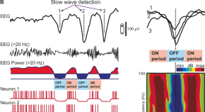
Sleep-like cortical dynamics during wakefulness and their network effects following brain injury
In this Perspective, the authors propose that brain injury can result in sleep-like slowing of cortical EEG waves during wakefulness. The generation of these dynamics and their effects on brain networks and behavior are discussed, as well as future directions for neuromodulation.
- Marcello Massimini
- Maurizio Corbetta
- Simone Sarasso
Analyzing brain-activation responses to auditory stimuli improves the diagnosis of a disorder of consciousness by non-linear dynamic analysis of the EEG
- Fanshuo Zeng
Reducing the percentage of surviving infants with acute symptomatic seizures discharged on anti-seizure medication
- Anne Marie Nangle
- Elizabeth K. Sewell
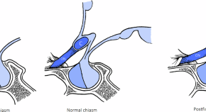
Neuro-ophthalmic evaluation and management of pituitary disease
- Michael T. M. Wang
- Juliette A. Meyer
- Helen V. Danesh-Meyer
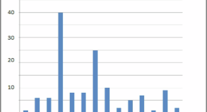
Normative longitudinal EEG recordings during sleep stage II in the first year of age
- Thalía Harmony
- Gloria Otero-Ojeda
- Lourdes Cubero-Rego
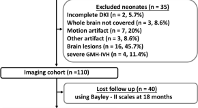
Associations between diffusion kurtosis imaging metrics and neurodevelopmental outcomes in neonates with low-grade germinal matrix and intraventricular hemorrhage
- Chunxiang Zhang
- Meiying Cheng
News and Comment
Non-invasive deep brain stimulation: interventional targeting of deep brain areas in neurological disorders.
A non-invasive technique using transcranial electrical stimulation offers an improvement in focality over other non-invasive techniques, presenting an opportunity to target deep brain structures for the treatment of neurological disorders.
- Friedhelm C. Hummel
- Maximilian J. Wessel
Ibogaine therapy in TBI
- Leonie Welberg
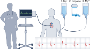
An ancient psychedelic for traumatic brain injury
In military veterans with traumatic brain injury, treatment with ibogaine plus magnesium led to dramatic clinical improvements and a favorable safety profile; further studies with state-of-the art safety monitoring will be crucial to unlocking the potential benefits of this psychedelic compound.
- David L. Brody
- Shan H. Siddiqi
Is erythropoietin beneficial and safe as an adjunctive therapy to therapeutic hypothermia in newborns with hypoxic ischemic injury?
- Abigail L. Melemed
- Jonathan L. Slaughter
- Sara Conroy
Clinical outcomes evolve years after traumatic brain injury
The TRACK-TBI LONG study has shown that outcomes are highly variable in the 7 years after traumatic brain injury (TBI). Although many patients remain stable, almost one-third experience declines in cognitive, psychiatric and functional state. These findings suggest that TBI is a chronic disease and that its management should change accordingly.
- David J. Sharp
- Neil S. N. Graham
Cytomegalovirus link to concussion changes
- Sarah Lemprière
Quick links
- Explore articles by subject
- Guide to authors
- Editorial policies
Most Popular Articles : The Journal of Head Trauma Rehabilitation
- Subscribe to journal Subscribe
- Get new issue alerts Get alerts
Secondary Logo
Journal logo.
Most Popular Articles
Incog 2.0 guidelines for cognitive rehabilitation following traumatic brain injury, part iv: cognitive-communication and social cognition disorders.
Journal of Head Trauma Rehabilitation. 38(1):65-82, January/February 2023.
- Abstract Abstract
- Permissions
Go to Full Text of this Article
INCOG 2.0 Guidelines for Cognitive Rehabilitation Following Traumatic Brain Injury: What's Changed From 2014 to Now?
Journal of Head Trauma Rehabilitation. 38(1):1-6, January/February 2023.
INCOG 2.0 Guidelines for Cognitive Rehabilitation Following Traumatic Brain Injury, Part I: Posttraumatic Amnesia
Journal of Head Trauma Rehabilitation. 38(1):24-37, January/February 2023.
INCOG 2.0 Guidelines for Cognitive Rehabilitation Following Traumatic Brain Injury: Methods, Overview, and Principles
Journal of Head Trauma Rehabilitation. 38(1):7-23, January/February 2023.
INCOG 2.0 Guidelines for Cognitive Rehabilitation Following Traumatic Brain Injury, Part II: Attention and Information Processing Speed
Journal of Head Trauma Rehabilitation. 38(1):38-51, January/February 2023.
INCOG 2.0 Guidelines for Cognitive Rehabilitation Following Traumatic Brain Injury, Part III: Executive Functions
Journal of Head Trauma Rehabilitation. 38(1):52-64, January/February 2023.
Silent Struggles: Traumatic Brain Injuries and Mental Health in Law Enforcement
Journal of Head Trauma Rehabilitation. : August 05, 2024
INCOG 2.0 Guidelines for Cognitive Rehabilitation Following Traumatic Brain Injury, Part V: Memory
Journal of Head Trauma Rehabilitation. 38(1):83-102, January/February 2023.
Fatigue After Traumatic Brain Injury: A Systematic Review
Journal of Head Trauma Rehabilitation. 37(4):E249-E257, July/August 2022.
Pharmacological Treatment of Agitation and/or Aggression in Patients With Traumatic Brain Injury: A Systematic Review of Reviews
Journal of Head Trauma Rehabilitation. 36(4):E262-E283, July/August 2021.
2024 NABIS Conference on Brain Injury Abstracts
Journal of Head Trauma Rehabilitation. 39(4):E247-E334, July/August 2024.
Long-term Participation and Functional Status in Children Who Experience Traumatic Brain Injury
Journal of Head Trauma Rehabilitation. 39(4):E162-E171, July/August 2024.
Understanding Traumatic Brain Injury in Females: A State-of-the-Art Summary and Future Directions
Journal of Head Trauma Rehabilitation. 36(1):E1-E17, January/February 2021.
Summary of the Centers for Disease Control and Prevention’s Self-reported Traumatic Brain Injury Survey Efforts
Journal of Head Trauma Rehabilitation. : July 22, 2024
Identifying and Predicting Subgroups of Veterans With Mild Traumatic Brain Injury Based on Distinct Configurations of Postconcussive Symptom Endorsement: A Latent Class Analysis
Journal of Head Trauma Rehabilitation. 39(4):247-257, July/August 2024.
Altered Oculomotor and Vestibulo-ocular Function in Children and Adolescents Postconcussion
Journal of Head Trauma Rehabilitation. 39(4):E237-E246, July/August 2024.

Neuromodulatory Interventions for Traumatic Brain Injury
Journal of Head Trauma Rehabilitation. 35(6):365-370, November/December 2020.
Normative Data for the Fear Avoidance Behavior After Traumatic Brain Injury Questionnaire in a Clinical Sample of Adults With Mild TBI
Journal of Head Trauma Rehabilitation. 36(5):E355-E362, September/October 2021.
Characterization and Treatment of Chronic Pain After Traumatic Brain Injury—Comparison of Characteristics Between Individuals With Current Pain, Past Pain, and No Pain: A NIDILRR and VA TBI Model Systems Collaborative Project
Journal of Head Trauma Rehabilitation. 39(1):5-17, January/February 2024.
INCOG 2.0 Guidelines for Cognitive Rehabilitation Following Traumatic Brain Injury, Part III: Executive Functions: Erratum
Journal of Head Trauma Rehabilitation. 39(2):159, March-April 2024.
Colleague's E-mail is Invalid
Your message has been successfully sent to your colleague.
Save my selection
Traumatic Brain Injury and Treatment of Behavioral Health Conditions
Information & authors, metrics & citations, view options.
An alteration in brain function, or other evidence of brain pathology, caused by an external force. External forces include the head being struck by an object; the head striking an object; the head accelerating or decelerating without direct external trauma (as occurs in shaken baby syndrome); a foreign body penetrating the brain; or energy generated from events such as a blast or explosion. ( 5 )
What Is TBI?
Tbi and behavioral health problems, why would tbi cause behavioral health problems, tbi in treatment of behavioral health conditions, workforce implications, conclusions, information, published in.
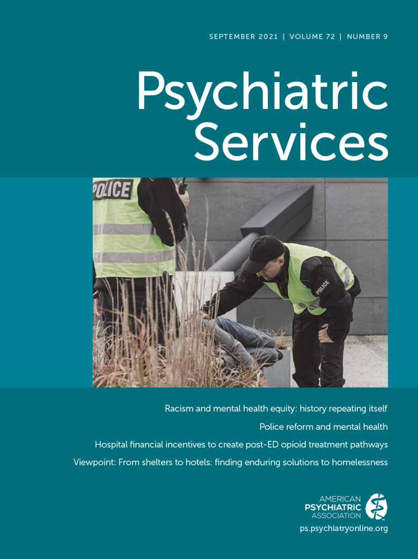
- traumatic brain injury
- neurobehavioral manifestations
- mental health services
- substance use disorders
Competing Interests
Export citations.
If you have the appropriate software installed, you can download article citation data to the citation manager of your choice. Simply select your manager software from the list below and click Download. For more information or tips please see 'Downloading to a citation manager' in the Help menu .
| Format | |
|---|---|
| Citation style | |
| Style | |
To download the citation to this article, select your reference manager software.
There are no citations for this item
View options
Login options.
Already a subscriber? Access your subscription through your login credentials or your institution for full access to this article.
Purchase Options
Purchase this article to access the full text.
PPV Articles - Psychiatric Services
Not a subscriber?
Subscribe Now / Learn More
PsychiatryOnline subscription options offer access to the DSM-5-TR ® library, books, journals, CME, and patient resources. This all-in-one virtual library provides psychiatrists and mental health professionals with key resources for diagnosis, treatment, research, and professional development.
Need more help? PsychiatryOnline Customer Service may be reached by emailing [email protected] or by calling 800-368-5777 (in the U.S.) or 703-907-7322 (outside the U.S.).
Share article link
Copying failed.
PREVIOUS ARTICLE
Next article, request username.
Can't sign in? Forgot your username? Enter your email address below and we will send you your username
If the address matches an existing account you will receive an email with instructions to retrieve your username
Create a new account
Change password, password changed successfully.
Your password has been changed
Reset password
Can't sign in? Forgot your password?
Enter your email address below and we will send you the reset instructions
If the address matches an existing account you will receive an email with instructions to reset your password.
Your Phone has been verified
As described within the American Psychiatric Association (APA)'s Privacy Policy and Terms of Use , this website utilizes cookies, including for the purpose of offering an optimal online experience and services tailored to your preferences. Please read the entire Privacy Policy and Terms of Use. By closing this message, browsing this website, continuing the navigation, or otherwise continuing to use the APA's websites, you confirm that you understand and accept the terms of the Privacy Policy and Terms of Use, including the utilization of cookies.
An official website of the United States government
The .gov means it’s official. Federal government websites often end in .gov or .mil. Before sharing sensitive information, make sure you’re on a federal government site.
The site is secure. The https:// ensures that you are connecting to the official website and that any information you provide is encrypted and transmitted securely.
- Publications
- Account settings
Preview improvements coming to the PMC website in October 2024. Learn More or Try it out now .
- Advanced Search
- Journal List
- Biomedicines

Understanding Acquired Brain Injury: A Review
Liam goldman.
1 Department of Neurosurgery, Medical College of Georgia, Augusta University, Augusta, GA 30912, USA
Ehraz Mehmood Siddiqui
2 Department of Pharmacology, Buddha Institute of Pharmacy, CL-1, Sector 7, Gorakhpur 273209, India
Andleeb Khan
3 Department of Pharmacology and Toxicology, College of Pharmacy, Jazan University, Jazan 45142, Saudi Arabia
Sadaf Jahan
4 Medical Laboratories Department, College of Applied Medical Sciences, Majmaah University, Majmaah 15341, Saudi Arabia
Muneeb U Rehman
5 Department of Clinical Pharmacy, College of Pharmacy, King Saud University, Riyadh 11451, Saudi Arabia
Sidharth Mehan
6 Neuropharmacology Division, Department of Pharmacology, ISF College of Pharmacy, Moga 142001, India
Rajat Sharma
7 Center for Undergraduate Research and Scholarship, Augusta University, Augusta, GA 30912, USA
Stepan Budkin
8 The Graduate School, Augusta University, Augusta, GA 30912, USA
Shashi Nandar Kumar
9 Environmental Toxicology Laboratory, ICMR-National Institute of Pathology, New Delhi 110029, India
Ankita Sahu
10 Tumor Biology Lab, ICMR-National Institute of Pathology, Safdarjung Hospital Campus, New Delhi 110029, India
Manish Kumar
Kumar vaibhav.
11 Department of Oral Biology and Diagnostic Sciences, Dental College of Georgia, Augusta University, Augusta, GA 30912, USA
Associated Data
Not applicable.
Any type of brain injury that transpires post-birth is referred to as Acquired Brain Injury (ABI). In general, ABI does not result from congenital disorders, degenerative diseases, or by brain trauma at birth. Although the human brain is protected from the external world by layers of tissues and bone, floating in nutrient-rich cerebrospinal fluid (CSF); it remains susceptible to harm and impairment. Brain damage resulting from ABI leads to changes in the normal neuronal tissue activity and/or structure in one or multiple areas of the brain, which can often affect normal brain functions. Impairment sustained from an ABI can last anywhere from days to a lifetime depending on the severity of the injury; however, many patients face trouble integrating themselves back into the community due to possible psychological and physiological outcomes. In this review, we discuss ABI pathologies, their types, and cellular mechanisms and summarize the therapeutic approaches for a better understanding of the subject and to create awareness among the public.
1. Introduction
Acquired Brain Injury (ABI) is an umbrella term encapsulating its two main categories: Traumatic Brain Injury (TBI) or Non-Traumatic Brain Injury (Non-TBI) [ 1 ]. TBI is an external traumatic event in which injury to the brain is sustained, while Non-TBI occurs due to an internal disease process that also leads to damaged brain tissue. Causes of TBI include motor vehicle accidents, falls, sports-related injury, and violence, whereas Non-TBI could be triggered by a stroke, neoplasm, infection, and anoxia [ 1 ]. Clinical outcomes of both categories of ABI vary, depending on the specific disease process and the premorbid circumstances such as age, genetics, and socioeconomic background. Risk rates for TBI are the greatest in the elderly at and above 75 years, and male individuals are at greater odds of getting TBI [ 2 , 3 , 4 , 5 ]. The vectors of brain damage in both TBI and Non-TBI include vascular abnormalities, broad axonal injury, focal or disseminated atrophy, and neuronal circuit disruption [ 6 ] ( Figure 1 ).

ABI: Etiopathology, classifications, the brain region affected, and related complications. The pictorial presentation of ABI describes its type (purple) and etiology of the disease (in orange texts). ABI is mainly divided into TBI and Non-TBI injuries. Non-TBI can arise from tumors, vessel occlusion, infection, or alcohol consumption. ABI can affect different regions of the brain depending on impact, insult, infection, or blockage (shown in pink) and may show related signs and symptoms (depicted in green). HR: Heart rate.
ABI describes a wide range of diseases, establishing it as a vastly important area in medicine and public health. According to the Centre for Disease Control and Prevention (CDC), TBI is one of the major groups of ABI and is a principal cause of mortality and lifelong disability [ 7 , 8 ]. As per CDC reports (2006–2014), the frequency of TBI-related hospitalizations, emergency department visits, and deaths had increased by 53 percent [ 9 ]. In 2013, roughly 2.8 million TBI cases occurred in the United States of America. Among the 2.5 million emergency department visits, there were approximately 300,000 TBI hospitalizations and about 60,000 deaths [ 2 , 10 ]. It is important to keep in mind that these numbers only refer to one-half of the diseases associated with ABI. Non-TBI also plays a large role in the number of individuals ending up in the hospital. The CDC reports that every year, approximately 800,000 people will have a stroke and, in 2018, one in every six cardiovascular-disease-related deaths was due to stroke [ 11 , 12 ]. These epidemiological data on ABIs elucidate the necessary interventions that hospitals and researchers need to accomplish to serve the large extent of individuals affected by ABI.
While both TBI and Non-TBI carry many different disease processes and medical problems ( Figure 2 ), the patients usually receive treatment and rehabilitation in the same facilities in the hospital. This is important to mention because demographic characteristics of TBI and Non-TBI vary considerably. For example, the Toronto Rehabilitation Institute demonstrated that the patient population for TBI compared to Non-TBI were significantly younger, tended to be male, and lived in metropolitan areas [ 13 ]. In addition, the global population is aging, so leaders in the medical profession need to anticipate larger demand for units and specially trained staff to treat patients with TBI and Non-Traumatic Brain Injuries with possible comorbidities [ 14 , 15 ]. Nevertheless, to ensure exceptional clinical outcomes for patients with ABI, physicians, and nurses must be able to provide personalized and specific treatments to the patients. To achieve that, a good understanding of ABI and its pathology in different categories is preferred. Therefore, the present review aims to give a comprehensive and clear description of ABI, its types, mechanism, and treatment strategy.

Poor outcomes post-ABI. A variety of parameters related to the ABI mechanism play a role in predicting the outcome. ABI can develop as a result of a stroke or disease or an iatrogenic cause, and there is some indication that those who suffer from a head injury are a self-selecting group, with poor attention, impulsivity, and overactivity being associated with poor road-crossing skills. These may interact with other premorbid characteristics that are predictors of poor post-injury prognosis.
2. Acquired Brain Injury and Its Types
As mentioned above, ABI is a broad classifying term encompassing any non-congenital brain injury; therefore, ABI is inherently diverse in the populations it affects, in the mechanisms by which brain injury ensues, and in prognosis. The following paragraphs break down the different types of TBI and Non-Traumatic Brain Injuries that make up ABIs ( Figure 1 ).
2.1. Traumatic Brain Injuries (TBIs)
TBI arises because of a hit or jolt to the brain and comprises mild to severe injury. TBI patients show symptoms such as unconsciousness, confusion, nausea, dizziness, headache, or incoordination and receive symptomatic and stabilizing treatment. Patients keep visiting clinics with chronic symptoms even after weeks or months post-initial traumatic experiences. If either symptoms continue or neurologic impairments appear, routine radiological re-imaging may be required to assess the situation [ 16 ]. There is a comprehensive review published by our group that might be of interest to the TBI audience to obtain more insight [ 17 ].
2.1.1. Concussion
A concussion is one of the most widely recognized forms of TBI. It occurs due to a sudden strike or whip to the head that causes the brain to bounce or twist within the skull. Symptoms can range from minor confusion and disorientation to complete amnesia, nausea, vomiting, and loss of consciousness [ 18 ]. These symptoms occur due to abnormal brain movement upon impact, which at the molecular level disrupts neuronal cell membranes and axonal stretching. This, in turn, causes the extensive flux of ions across neuronal membranes resulting in diffuse waves of depolarization, which precipitate the classic concussion symptoms [ 18 ]. In 2006, a study on concussion epidemiology in Canada noted that 110 individuals per 100,000 had had a concussion within the previous year [ 19 ]. In 2014, 2.87 million cases of TBI in the United States were recorded by the CDC, and of those, 812,000 cases were children diagnosed with concussion alone or in combination with other injuries [ 2 ]. Similarly, according to a US study carried out in 2017, approximately 19.5% of adolescents (in grades 8–12) reported a minimum of one concussion, while 5.5% had more than one concussion in their lives [ 20 ].
2.1.2. Skull Fractures
A skull fracture results from any impact to the head that surpasses the bone’s capability to withstand the pressure. Although a fracture of the skull itself is not a brain injury, over 75% of all skull fractures are associated with some form of brain injuries, such as intracranial hemorrhages or subdural or epidural hematomas [ 21 ]. Fractures that occur at the basilar skull are more problematic as this area of the skull harbors essential areas of the brain that allow us to eat, breathe, and walk [ 22 ].
2.1.3. Epidural or Subdural Hematomas and Subarachnoid Hemorrhage
In general, a hematoma is a bruise, and a hemorrhage is a bleeding blood vessel. In the case of TBI, it is called an epidural or subdural hematoma if the insult occurs above or below the dura matter. Hematomas mostly occur from blunt force trauma to the head and are typically found in the temporal brain; however, they may also occur from a penetrating TBI or spontaneously (the spontaneous type would not be considered TBI) [ 23 ]. A subarachnoid hemorrhage is divided into two groups: aneurysmal vs. non-aneurysmal. The non-aneurysmal hemorrhage most often occurs due to blunt force trauma to the brain or sudden acceleration changes. Epidural hematomas account for roughly 2% of injuries to the head and 5–15% of fatal head injuries. Subdural hematomas are more common, with an estimated rate of 5–25% in patients with head trauma [ 21 ].
2.1.4. Penetrating Brain Injury
Penetrating brain injuries can be caused by assaults, collisions, or even suicide attempts [ 24 ] and may be defined from mild to severe TBI based on the Glasgow coma scale (GCS). Following a patient’s preliminary examination, neurosurgical examination starts with a clinical exam and documentation of raised intracranial pressure (ICP). In the case of suspected arterial or venous injury, a CT scan is the first choice for diagnosis. As the possibility of post-TBI epilepsy is 45–53%, a prophylactic anticonvulsant is given to patients [ 25 ].
2.2. Non-Traumatic Brain Injuries (Non-TBI)
2.2.1. infections.
Due to the multiple layers of protection from the skull, meninges, and the blood-brain barrier (BBB), the brain is relatively repellent to any pathogenic invaders [ 26 , 27 ]. However, when bacteria and other pathogens breach the brain’s defenses, the damage can be devastating. Two major types of brain infections are meningitis and encephalitis. Meningitis occurs when a bacterial agent infects the meninges and encephalitis is the infection of the brain tissue itself [ 28 , 29 ]. Approximately 1.2 million cases of meningitis occur globally each year [ 30 ], while the incidence of encephalitis infection tends to vary between studies, but the 2019 census estimated 1.4 million cases with 89,900 deaths and 4.80 million DALYs [ 31 ].
2.2.2. Anoxia
The brain needs a lot of oxygen and energy in the form of glucose. Anoxic brain injury results when the brain is completely denied of oxygen in incidences such as drowning, heart attack, carbon monoxide poisoning, and much more. As a result, the metabolic homeostasis of the brain is destroyed resulting in major neuronal injury and cell death. Since there are many different causes of anoxic brain injury, rates are hard to gauge [ 32 ].
2.2.3. Stroke
There are two main types of stroke: ischemic and hemorrhagic. Ischemic strokes result from occluded cerebral arteries, which prevent nutrient-rich blood from supplying the surrounding brain tissue. This results in permanent tissue damage. Transient ischemic attack (TIA), also referred to as a mini ischemic stroke, only lasts for a short amount of time. Ischemia that affects more than two-thirds of the middle cerebral artery (MCA) territory is termed malignant cerebral infarction (MCI) and causes space-occupying edema and neurological deterioration [ 33 ]. Swelling and symptomatology peak in the first 48 h after a stroke. The first step in treatment is to reduce risk factors and keep ICP under control. Although there are no precise surgical recommendations, a hemicraniectomy is generally recommended [ 34 ]. Hemorrhagic strokes result from cerebral artery leakage into the brain, causing elevated ICP and cellular damage [ 35 ]. The incidence of stroke among adults aged between 35 to 44 years is roughly 30 to 120 per 100,000 per year. This number increases drastically for individuals aged between 65 to 74 years, where the yearly incidence is about 670–970 per 100,000 [ 36 ]. A detailed account of brain hemorrhage for further reading can be found here [ 37 ].
2.2.4. Alcohol and Drug Use
The usage and abuse of alcohol and drugs are highly prevalent in modern-day lifestyles estimating a high lifetime risk for either drug or alcohol abuse and dependence [ 38 ]. There are many mechanisms through which drugs and alcohol can have negative effects on the normal functioning of the brain. These include disturbing nutrient distribution to brain tissue, direct cellular damage, altered chemical homeostasis of the brain, and hypoxia [ 38 , 39 ].
2.2.5. Neoplasm
In a similar way to anoxia and infectious Non-TBI, brain cancers (neoplasm) are vastly diverse in pathophysiology and epidemiology. Gliomas are the most prevalent class of brain neoplasm, accounting for roughly 78% to 80% of all malignant brain tumors. These cancers stem from the supporting neuronal cells of the brain called glia. Gliomas include astrocytomas, ependymomas, glioblastoma multiforme, medulloblastomas, and oligodendrogliomas [ 40 ].
Meningiomas are the most prevalent primary tumors and are also classified as ABIs [ 41 ]. Patients with genetic predispositions to disorders, such as neurofibromatosis type 2 or multiple endocrine neoplasia type 1, are more likely to develop meningioma [ 42 ]. The preponderance is asymptomatic and histologically benign [ 43 ]. Initially, generalized symptoms (nausea, headache, or altered mental status) may be present, with localized neurological abnormalities developing later [ 44 ]. In situations with subtotal meningioma extraction, adjuvant therapy in combination with postoperative radiation may be recommended. Patients with meningioma have a good prognosis, albeit those with a higher WHO grade or partial resection have a higher chance of continuation [ 45 ].
3. Mechanism of ABI
A sort of physical trauma from an external entity may lead to a brain injury. The medical field has acquired tremendous success in the treatment of head injuries over the last few decades. A clearer understanding exists of the causes of tissue damage and the biophysical, biochemical, or physiological repercussions that culminate in a variety of clinical manifestations such as scalp laceration, syncope, and progression to a persistent vegetative state [ 46 , 47 , 48 , 49 , 50 , 51 ]. Various sorts of pathologies, such as skull fracture, hematoma (intracerebral, epidural, subdural, or intraventricular), as well as different types of contusion and brain injuries, could be recognized and their clinical and functional repercussions could be defined by contemplating the mechanisms of injury to the head [ 50 ].
3.1. Biophysical Mechanism of ABI
The physical characteristics of the intruding substance, such as density, size, speed, and length of loading, determine how much energy is delivered to the cranium in ABIs [ 52 ]. When a harmful energy burden or mechanical response is exerted to the head, the length of the energy loading will be the first determinant to determine the severity of the injury [ 53 ]. This period has been set between 50 and 200 milliseconds. Static loads are defined as those lasting more than 200 milliseconds, while dynamic loads are defined as those lasting less than 200 milliseconds [ 54 , 55 ]. Static injuries are extremely rare and mainly occur when the head is ensnared between two hard objects. These enormous weights may cause distortion and injury to the skin and bones. The dynamic load could be caused by the passage of energy to cerebral tissue via impetuous loads, which are variable in speed. When the head does not receive direct impact but is put into motion as a result of an impulse generated by a force exerted on different parts of the body, this is known as impulsive loading [ 56 ]. In such cases, the injury is caused by inertial shifts in the head. In the next category, called impact loads, when an infringing object strikes the head, it may cause tissue injury in-depth, depending on the surface area, density, size, and speed of the object. It has the potential to alter the head’s pace and induce acceleration or deceleration and may cause inertial shifts in the head too.
The influence of an object on the head can cause changes in the tissue’s arrangement, including the skin, bone, and deep frameworks. If the adjustment is greater than the tissue’s elasticity, it will lead to permanent disfigurement, skin laceration, or a bone fracture. With higher weights, the aggravating agent may produce depressed skull fractures and destruction of underlying tissue, such as the dura, brain, and arteries. This often results in an epidural hematoma, subdural hematoma, contusion, or intracerebral hemorrhage. Perforation and permeation may occur in more extreme situations, specifically at fast speeds, and with small agent sizes. The transmission of energy to the skull and cerebral tissue may be related to the collision. The brain tissues distort, and contusion could be because of this energy burden [ 57 , 58 ].
3.2. Injury to the Tissues
Tissue distortion is caused by deformation, shock waves, and acceleration/deceleration, which all impose energy on the tissue. These can cause damage to the tissues in the skull, which include neural components, vessels, and bone. Injuries arise when the stress applied to the tissue exceeds the threshold. The physical properties of tissues, the amount of energy, the length of energy loading, and the magnitude of the load all influence their endurance. More intense activities, while still within the continuum, are at the brink of physiologic endurance and, if repeated, will cause progressive or even acute brain malfunction. In normal physiological conditions, the tissues’ physical tolerance to injury is substantially lower, resulting in a variety of outcomes depending on the implicated components of pathological injury [ 59 ].
3.2.1. Primary and Secondary Injuries
The mechanism that causes the initial injury is a direct outcome of the delivered energy to the head [ 17 , 60 ]. They may cause additional injuries as a result of themselves, either as sequelae of the original event or by exacerbating it, resulting in secondary injuries, the most prevalent of which are hypotension and hypoxia. Secondary injury can include excitotoxicity, free radical formation, mitochondrial dysfunction, induction of damaging intracellular enzymes, and other pathways inside the wounded neural tissues, all of which can cause continued system dysfunction ( Figure 3 ) [ 17 , 61 , 62 , 63 ]. Certain tertiary damages are also included, which are frequently secondary results of the head’s energy loading. This includes electrolyte imbalance due to renal issues, various types of cardiac abnormalities, hepatic insufficiency, and so on.

Schematic representation of pathophysiology of ABI. BBB dysfunction caused by injury allows the transmigration of activated leukocytes into the injured brain parenchyma, which is facilitated by the upregulation of cell adhesion molecules. Activated leukocytes, microglia, and astrocytes produce ROS and inflammatory molecules such as cytokines and chemokines that contribute to demyelination and disruption of the axonal cytoskeleton, leading to axonal swelling and accumulation of transport proteins at the terminals. On the other hand, excessive accumulation of glutamate and aspartate neurotransmitters in the synaptic space due to spillage from severed neurons activates NMDA and AMDA receptors located on post-synaptic membranes, which allow the production of ROS. As a result of mitochondrial dysfunction, molecules such as apoptosis-inducing factor (AIF) and cytochrome c are released into the cytosol. These cellular and molecular events including the interaction of Fas with its ligand Fas ligand (FasL) ultimately lead to caspase-dependent and -independent neuronal cell death. BBB: blood-brain barrier; NMDA: N-methyl-D-aspartate receptor; AMPA: α-amino-3-hydroxy-5-methyl-4-isoxazole propionic acid receptor; ROS: Reactive oxygen species; Cyt c: Cytochrome c; ICP: Intracranial pressure; AIF: Apoptosis-inducing factor.
Various types of clinical cases can be distinguished based on the above-mentioned aspects in the formation of a head injury. It can begin with a bone injury, since prolonged static loading causes a change in the normal structure of the skull, ultimately leading to a fracture when the flexibility of the bone toleration is exceeded. The amount of force and the timing of the fracture determine the severity of the crack. When a large load is applied, the entire skull is severed into segments, and the brain tissue is ruptured, causing it to leak from the punctured nose, ear canals, and scalp. The sufferer may present with severe impairment of cerebral and brain stem function. Death is often the result [ 64 , 65 , 66 ].
If the injury occurs in an acoustic region, neurological deficits may occur as a result of damaged brain function. These could be the injury’s direct or major consequences. There are certain further occurrences of the above-mentioned events, which are referred to as secondary traumatic effects. Various types of intracranial hematomas, as well as intraventricular hematomas and brain tissue contusions, can occur from injury to the cerebrovasculature in the affected areas. These particulate lesions can cause a mass effect, as well as an upsurge in ICP and brain herniation [ 17 , 65 , 67 , 68 ]. As a primary injury, brain laceration may predispose the sufferer to convulsions and epilepsy [ 69 , 70 ].
Infections of the bone and cerebral contents are another serious result of this type of injury, as the overlaying skin is punctured, allowing bacteria to enter the deeper structures. These later occurrences are also secondary impacts. Lesions can occur as a result of the expansion of a hematoma or contusion, or as a result of the mass effect caused by various subsequent effects of damage, such as edema around the lesions. Cerebral herniation, concussions, diffuse axonal damage, subdural hemorrhage, intracerebral hemorrhage, and intraventricular hemorrhages are some of the other problems [ 71 , 72 , 73 ]. Severe injuries can range from a short period of disorientation and cognitive impairment to concussion or loss of consciousness to a long-term coma or persistent vegetative state (PVS) due to extensive harm to brain neurons and axons [ 17 , 74 ].
3.2.2. Prenatal and Birth Damage
Early prenatal injury can result in the embryo’s mortality. On other hand, insufficient growth (agenesis of the corpus callosum or anencephaly) or abnormal development (lissencephaly or microcephaly) could be the possible outcomes of late injuries [ 75 , 76 , 77 , 78 ]. During a period of heightened development, trauma (such as fetal stroke in the womb or damage to the mother) will result in more structural issues than when progression has slowed or has ended. Intricate stent delivery or hypoxia may result in birth failure [ 79 , 80 , 81 , 82 ].
3.2.3. Post-natal Injury
Pediatric patients may have acquired ABI from metabolic disturbances (phenylketonuria), systemic illness (sickle cell disease, diabetes), trauma, central nervous system tumors, infections (meningitis or encephalitis), toxins (use of alcohol or anticonvulsants during pregnancy), and clinical treatment such as radiotherapy or chemotherapy for leukemia [ 83 , 84 , 85 , 86 , 87 , 88 , 89 ].
3.2.4. Injuries in Adulthood
According to recent CDC data, people aged 75 years or older had the highest rate of hospitalizations (325 of total TBI-related hospitalizations) and mortality (28% of TBI-related deaths) [ 90 , 91 ]. The CDC further stated that males are two times more likely to get TBI-related hospitalization and have three times higher mortality than females [ 90 , 91 ]. Alcohol intake is identified as a major risk factor for TBIs, with effects on assessment, intensity, and prognosis. It was discovered that 16 percent of brain injury patients aged 15 and above were intoxicated at the time of injury. The alcohol group had a fatality rate of 14.5 percent compared to 9 percent in the non-alcoholic group [ 92 ]. Individuals with an older ABI have a higher chance of poorer physical, intellectual, and psychosocial outcomes, as well as a lengthier recovery period and more comorbidities. The major cause of brain injuries that comes under this category is fall. More than half of all fatal falls and 8% of nonfatal fall-related hospitalizations were caused by these brain injuries. ABIs have the highest incidence of death and hospital admittance among fall-related injuries in adults and older adults during the first year after the injury. Furthermore, even after controlling for age and gender, there are rising tendencies in the incidence and mortality of trauma-induced ABIs in older persons. According to several studies, those who take anti-arrhythmic medications are more prone to suffer from brain damage. Several studies show that men had a higher chance of serious brain injuries during a fall than women, despite the possibility of a reverse relationship with nonfatal brain injuries [ 92 , 93 ].
The number of elderly persons hospitalized for a fracture has decreased over the last decade, whereas the percentage of those with a TBI, subarachnoid hemorrhage, and/or, subdural, in particular, has risen dramatically [ 94 ]. TBIs are becoming more common, which appears to be linked to the increased use of anticoagulants and antiplatelet medicines like clopidogrel and warfarin. Chronic illnesses related to equilibrium disturbance (Stroke and Parkinson’s disease), scenarios of falls likely to result in an ABI, and risky behaviors may happen more frequently in men, in contrast to the use of anticoagulants and antiplatelet drugs. There is a need for more investigation into the underlying principles [ 95 , 96 ]. It is reasonable to assume that elderly adults with chronic diseases that affect the joints, nervous system, cardiac system, and cognition are at a higher risk of falling and developing ABIs. These may also be exacerbated by a lack of visual perception and visuo-motor reflexes [ 97 , 98 ].
3.3. Physiological Mechanisms of ABI
There are different mechanisms that arise from primary and secondary injury and contribute to ABI pathology ( Figure 3 ). We briefly described the pathological events here to understand the pathology of ABI.
3.3.1. Excitotoxicity
Glutamate, an excitatory amino acid neurotransmitter is primarily responsible for triggering cellular damage during brain ischemia. It has a multifaceted role in synaptic plasticity, brain development and maturation, axon guidance, and general neuronal growth [ 17 , 99 ]. In ABI, restricted blood flow to the brain diminishes energy reserves and causes membrane depolarization, thus leading to the reduced uptake of glutamate from the surroundings. Under stable conditions, glutamine activates multiple receptors such as N-methyl-D-aspartic acid (NMDA), kainic acid receptors, and alpha-amino-3-hydroxy-5-methylisoxazole-4-propionate (AMPA), while its clearance is managed by active ATP-dependent transporters [ 100 , 101 ]. During ABI, glutamine triggers the activation of sodium channels (causes brain swelling), calcium channels (causes neuronal death), and intracellular catabolic enzyme activity via glutamate receptors thus leading to cell death, which further cascades into the generation of oxygen free radicals, membrane depolarization, and intracellular toxicity leading to brain injuries [ 102 , 103 ]. Preclinical studies suggest a protective effect of suppressing NMDA and AMPA receptors post-ABI but have undesirable side effects [ 104 , 105 ]. To overcome this, Memantine (partial NMDA antagonist) was tested, along with death-associated protein kinase and calcium-calmodulin-dependent protein kinase, and showed potential therapeutic efficacy without many side effects [ 100 , 106 ]. Another dopaminergic agonist, Amantadine was found to be promising in several brain injuries. It triggers the dopamine release in neurons and delays the reuptake of dopamine by neural cells and also inhibits the NMDA receptor signaling, thus proving its potential effect in the brain injuries [ 107 , 108 , 109 , 110 ]. A non-psychotropic cannabinoid (Dexanabinol), which acted as a potent NMDA receptor antagonist was also reported to have a potential effect in glutamate injury, but also showed unwanted side effects that impaired normal brain functioning [ 37 , 111 ]. Additionally, metabotropic glutamate receptors (mGluRs) were also reported to express a promising response in retarding excitability thus hindering excitotoxicity [ 112 ].
3.3.2. Oxidative Stress
A possible precursor to the pathogenesis of cerebral injury has been identified as oxidative stress. Several reactive oxygen species (ROS), such as superoxide, hydrogen peroxide, hydroxyl radicals, and per hydroxyls can be generated, followed by the development of several reactive nitrogen species (RNS), which can cause brain tissue damage through a variety of cellular and molecular pathways [ 17 , 37 , 102 ]. The reaction of nitric oxide along with superoxide forms peroxynitrite, which can also bind to DNA directly, altering its structural integrity and causing cell damage as well as apoptosis [ 17 , 113 ]. These highly reactive radicals can degrade nucleic acids, proteins, and lipids, leading to neuronal cell death. Edaravone and NXY-059, two promising antioxidants, were used to treat stroke but did not produce significant effects [ 102 , 114 , 115 ]. Polyethylene glycol (PEG)-conjugated SOD (PEG-SOD or pegorgotein) was reported to have promising effects in several studies but failed in a larger phase III clinical trial [ 17 ]. Another study with lecithinized superoxide dismutase (PC-SOD) showed that it inhibited secondary neuronal loss after brain injury and enhanced survival rates [ 116 ]. As a result, novel therapeutic strategies for minimizing the devastation caused by ROS are desired. Incipient interventions, such as modulation of transient receptor potential melastatin-2 channels or poly (ADP ribose) polymerase-1 control endogenous facilitators of oxidative stress [ 117 , 118 ]. More investigation into the neurological effects of oxidative stress could lead to new targeted therapies for the reduction of several brain injuries, especially ABIs [ 118 ].
3.3.3. Acidosis
When mitochondrial respiration is disrupted, acidosis may arise as a result of lactate buildup in the cells. Acid-sensing ion channels (AICs) are activated by protons and serve as pH sensors in the body. They are amiloride-sensitive cation ports that relate to the epithelial sodium group and enable calcium and sodium to enter neurons [ 37 , 119 ]. About six AIC domains have been reported, with AIC1a, AIC2a, and AIC2b being expressed in the brain and spinal cord. AIC1a and AIC2s are present in high-synaptic-density areas of the brain to help with excitatory signaling and are involved in several brain injuries [ 119 ]. With their activation, neuronal cell death occurs through sodium, zinc, and calcium influx into the cell. In experimental stroke models, the inhibition of AIC1a has a longer therapeutic window, which was much more effective than currently available drug therapies [ 100 ].
3.3.4. Inflammation
Inflammation may sometimes lead to brain injuries of several types including ABI [ 120 ]. Conversely, the pathogenesis of ABIs is further complicated by inflammation [ 121 , 122 ]. During brain injuries, there is an intense and long-lasting inflammatory response that includes the activation of microglia, development of pro-inflammatory mediators, and penetration of different kinds of immune cells into the brain tissue [ 17 , 123 ]. Cytokines such as interleukin IL-6, IL-1β, tumor necrosis factor-alpha (TNFα), transforming growth factor beta (TGFβ), and chemokines such as monocyte chemoattractant protein-1 (MCP-1) and cytokine-induced neutrophil chemoattractant play an important role in the pathogenesis of inflammation in neuronal cells. Depending on the type of inflammatory response and when it happens, the immune response in the brain might have a variety of outcomes [ 17 , 102 ]. While chronic inflammatory activities may contribute to secondary ABIs and more prolonged detrimental events, inflammation early point may be beneficial. However, elucidating the exact mechanisms of inflammatory responses is challenging, as it is a diverse set of perceptions involving inflammatory cellular components, all of which may be harmful or beneficial [ 124 ]. Broad-spectrum blockers of inflammation (AT1 receptor blockers, PPAR gamma blockers, beta-blockers, etc.), not shockingly, minimize neuronal cell damage [ 125 , 126 , 127 ]. The lack of systematic implementation progress highlights the need for a deeper knowledge of the numerous molecular and cellular pathways after inflammation. In addition, a better understanding of the different structural profiles of diverse inflammatory mediators is needed.
3.3.5. Tauopathies
Abnormal aggregation of tau proteins inside brain cells leads to several disorders including ABI [ 128 , 129 , 130 ]. The concentrations of a particular tau protein in brain tissue, CSF, and serum change in ABI pathogenesis ( Figure 4 ) [ 131 , 132 ]. The events that lead to tau release can be numerous and complicated, as can the types of modified tau species. Tau’s basic role is to promote microtubule flexion and saturation, which is dependent on its post-translational modifications [ 133 , 134 , 135 ]. When Tau attaches to microtubules with a poor or no phosphorylation state; microtubule flexion is hindered; phosphorylated tau has a low potential for microtubules ( Figure 1 ) [ 136 , 137 ]. Alternative splicing, which results in different-sized tau isoforms, might be another significant tau modulation [ 138 , 139 , 140 ]. Tau’s capacity to disperse amongst cells is also steered by its accumulation feature [ 141 , 142 ]. Oligomeric tau species disperse between cells, whereas integrated insoluble tau does not [ 143 , 144 ]. Tau spreads because of illness, and this property may express pathophysiological conditions triggered by ABI [ 145 , 146 ].

Expected molecular mechanism of brain injury on tau in the nervous system. Neurons, glia, oligodendrocytes, and blood vessels are damaged by the impact load that arises after a head injury. Injury to some or more of these cells causes intracellular unfolding, which causes the entire device to malfunction. Tau, which is highly correlated with microtubules, is abundant in axons. Impact forces devastate cell membrane integrity, as well as the microtubule framework in the axon. Tau disengages from the microtubule as it becomes unstable. Tau would then be misfolded, phosphorylated, develop a porous oligomeric conformation, accumulate, or disperse in a dysfunctional pathway. Tau may also invade other neighboring cells (glia, serum, or CSF) as it spreads.
Due to the extreme sudden TBI-induced protein abundance, protein catabolic pathways such as autophagy and proteasomal degradation may become exhausted [ 147 , 148 , 149 ]. When the plasma membrane of a compromised cell is disrupted, leftover cytoplasmic proteins such as tau which leave the cell can be absorbed by neighboring cells, confirming trophic rearrangement [ 149 , 150 ]. Tau can cross into the cerebrovasculature, and CSF, relying on where the weakened, tau-releasing cell is located, which further tends to contribute to brain injuries [ 151 , 152 , 153 ].
4. Injury and Outcome
Issues may occur as a result of an ABI in a variety of ways, either directly from the injured brain or implicitly from the response of an individual to the injury. Due to changing perceptions, family factors (pre-injury family functioning and managing), psychological background, as well as social exclusion lead to an abrupt cessation of current issues [ 154 ] ( Figure 2 ).
The foreseen consequence is aided by a variety of factors related to the mechanism of insult. Given a severe brain injury, the duration and extent of coma with a duration of post-traumatic amnesia for less than 20–30 min and with a level of the coma of 12 or less on the Glasgow Coma Scale are common characteristics of an ABI [ 155 ]. The magnitude of the damage and the functional loss normally has a dose-response correlation. Many adults and infants with traumatic complications do better than expected, whereas those with relatively minor injury issues can encounter problems. There might be conflicts between parents and professionals. Even if the injuries are minor, it is crucial not to ignore the complaints [ 96 ]. Brain injuries can attribute to low concentration, impulsivity, and overactivity which can associate with other comorbid parameters that indicate poor post-injury performance. Learning difficulties that exist pre- and post-trauma can put many individuals of discrete ages at a heightened risk, exacerbating difficulties. The most critical thing to evaluate is the observed change in behavior or educational progression [ 156 ].
4.1. Physical Outcomes
Aside from apparent gross motor problems, disabilities in the brain may have a significant effect on intellectual and behavioral performance. Sensory loss, weakness, tremors, seizures, excessive sweating, intermittent vision issues, ground abnormalities, and hearing problems are also possible side effects. All of these factors affect well-being, social interactions, and self-esteem [ 157 ]. Further longitudinal studies investigating these features of ABI are needed to uncover underlying mechanisms.
4.2. Cognitive Outcomes
Mental manifestations are among the most obscure but recurrent issues which can lead to a variety of information-processing skills—thought, speed, as well as the ability to react to tasks—being slowed down. High levels of impulsivity and impaired judgment are normal, and they have important long-term consequences [ 158 ]. Verbal communication issues may not be apparent, but there may be impaired language abilities in word searching, interpretation, and comprehension [ 159 ]. Reading, painting, structural skills, and job performance can be challenging for patients with ABIs, as well as activities and knowing physical differences. Cognition and listening abilities are often harmed. The ability to prepare, specify objectives, coordinate, and implement a plan to achieve an intended goal are examples of executive skills. This also includes expertise in efficiency surveillance and planning. Any of these issues can affect individuals or some combination of them. The severity of these functional disabilities is determined not only by the extent of the injury but also by the age at the time of concussion [ 160 , 161 ].
4.3. Educational Outcomes
Given the neuropsychological impact, the majority of rehabilitation emerges in the starting years, but developmental problems continue and can worsen. After the injury, issues about a lack of improvement in learning, speaking, and reading, as well as an inability to understand intelligent concepts and conceptualization [ 162 ]. More research is in demand to explore the underlying mechanisms.
4.4. Emotional and Behavioral Outcomes
Unidentified cognitive episodes and a lowered self-image linked to an understanding of logical and technical difficulties can suggest behavioral and emotional issues [ 163 ]. Making rude remarks about others may be connected with impulsivity and this behavior can be incredibly embarrassing and disconcerting for other people in society [ 164 ]. Elevated anxiety, rage, utterances of violence, fatigue, and inertia are the common and normal parts of the recovery stage and can last a significant amount of time. This is marked by a lack of enthusiasm and interest in everything, as well as difficulties maintaining focus and working at a fast pace, as well as passively carrying out recommendations rather than initiating activity. Both the extent of the incident and pre-existing symptoms can be linked to oppositional defiant disorder [ 165 , 166 ]. This is frequently linked to the realization of a lack of skill in certain things and being able to cope with everyday life less well [ 161 ]. The assumption by patients that they will be able to catch up with things soon can cause a lot of anxiety. Patients may be conscious of actual or potential losses, regardless of the circumstances of ABIs. Serious personal trauma can be humiliating and have a significant effect on one’s self-esteem. Fear of failure might be really serious, and it could be the origin of depression and anxiety [ 161 ]. Post-traumatic stress disorder can occur even though there is no continuous recollection of head trauma [ 167 ].
5. Pre-Existing Medications
Nimodipine, triamcinolone, polyethylene glycol-conjugated superoxide dismutase, and mild hypothermia have all shown positive results in phase II clinical trials [ 162 ]. Excitatory amino acid inhibitors, calcium channel blockers, NMDA receptor antagonists, corticosteroids, free radical scavengers, magnesium sulfate, and growth factors have all been used in preclinical research to evaluate the therapeutic effects of drugs in various animal models [ 168 , 169 ]. Regretfully, none of the formulations or methods that have been examined in phase III trials have shown to be successful [ 167 , 170 ]. Mannitol has been shown to help reduce brain swelling after a brain injury [ 171 ]. However, its efficacy in the long-term treatment of serious TBI is unknown. Inordinate mannitol injection has been shown to be dangerous, as mannitol passes through the circulation and the brain, increasing pressure inside the skull and worsening internal brain injuries [ 171 ]. A new meta-analysis backs up earlier reports that hypothermic treatment is a good cure for brain injuries in some situations. Health professionals should continue to use vigilance when assessing hypothermia for TBI care before more data from well-conducted trials become clear [ 172 ]. After an extreme brain injury, elevated ICP is still the leading cause of disability and death. When estimated within any intracranial space, an accelerated ICP is usually characterized as 15–20 mmHg [ 173 ]. Raised ICP has been linked to increased mortality and morbidity after extreme brain injuries. A rise in brain size at the cost of one or more intracranial resources is the cause of high ICP [ 174 ]. In ABI, increased ICP is caused by mass lesions, edema, and increased cerebral blood flow. Fortunately, there is no proof to substantiate the regular use of decompressive craniotomy in any brain injuries in adults with high ICP to increase survival and the standard of living [ 175 ]. A decompressive craniotomy can be a valuable choice when optimum medical care has failed to stop ICP. One randomized trial of decompressive craniotomy (DECRA) with extreme brain injury is currently underway, which could provide more information on the procedure’s effectiveness in adults [ 175 ].
6. Plausible Drug Therapies
The S100B protein is a member of a phenotypic family of low molecular weight calcium-binding S100 proteins that are primarily developed by glial cells. It also functions as a neurotrophic agent and a neuronal protection protein [ 176 ]. Excess supply of S100B by triggered glia, on the other hand, may exacerbate neuroinflammation and cause neuronal disruption. The brain and the serum S100B levels are scarcely associated, with serum concentrations largely determined by the blood-brain barrier’s consistency contrary to the amount of S100B in the brain [ 177 ]. Cerebrospinal S100B can be valuable as an indicator of consequence in adults with serious brain injury. Long-term functional restoration after ABI was shown to be aided by intraventricular S100B implementation [ 178 , 179 ]. Five weeks after brain injury, it significantly boosted hippocampal neurogenesis. The Morris water maze used to test spatial learning capacity on days 30–34 after injury, showed that an S100B injection improved cognitive efficiency [ 180 , 181 ]. S100B has not been used for the clinical care of any brain injury. S100B was used in a clinical trial called S100B as a Pre-Head CT Scan Screening Test After Mild TBI ( {"type":"clinical-trial","attrs":{"text":"NCT00717301","term_id":"NCT00717301"}} NCT00717301 ) to see whether a serum can anticipate traumatic anomalies on a brain CT scan after a mild TBI. A change in serum S100B suggested whether the patient’s neurological condition had improved or deteriorated [ 182 ]. Finally, surgical therapy resulted in lower levels of S100B. Serum S100B protein represented the seriousness of the injury and aided in the prediction of outcomes after a serious brain injury [ 183 ]. S100B was also useful in determining the effectiveness of treatment following a serious TBI [ 184 ].
6.2. Statins
Statins, which are powerful inhibitors of cholesterol synthesis, can also help people with brain injuries [ 185 ]. Many of its effects, such as increased NO bioavailability, immunomodulatory activities, improved endothelial function, antioxidant properties, upregulation of endothelial nitric oxide synthase, suppression of inflammatory responses, and platelet actin reduction are cholesterol-independent [ 186 ]. Simvastatin treatment significantly increased Akt, cAMP response element-binding proteins (CREB) phosphorylation, and GSK-3; amplified the production of BDNF and VEGF in the dentate gyrus (DG); enhanced tissue regeneration in the DG; and improved cognitive and memory restoration [ 187 ]. In rats with traumatic brain injury, atorvastatin injection decreased cognitive brain abnormalities, enhanced neuronal survival and synaptogenesis in the glioma parameter range and the CA3 areas of the hippocampus, and promoted angiogenesis in these areas [ 188 ]. Pre-treatment of rats with lovastatin enhanced mental outcomes and decreased the severity of brain injury, with a concurrent decline in serum concentrations of TNF-α and IL-1β mRNA and protein [ 189 ]. In addition, statin therapy increased cerebral hemodynamics in mice after a severe brain injury [ 190 ]. Statins helped animals regain their spatial memory quickly after a brain injury. A double-blind controlled clinical trial was conducted on 21 patients with TBI (aged 16 to 50 years) who had Glasgow Coma Scale scores of 9 to 13 and intracranial deposits as evidenced by a computed tomography (CT) scan [ 191 ]. Despite the overwhelming usefulness of statins, their desirable healthcare quality profile, and comprehensive preclinical research showing both neurorestoration and neuroprotection, further clinical trials are needed to assess statins’ neuroprotective and neurorestorative properties after any type of brain injury [ 192 ].
6.3. Role of Phytochemicals in Brain Injury
Plants develop metabolic systems that generate hazardous and/or antinociceptive bioactive molecules as a result of their static existence and exposure to herbivores and other pathogens [ 193 , 194 ]. Among phytochemicals, sulforaphane (isothiocyanato-4-(methylsulfinyl)-butane) has been demonstrated to have neuroprotective effects in several experimental paradigms. Sulforaphane has shown to have a protective effect on the neurological disorder and reduces Aβ1-42-induced inflammation via nuclear factor erythroid-2-related factor 2 (Nrf2) signaling [ 195 , 196 ]. The putrid phytonutrients have many other effects, particularly neuroprotective, anti-proliferative, neurogenerative, anti-microbial, and allelopathic properties, in addition to their bitter taste [ 197 ]. The majority of phytochemicals formed by plants to contend with environmental factors are classified as alkaloids, phenolics, or terpenoids. There are a variety of functions and ecological positions shared by these three classes [ 198 ]. Other phytonutrients, on the other hand, readily penetrate the brain when consumed (or inhaled), where they have the capability of altering brain functions, as psychoactive phytochemicals like cannabinoids, psilocybin, and mescaline have shown [ 199 ]. These behavior-altering phytonutrients are extremely potent, acting at subtherapeutic levels and connecting explicitly to particular neurotransmitter receptors. Neuroactive phytonutrients found in widely eaten fruits, vegetables, and nuts, on the other hand, contain a bitter or sour taste that is usually well-tolerated [ 200 , 201 ]. Nephrotoxic phytochemicals are found in the body at concentrations much lower than their harmful level in the volume usually ingested, which explains their positive benefits [ 202 ]. Curiously, many phytochemicals are synthesized using cytochrome p450 (CYP450) enzymes [ 203 ]. It is worth noting that phytochemicals have emerged to stimulate all of the same cellular processes in mammalian cells as they have in plants. Signaling pathways involving Nrf2, SIRT1, and AMPK that developed in insects and other herbivores before humans in response to phytochemicals have been retained in human neurons [ 204 , 205 , 206 ]. Several of the compounds incorporated in the skins of fruits, according to new research, will boost cognitive function and safeguard against cognitive dysfunction in animal models of dementia, Alzheimer’s disease, and many other neurodegenerative diseases [ 207 ]. Image recognition ability was improved in old rats fed a blueberry-augmented diet, and administering green tea catechins to mice alleviated age-related contextual memory formation decline [ 208 ]. By optimizing the expression of the transcription factor CREB, both blueberry and green tea phytochemicals can strengthen cognitive performance [ 209 , 210 ]. Caffeine, the most commonly consumed psychoactive phytochemical, has been shown to improve cognitive performance by enhancing intracellular calcium and cyclic AMP levels, which stimulate kinases that phosphorylate and thus activate the cAMP-response element binding protein (CREB) [ 211 ].
6.4. Magnesium
Magnesium’s impact on calcium channels, NMDA receptors, and neuron membranes makes it a potential clinical weapon [ 212 ]. In animal studies, magnesium has been shown to improve conditions such as intellectual and sensorimotor control after a brain injury [ 213 ]. Furthermore, because of the lack of side effects and proportional effectiveness to corticosteroids, the magnesium sulfate approach has proved to be the most appropriate move [ 214 ]. Clinical trials with patients with mild or extreme TBI who reported to a level-1 community trauma unit and were assigned randomly one of two magnesium doses or placebo within 8 h of injury continued for 5 days in a double-blind study. Consistent magnesium infusions for 5 days given to patients within 8 h of a mild or extreme TBI displayed less significant effects [ 215 ].
6.5. Barbiturates
ICP is a risk factor for extreme ABI, and it is linked to a high risk of death. Barbiturates (pentobarbital and thiopental) are thought to lower ICP by preventing cerebral proliferation, which lowers cerebral physiological requirements and blood volume [ 174 , 216 , 217 ]. Barbiturates also lower blood pressure and can thus have an adverse impact on cerebral blood flow [ 218 ]. In one analysis, pentobarbital was considered less efficient than mannitol in lowering the ICP. In 25% of ABI patients, barbiturate therapy causes a drop in blood pressure. Any lowering ICP impact on cerebral blood flow would be compensated by this hypotensive effect [ 219 ]. Despite the fact that barbiturate coma is the secondary treatment for post-traumatic adjuvant ICP, and persistent hypotension is the most common side effect of it, recent studies indicate that low-dose corticosteroid therapy can be used in a fraction of patients to prevent hypotension [ 220 ]. ABI patients, who are plunged into a barbiturate coma, are more likely to experience adrenal insufficiency [ 221 , 222 ]. Some ABI patients who received barbiturates experienced adrenal dysfunction and needed higher concentrations of norepinephrine to manage cerebral blood flow than those who did not receive barbiturates [ 169 , 223 ].
6.6. Glutamate Receptor Antagonist
Neuronal cells, as a result of TBI, may become excitotoxic, which is when there is a buildup of the excitatory neurotransmitter, glutamate. This results in the overactivation of glutamate receptors causing brain damage to occur at multiple levels such as loss of the blood-brain barrier (BBB), neuron cell membrane integrity, cerebral edema, and cell death [ 224 ]. To quell the excitotoxic effects of glutamate, researchers have tried introducing a glutamate receptor antagonist (Dizoclipine), in rats with TBI [ 225 ] or topiramate in an epilepsy model in rodents [ 226 ]. The receptor antagonist was shown to alleviate continued brain damage in rodents; however, the drug was associated with an inadequate therapeutic window [ 227 , 228 ]. This research, although in its infancy, shows that glutamate receptors could be a viable target for TBI therapy.
6.7. Antioxidants
Another prominent process of secondary brain injury in TBI is through the presence of free radicals in the cerebral tissue. There are many complex mechanisms through which the injured brain produces free radicals; however, in TBI, the balance of oxidants and antioxidants is shifted [ 229 ]. The shifted balance toward oxidant production results in increased membranous lipid peroxidation, oxidized proteins, DNA damage, and mitochondrial respiration leading to neuronal cell death [ 17 , 37 , 230 ]. Researchers have demonstrated that melatonin and N-acetylserotonin have anti-inflammatory, antioxidant, and anti-apoptotic effects [ 231 ]. The administration of melatonin was shown to up-regulate antioxidant enzymes in rodent studies, which could provide a possible neuroprotective effect in humans [ 232 ].
6.8. Targeting Inflammation
Upon receiving a TBI, the immune system is already at work. Drugs such as dexamethasone, pioglitazone, indomethacin, or ibuprofen have shown to have prominent effects on ABI-induced inflammation [ 233 , 234 ]. Damage-associated molecular patterns (DAMPs), chemokines, and cytokines flush through the neural tissue, recruiting armies of white blood cells to help clean up the injury. While the intent of the WBCs is to save the brain from further disastrous damage, activated microglia (macrophages in the brain) release reactive oxidative species and excitatory neurotransmitters that contribute to further cell death [ 235 ]. Researchers are targeting inflammatory cytokines such as IL-1β and TNF-α. These studies show some promise as they were able to reduce neurologic damage and even improve cognition and motor ability [ 236 ]. Another avenue of research of neurologic immunology in TBI is through the endocannabinoid system, which has been shown to play an essential role in the homeostasis of the cell and may play a prominent role in cellular repair mechanisms after or during disease processes [ 237 ]. Endocannabinoid clinical trials failed to show significant protective effects; however, tests of synthetic endocannabinoid receptors showed some therapeutic promise in rodent studies [ 17 , 238 ].
6.9. Programmed Cell Death Inhibitors
Apoptosis of neuronal cells is another result of TBI and is considered a poor prognostic factor. Studies are targeting different areas of the apoptotic cascade. The design of drugs that inhibit cyclin-dependent kinases (regulators of the cell cycle) showed potential therapeutic outcomes as they slowed the progression of neuron death and improved health outcomes in TBI mice [ 239 ]. Another area of the programmed cell death pathway researchers are targeting for therapies is that of caspase-dependent apoptosis. Caspase-3 and 12 inhibitors (peptide-based inhibitors such as z-VAD-fmk, and qVD-oph and the small molecule inhibitors, IDN-6556 and p35) have improved health outcomes in a hemorrhagic and TBI model of rodents and can be used as an effective therapy due to its wide therapeutic window [ 17 , 240 ].
7. Future Prospective
Because of the breadth of diseases and pathophysiological mechanisms, ABI encompasses there is a range of different therapeutic options specified for each disease process. These treatments range from chemotherapies to surgical interventions. However, when it comes to TBI, contemporary treatment options are limited due to the innate complexity of TBI pathophysiology [ 241 ]. Due to this complicated nature of TBI care, modern-day intervention plans are generalized approaches that may be able to address the primary brain damage (occurring due to direct brain damage after immediate impact) but customarily fail to impede secondary neuronal tissue damage (damage that continues months to years after the traumatic episode) from the brain’s response to the traumatic event. The following covers the current treatments for TBI and their strengths and weaknesses, in addition to identifying promising future therapies.
7.1. ICP Monitoring and Management
ICP is the pressure that results from the closed system of the craniospinal compartment. Increasing pressure can produce disastrous amounts of stress on the brain tissue causing neuronal damage and possible brain herniation. Within the skull, there is a precious balance in secretion, composition, and volume of CSF [ 242 ]. Increased ICP is a pervasive result of TBI and is a significant result of secondary brain injury; therefore, monitoring and managing its levels is established as a critical aspect of TBI treatment. There are multiple methods healthcare providers use to monitor ICP. Computed tomography (CT) scans are often used to visualize the increase in pressure while intraventricular catheters are the “gold standard” for ICP monitoring. The catheter is usually surgically placed into the lateral ventricle where a standard pressure transducer will monitor pressure changes [ 243 ]. The catheter can also double as a drain, which can be used for the therapeutic draining of the intracranial space or, as mentioned above, for diagnostic objectives [ 244 ]. In addition to catheter use as a means of ICP therapy, head elevation is used to displace much of the CSF from the cranium and encourage venous return to the heart. With head elevation, ICP may be reduced without the disruption of cerebral blood flow [ 245 ]. Hyperventilation is another means through which ICP can be reduced medically. Hyperventilation lowers the ICP by increasing the intraarterial CO 2 partial pressure which signals the sympathetic nervous system to vasoconstriction; however, it is only used for brief periods, when the brain tissue is under stress [ 246 ].
7.2. Medically Induced Coma
The brain is the single largest organ in the form of glucose consumer in the body. Accounting for roughly 2% of the body’s weight, it nonetheless consumes 20% of the body’s glucose [ 247 ]. This shows how active the human brain is. In traumatic episodes, it is important to preserve its function by reducing the metabolic demand of the brain. To do this, physicians often administer benzodiazepines or infuse barbiturates to induce a coma in the patient. This can save brain tissue from excitotoxic events and seizures, saving a great amount of neuronal tissue [ 246 , 248 ].
7.3. Surgical Intervention
As mentioned above, hematomas and hemorrhages are often associated with TBI. If the significant mass effect from a hematoma or bleeding is appreciated in imaging, then surgery is warranted. A hematoma may continue to grow, which could apply large amounts of pressure to the brain tissue resulting in neuronal death. If the hematoma begins to expand rapidly this is considered a neurosurgical emergency and pressure must be relieved via decompressive craniotomy [ 244 ].
ICP monitoring and management, medically induced comas, and surgical interventions are adequate means of immediate therapy; however, their limitation is that they fail to address the secondary effects of TBI. These effects can last from months to years and can result in neuronal cell dysfunction and neurodegeneration. There are currently little to no therapies that adequately address the main pathophysiologic mechanisms of secondary brain damage; nevertheless, many researchers are working to address this aspect of TBI treatment by addressing the different aspects of disease phenomenon such as excitotoxicity, oxidative stress, inflammation, and programmed cell death [ 17 ].
7.4. Remote Ischemic Conditioning as an Adjuvant Therapy
Remote Ischemic Conditioning (RIC) is a non-invasive adjuvant therapy particularly useful in treating ischemic and hemorrhagic injuries [ 249 , 250 ]. Recent studies validated the therapeutic effect of RIC on the treatment of several brain disorders such as focal ischemia [ 251 , 252 ], acute ischemic stroke [ 253 , 254 ], aneurysmal sub-arachnoid hemorrhage [ 255 , 256 , 257 ], and intracranial arterial stenosis [ 258 ] and in the prevention of stroke-associated pneumonia [ 259 ]. It consists of repeated cycles of temporary ischemia-reperfusion in the arms or legs. The procedure involves a manual or electronic tourniquet, which applies a pressure of 30 mm of Hg above the systolic blood pressure to establish repeated cycles of occlusion and reperfusion [ 249 , 250 ].
The principle of RIC, as both pre-and post-conditioning, has been validated in different in vitro, preclinical, and clinical studies in distinct disease models such as myocardial, pulmonary, and endothelial injury [ 260 , 261 ]. Multiple mechanisms have been put forward to explain the therapeutic effect of RIC and might involve the release of humoral factors such as nitric oxide or biogenic amines such as ornithine, glycine, kynurenine, spermine, carnosine, and serotonin [ 262 ]. These factors modulate the systemic immune response by regulation neutrophils activation [ 263 , 264 ], macrophage polarization [ 265 ], and/or T cell activation [ 266 ]. These mediators are also transported through the bloodstream towards the site of injury, where they attenuate disease progression by regulating multiple pathways at cellular and molecular levels [ 267 ]. This involves changes in mitochondrial metabolism characterized by a reduction in the levels of glycerol, a decrease in the lactate/pyruvate ratio, and a reduction in the rate of ATP depletion through the regulation of KATP channels [ 268 ]. At the molecular levels, it regulates distinct pathways such as AMPK [ 269 ], opioid pathway [ 270 ], Notch signaling [ 271 ], and peroxisome proliferator-activated receptor (PPAR) gamma [ 272 ]. RIC also regulates gene expression at the site of injury at both genetic and epigenetic levels. It downregulates the expression of genes associated with the regulation of metabolism, molecular transport, oxidative stress, and cell cycle regulation [ 273 ]. In the case of brain injury, its clinical relevance produced results in several clinical trials and was discussed in detail in the recently published review article by Baig S et al. [ 249 ]. While the research on the effect of the RIC on ABI is still in its infancy, further progress in this area requires investigating which patient groups respond best to RIC, identifying the optimal protocol such as dose and duration of therapy, and establishing biological and radiological biomarkers of the conditioning response.
7.5. Elastin Derived Peptides in Acquired Brain Injury
Elastin-derived peptides (EDPs), the fragmented product of elastin protein, have been implicated in the progression of neurological degeneration during acquired brain injury. EDPs are detectable in CSF and blood in healthy people and patients after ischemic injury [ 274 , 275 ]. Elastin is the major structural matrix protein found on the surface of arteries, lung tissue, cartilage, elastic ligaments, brain vessels, and skin. Due to extensive crosslinking, elastin is highly insoluble and has a long half-life [ 276 , 277 ]. However, under normal and disease conditions, it is degraded by serine protease (also known as elastase) [ 278 ], cathepsins [ 279 ], and matrix metalloproteases (MMPs) particularly MMP-2, -3, -7, -9, -10, and -12 [ 280 , 281 ].
During brain injury, elastase is released from interstitial and inflammatory cells, and together with cathepsins and MMPs degrades elastin to release EDPs [ 276 , 282 ]. EDPs bind to the cell-surface protein complex consisting of elastin-binding protein (EBPs), cathepsin A, and neuraminidase (Neu1) [ 283 , 284 ]. Another EDP receptor, Galactin-3, is expressed in inflammatory cells and is associated with tumor progression and metastasis [ 285 , 286 , 287 ]. Other less characterized EDP-binding proteins are integrins αvβ3 and αvβ5 [ 288 , 289 ]. The immune cells recognize EDPs as a foreign antigen and produce anti-elastin antibodies that result in an autoimmune reaction as seen during psychiatric diseases and other neurodegenerative disorders such as Alzheimer’s disease [ 290 , 291 , 292 ].
The binding of EDPs to their receptors in astrocytes activates various intracellular signaling pathways such as peroxisome proliferator-activated receptor gamma (PPARγ) [ 293 ] and AHR [ 294 ]. Together, these pathways regulate cellular activities such as cytotoxicity, apoptosis, cell proliferation, and metabolism [ 295 ]. Limited studies have investigated the effect of EDPs on nervous system cells, particularly in astrocytes. An in vitro study suggested that astrocytes express EBPs, which might be involved in the process of astrocytoma invasion [ 296 ]. In the mouse cortical glial cells, EDPs peptide decreased the expression of Mmp-2 and Mmp-9 , whereas it increased the expression of Timp-2 , Timp-3 , and Timp-4 mRNA indicating its inhibitory effect on neovascularization [ 297 ]. The EDPs also decreased NO production and increased ROS production in astrocytes [ 298 ]. Further studies suggested that EDPs reduce the proliferation of undifferentiated neuroblastoma cells, thereby promoting aging which may underlie several neurodegenerative diseases [ 299 ].
Taken together, the studies so far indicate pro-inflammatory and anti-angiogenic effects of EDPs in brain injury. How EDPs levels in different pre-clinical and clinical ABI models affect the outcome of the diseases is worth further investigation.
7.6. Stem Cell—Contemporary Clinical Trial
Early studies of stem cell therapies in brain injury focus on the restoration and rehabilitation of damaged neuronal connections. Recently, the paradigm of stem cell therapy has shifted from the reconstruction of neural networks to the reduction of the secondary immune response, responsible for secondary brain injury and neurodegeneration [ 300 ]. Studies showed that hematopoietic stem cells attenuated proinflammatory markers like TNF-α and IL-β. In turn, this resulted in lower immune cell infiltration of the brain and decreased activation of microglia [ 301 ]. In cellular therapy studies, researchers found that the infusion of multipotent adult progenitor cells (MAPCs) after TBI in rodents achieved some level of neuroprotection from the secondary immune response. The results showed that MAPCs led to the efflux of IL-4 and IL-10 cytokines which stimulated the M2-microglial phenotype and MAPCs led to modulated microglial activity in neural tissue [ 302 ]. Currently, the same researchers are carrying out clinical trials with the use of mononuclear cells derived from bone marrow as a treatment for TBI. In phase 1, the bone marrow mononuclear cells were delivered intravenously to pediatric patients with a severe traumatic brain injury which showed some central nervous system preservation. Phase 2 clinical trials were completed in adults with the same functional, structural, and neurocognitive outcomes measured as in phase 1 [ 302 ].
The new developments of different therapies, targeting different aspects of the complex nature of TBI are important as they will help save the lives of millions worldwide. These possible treatment options will also give physicians more options in treatment which may be personalized to the patient’s specific injury and bodily response to said damage.
8. Conclusions
The established medical treatment of ABI patients consists primarily of advanced prehospital treatment, comprehensive clinical care, and long-term recovery; however, there is no scientifically validated successful management of neuroprotective agents to prevent subsequent injury or improve healing. The massive impact of ABI, on the other hand, clearly demonstrates the need for certain neuroprotective and/or neurorestorative strategies or therapies.
Combining therapies can lead to improved results. Several agents and cells or even other strategies may be used in these possible permutations. Inadequacies in clinical trial designs and analyses can affect the outcome. In new clinical trials, a more responsive interpretation of the outcome is needed with substitute performance indicators and new forms of outcome analyses. Certain phytochemicals’ potential to stimulate certain beneficial stress response mechanisms that exercise and energy restriction suggests that they can enhance brain performance and reduce the risk of neurodegenerative diseases. These neurodegenerative diseases, including Alzheimer’s and Parkinson’s disease, show that oxidative stress, reduced immune bioenergetics, mitochondrial function, and the aggregation of protein aggregates all play a role in the malfunction and continued degeneration of the brain. There are mounting results showing that neurohormetic phytochemicals have the capability to stimulate mechanisms that resist or restore oxidative damage, boost bioenergetics, and improve the elimination of proteopathic proteins like amyloid peptide and synuclein, which is helpful in improving the patient’s condition. Additionally, stem cell therapy and non-invasive RIC showed promise in improving ABI. However, extensive and robust research is needed to investigate a number of significant, unanswered questions about the underlying neural mechanisms of action of particular phytochemicals, as well as their therapeutic effectiveness in animal experimental models and human beings. Further advancement of scientific proof therapies and application of these recommendations is likely to increase the likelihood of quantitatively effective agents showing promising effects in potential clinical trials.
Funding Statement
This study was supported in parts by AURI research fund and by grant from the National Institutes of Neurological Disorders and Stroke NS114560.
Institutional Review Board Statement
Informed consent statement, data availability statement, conflicts of interest.
The authors declare no conflict of interest.
Publisher’s Note: MDPI stays neutral with regard to jurisdictional claims in published maps and institutional affiliations.

IMAGES
COMMENTS
Traumatic brain injury (TBI) has the highest incidence of all common neurological disorders, and poses a substantial public health burden. TBI is increasingly documented not only as an acute condition but also as a chronic disease with long-term consequences, including an increased risk of late-onset neurodegeneration. The first Lancet Neurology Commission on TBI, published in 2017, called for ...
Introduction. The first Lancet Neurology Commission on traumatic brain injury (TBI), 1 published in 2017, provided a comprehensive resource for subsequent research, clinical care, and policy development. The Commission did more than just collate data; it provided an integrated picture of TBI in 2017, identified gaps in knowledge, and presented recommendations to improve clinical care and ...
INTRODUCTION. Traumatic brain injury (TBI) is defined as changes in brain function caused by an external force. It is an important cause of death or severe physical and mental dysfunction, leading to social and economic losses [].Approximately 69 million new TBI cases have been reported worldwide each year [].About 480,000 new cases of TBI occur annually in Korea, and the total medical cost of ...
The aim of this study was to identify which drugs are currently being evaluated in clinical trials for TBI. A search of ClinicalTrials.gov was performed on 3 December 2021 and all clinical trials that mentioned "TBI" OR "traumatic brain injury" AND "drug" were searched, revealing 362 registered trials. Of the trials, 46 were ...
Several advances have been made in traumatic brain injury (TBI) research in 2021, further progressing our understanding of this complex condition and its treatment. In particular, considerable efforts are underway to identify biomarkers that can predict neuropathology and outcome. For instance, axonal injury is a hallmark pathology after TBI and a strong predictor of clinical outcome.
Several important developments in traumatic brain injury (TBI) research were reported in 2022. The Lancet Neurology Commission on TBI1 provided an update on progress in treatment after the publication of new observational studies from the International Traumatic Brain Injury Research initiative, alongside insights into individualisation of the care and prognosis of patients. TBI is now ...
Improving Traumatic Brain Injury Care and Research: A Report From the National Academies of Sciences, Engineering, and Medicine. Katherine Bowman, PhD 1; Chanel Matney, PhD 1; Donald M. Berwick, MD, MPP 2. Author Affiliations Article Information.
This update to the 2017 Commission presents advances and discusses persisting and new challenges in prevention, clinical care, and research. In LMICs, the occurrence of TBI is driven by road traffic incidents, often involving vulnerable road users such as motorcyclists and pedestrians. In HICs, most TBI is caused by falls, particularly in older ...
7 Altmetric. Metrics. The TRACK-TBI LONG study has shown that outcomes are highly variable in the 7 years after traumatic brain injury (TBI). Although many patients remain stable, almost one-third ...
Traumatic brain injury (TBI) presents in various forms ranging from mild alterations of consciousness to an unrelenting comatose state and death. ... SUBMIT PAPER. Close Add email alerts. You are adding the following journal to your email alerts. ... Shum D, Chan RC. Understanding traumatic brain injury: current research and future directions ...
Brain Injury publishes critical information relating to research and clinical practice, adult and pediatric populations. The journal covers a full range of relevant topics relating to clinical, translational, and basic science research. Manuscripts address emergency and acute medical care, acute and post-acute rehabilitation, family and vocational issues, and long-term supports.
Epidemiology and Classification of Traumatic Brain Injury. In the United States in 2014, 2.53 million emergency department visits, 288,000 hospitalizations, and 56,800 deaths were associated with documented TBI ().Additionally, there are millions of additional TBIs estimated annually, including injuries not formally diagnosed in hospital settings (56% of TBI in the emergency department ()) and ...
From 1988 to 2022, 697 journals published at least one article on TBI rehabilitation. The top 10 most productive journals have been identified, accounting for about 51.87% of the total publication. As shown in Table 2, Brain Injury published the largest number of articles, accounting for 15.56% of the total.
Major advances in clinical and preclinical research on traumatic brain injury were made in 2020, propelling this field closer towards its ultimate goal: targeted therapeutics that can improve outcomes for patients. A crucial advance has been the development of diagnostic and prognostic biomarkers, an effort expedited by two large observational ...
1 INTRODUCTION. Traumatic brain injury (TBI) is a significant contributor to global mortality and morbidity. Annually, over 50 million people worldwide are affected by brain trauma, with 10 million of them dying and almost 30 million suffering from long-term physical disabilities. 1, 2 The social and economic burden of TBI has placed it at the forefront of public attention, but decades of ...
Abstract. Background: Traumatic brain injury (TBI) is a leading cause of death and disability worldwide. It has been estimated that 64-74 million individuals experience TBI from all causes each year. Due to these variations in reporting TBI prevalence in the general population, we decided to perform a meta-analysis of published studies to better understand the prevalence of TBI in the ...
All published papers on traumatic brain injury were retrieved from the WoSCC Science Citation Index Expanded (SCI-Expanded) Edition on 31 December 2022, with the following search strategy: TS= ("traumatic brain injur*") AND Publication Date = (2000-01-01-2022-12-31). ... Network analysis of research on mild traumatic brain injury in US ...
Clinical outcomes evolve years after traumatic brain injury. The TRACK-TBI LONG study has shown that outcomes are highly variable in the 7 years after traumatic brain injury (TBI). Although many ...
In the past 20 years since The Lancet Neurology was launched, the number of publications about traumatic brain injury has steadily increased. The first paper published in the journal two decades ago was a Review on the use of bone marrow stromal cells to treat neural injury.1 In 2022, six original research Articles have been published in the journal, including two in this issue reporting on ...
The Journal of Head Trauma Rehabilitation is a leading, peer-reviewed resource that provides up-to-date information on the clinical management and rehabilitation of persons with traumatic brain injuries. Six issues each year aspire to the vision of "knowledge informing care" and include a wide range of articles, topical issues, commentaries and special features.
The year 2023 saw substantial advances in the field of traumatic brain injury, in terms of new pathophysiological insights, clinical evidence, and the development and strengthening of productive research networks. Importantly, the year also saw enhanced input from patient-facing organisations and individuals with lived experience of traumatic brain injury, both in research design and ...
Traumatic brain injury (TBI) presents in various forms ranging from mild alterations of consciousness to an unrelenting comatose state and death. In the most severe form of TBI, the entirety of the brain is affected by a diffuse type of injury and swelling. Treatment modalities vary extensively based on the severity of the injury and range from ...
In 2013, the most recent year for which U.S. data are available, traumatic brain injury (TBI) resulted in 2.8 million emergency department visits, hospitalizations, or deaths (), accounting for almost 2% of similar medical encounters in the United States.Among these encounters, approximately 89.3% of patients (N=2.5 million) were treated and released from emergency departments, another 10% (N ...
Acquired Brain Injury (ABI) is an umbrella term encapsulating its two main categories: Traumatic Brain Injury (TBI) or Non-Traumatic Brain Injury (Non-TBI) [ 1 ]. TBI is an external traumatic event in which injury to the brain is sustained, while Non-TBI occurs due to an internal disease process that also leads to damaged brain tissue.