- Type 2 Diabetes
- Heart Disease
- Digestive Health
- Multiple Sclerosis
- Diet & Nutrition
- Health Insurance
- Public Health
- Patient Rights
- Caregivers & Loved Ones
- End of Life Concerns
- Health News
- Thyroid Test Analyzer
- Doctor Discussion Guides
- Hemoglobin A1c Test Analyzer
- Lipid Test Analyzer
- Complete Blood Count (CBC) Analyzer
- What to Buy
- Editorial Process
- Meet Our Medical Expert Board

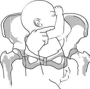
What Is Breech?
When a fetus is delivered buttocks or feet first
- Types of Presentation
Risk Factors
Complications.
Breech concerns the position of the fetus before labor . Typically, the fetus comes out headfirst, but in a breech delivery, the buttocks or feet come out first. This type of delivery is risky for both the pregnant person and the fetus.
This article discusses the different types of breech presentations, risk factors that might make a breech presentation more likely, treatment options, and complications associated with a breech delivery.
Verywell / Jessica Olah
Types of Breech Presentation
During the last few weeks of pregnancy, a fetus usually rotates so that the head is positioned downward to come out of the vagina first. This is called the vertex position.
In a breech presentation, the fetus does not turn to lie in the correct position. Instead, the fetus’s buttocks or feet are positioned to come out of the vagina first.
At 28 weeks of gestation, approximately 20% of fetuses are in a breech position. However, the majority of these rotate to the proper vertex position. At full term, around 3%–4% of births are breech.
The different types of breech presentations include:
- Complete : The fetus’s knees are bent, and the buttocks are presenting first.
- Frank : The fetus’s legs are stretched upward toward the head, and the buttocks are presenting first.
- Footling : The fetus’s foot is showing first.
Signs of Breech
There are no specific symptoms associated with a breech presentation.
Diagnosing breech before the last few weeks of pregnancy is not helpful, since the fetus is likely to turn to the proper vertex position before 35 weeks gestation.
A healthcare provider may be able to tell which direction the fetus is facing by touching a pregnant person’s abdomen. However, an ultrasound examination is the best way to determine how the fetus is lying in the uterus.
Most breech presentations are not related to any specific risk factor. However, certain circumstances can increase the risk for breech presentation.
These can include:
- Previous pregnancies
- Multiple fetuses in the uterus
- An abnormally shaped uterus
- Uterine fibroids , which are noncancerous growths of the uterus that usually appear during the childbearing years
- Placenta previa, a condition in which the placenta covers the opening to the uterus
- Preterm labor or prematurity of the fetus
- Too much or too little amniotic fluid (the liquid that surrounds the fetus during pregnancy)
- Fetal congenital abnormalities
Most fetuses that are breech are born by cesarean delivery (cesarean section or C-section), a surgical procedure in which the baby is born through an incision in the pregnant person’s abdomen.
In rare instances, a healthcare provider may plan a vaginal birth of a breech fetus. However, there are more risks associated with this type of delivery than there are with cesarean delivery.
Before cesarean delivery, a healthcare provider might utilize the external cephalic version (ECV) procedure to turn the fetus so that the head is down and in the vertex position. This procedure involves pushing on the pregnant person’s belly to turn the fetus while viewing the maneuvers on an ultrasound. This can be an uncomfortable procedure, and it is usually done around 37 weeks gestation.
ECV reduces the risks associated with having a cesarean delivery. It is successful approximately 40%–60% of the time. The procedure cannot be done once a pregnant person is in active labor.
Complications related to ECV are low and include the placenta tearing away from the uterine lining, changes in the fetus’s heart rate, and preterm labor.
ECV is usually not recommended if the:
- Pregnant person is carrying more than one fetus
- Placenta is in the wrong place
- Healthcare provider has concerns about the health of the fetus
- Pregnant person has specific abnormalities of the reproductive system
Recommendations for Previous C-Sections
The American College of Obstetricians and Gynecologists (ACOG) says that ECV can be considered if a person has had a previous cesarean delivery.
During a breech delivery, the umbilical cord might come out first and be pinched by the exiting fetus. This is called cord prolapse and puts the fetus at risk for decreased oxygen and blood flow. There’s also a risk that the fetus’s head or shoulders will get stuck inside the mother’s pelvis, leading to suffocation.
Complications associated with cesarean delivery include infection, bleeding, injury to other internal organs, and problems with future pregnancies.
A healthcare provider needs to weigh the risks and benefits of ECV, delivering a breech fetus vaginally, and cesarean delivery.
In a breech delivery, the fetus comes out buttocks or feet first rather than headfirst (vertex), the preferred and usual method. This type of delivery can be more dangerous than a vertex delivery and lead to complications. If your baby is in breech, your healthcare provider will likely recommend a C-section.
A Word From Verywell
Knowing that your baby is in the wrong position and that you may be facing a breech delivery can be extremely stressful. However, most fetuses turn to have their head down before a person goes into labor. It is not a cause for concern if your fetus is breech before 36 weeks. It is common for the fetus to move around in many different positions before that time.
At the end of your pregnancy, if your fetus is in a breech position, your healthcare provider can perform maneuvers to turn the fetus around. If these maneuvers are unsuccessful or not appropriate for your situation, cesarean delivery is most often recommended. Discussing all of these options in advance can help you feel prepared should you be faced with a breech delivery.
American College of Obstetricians and Gynecologists. If your baby is breech .
TeachMeObGyn. Breech presentation .
MedlinePlus. Breech birth .
Hofmeyr GJ, Kulier R, West HM. External cephalic version for breech presentation at term . Cochrane Database Syst Rev . 2015 Apr 1;2015(4):CD000083. doi:10.1002/14651858.CD000083.pub3
By Christine Zink, MD Dr. Zink is a board-certified emergency medicine physician with expertise in the wilderness and global medicine.
- Pregnancy Classes

Breech Births
In the last weeks of pregnancy, a baby usually moves so his or her head is positioned to come out of the vagina first during birth. This is called a vertex presentation. A breech presentation occurs when the baby’s buttocks, feet, or both are positioned to come out first during birth. This happens in 3–4% of full-term births.
What are the different types of breech birth presentations?
- Complete breech: Here, the buttocks are pointing downward with the legs folded at the knees and feet near the buttocks.
- Frank breech: In this position, the baby’s buttocks are aimed at the birth canal with its legs sticking straight up in front of his or her body and the feet near the head.
- Footling breech: In this position, one or both of the baby’s feet point downward and will deliver before the rest of the body.
What causes a breech presentation?
The causes of breech presentations are not fully understood. However, the data show that breech birth is more common when:
- You have been pregnant before
- In pregnancies of multiples
- When there is a history of premature delivery
- When the uterus has too much or too little amniotic fluid
- When there is an abnormally shaped uterus or a uterus with abnormal growths, such as fibroids
- The placenta covers all or part of the opening of the uterus placenta previa
How is a breech presentation diagnosed?
A few weeks prior to the due date, the health care provider will place her hands on the mother’s lower abdomen to locate the baby’s head, back, and buttocks. If it appears that the baby might be in a breech position, they can use ultrasound or pelvic exam to confirm the position. Special x-rays can also be used to determine the baby’s position and the size of the pelvis to determine if a vaginal delivery of a breech baby can be safely attempted.
Can a breech presentation mean something is wrong?
Even though most breech babies are born healthy, there is a slightly elevated risk for certain problems. Birth defects are slightly more common in breech babies and the defect might be the reason that the baby failed to move into the right position prior to delivery.
Can a breech presentation be changed?
It is preferable to try to turn a breech baby between the 32nd and 37th weeks of pregnancy . The methods of turning a baby will vary and the success rate for each method can also vary. It is best to discuss the options with the health care provider to see which method she recommends.
Medical Techniques
External Cephalic Version (EVC) is a non-surgical technique to move the baby in the uterus. In this procedure, a medication is given to help relax the uterus. There might also be the use of an ultrasound to determine the position of the baby, the location of the placenta and the amount of amniotic fluid in the uterus.
Gentle pushing on the lower abdomen can turn the baby into the head-down position. Throughout the external version the baby’s heartbeat will be closely monitored so that if a problem develops, the health care provider will immediately stop the procedure. ECV usually is done near a delivery room so if a problem occurs, a cesarean delivery can be performed quickly. The external version has a high success rate and can be considered if you have had a previous cesarean delivery.
ECV will not be tried if:
- You are carrying more than one fetus
- There are concerns about the health of the fetus
- You have certain abnormalities of the reproductive system
- The placenta is in the wrong place
- The placenta has come away from the wall of the uterus ( placental abruption )
Complications of EVC include:
- Prelabor rupture of membranes
- Changes in the fetus’s heart rate
- Placental abruption
- Preterm labor
Vaginal delivery versus cesarean for breech birth?
Most health care providers do not believe in attempting a vaginal delivery for a breech position. However, some will delay making a final decision until the woman is in labor. The following conditions are considered necessary in order to attempt a vaginal birth:
- The baby is full-term and in the frank breech presentation
- The baby does not show signs of distress while its heart rate is closely monitored.
- The process of labor is smooth and steady with the cervix widening as the baby descends.
- The health care provider estimates that the baby is not too big or the mother’s pelvis too narrow for the baby to pass safely through the birth canal.
- Anesthesia is available and a cesarean delivery possible on short notice
What are the risks and complications of a vaginal delivery?
In a breech birth, the baby’s head is the last part of its body to emerge making it more difficult to ease it through the birth canal. Sometimes forceps are used to guide the baby’s head out of the birth canal. Another potential problem is cord prolapse . In this situation the umbilical cord is squeezed as the baby moves toward the birth canal, thus slowing the baby’s supply of oxygen and blood. In a vaginal breech delivery, electronic fetal monitoring will be used to monitor the baby’s heartbeat throughout the course of labor. Cesarean delivery may be an option if signs develop that the baby may be in distress.
When is a cesarean delivery used with a breech presentation?
Most health care providers recommend a cesarean delivery for all babies in a breech position, especially babies that are premature. Since premature babies are small and more fragile, and because the head of a premature baby is relatively larger in proportion to its body, the baby is unlikely to stretch the cervix as much as a full-term baby. This means that there might be less room for the head to emerge.
Want to Know More?
- Creating Your Birth Plan
- Labor & Birth Terms to Know
- Cesarean Birth After Care
Compiled using information from the following sources:
- ACOG: If Your Baby is Breech
- William’s Obstetrics Twenty-Second Ed. Cunningham, F. Gary, et al, Ch. 24.
- Danforth’s Obstetrics and Gynecology Ninth Ed. Scott, James R., et al, Ch. 21.
BLOG CATEGORIES
- Pregnancy Symptoms 5
- Can I get pregnant if… ? 3
- Paternity Tests 2
- The Bumpy Truth Blog 7
- Multiple Births 10
- Pregnancy Complications 68
- Pregnancy Concerns 62
- Cord Blood 4
- Pregnancy Supplements & Medications 14
- Pregnancy Products & Tests 8
- Changes In Your Body 5
- Health & Nutrition 2
- Labor and Birth 65
- Planning and Preparing 24
- Breastfeeding 29
- Week by Week Newsletter 40
- Is it Safe While Pregnant 55
- The First Year 41
- Genetic Disorders & Birth Defects 17
- Pregnancy Health and Wellness 149
- Your Developing Baby 16
- Options for Unplanned Pregnancy 18
- Child Adoption 19
- Fertility 54
- Pregnancy Loss 11
- Uncategorized 4
- Women's Health 34
- Prenatal Testing 16
- Abstinence 3
- Birth Control Pills, Patches & Devices 21
- Thank You for Your Donation
- Unplanned Pregnancy
- Getting Pregnant
- Healthy Pregnancy
- Privacy Policy
- Pregnancy Questions Center
Share this post:
Similar post.

Episiotomy: Advantages & Complications

Retained Placenta

What is Dilation in Pregnancy?
Track your baby’s development, subscribe to our week-by-week pregnancy newsletter.
- The Bumpy Truth Blog
- Fertility Products Resource Guide
Pregnancy Tools
- Ovulation Calendar
- Baby Names Directory
- Pregnancy Due Date Calculator
- Pregnancy Quiz
Pregnancy Journeys
- Partner With Us
- Corporate Sponsors
Fetal Presentation, Position, and Lie (Including Breech Presentation)
- Key Points |
Abnormal fetal lie or presentation may occur due to fetal size, fetal anomalies, uterine structural abnormalities, multiple gestation, or other factors. Diagnosis is by examination or ultrasonography. Management is with physical maneuvers to reposition the fetus, operative vaginal delivery , or cesarean delivery .
Terms that describe the fetus in relation to the uterus, cervix, and maternal pelvis are
Fetal presentation: Fetal part that overlies the maternal pelvic inlet; vertex (cephalic), face, brow, breech, shoulder, funic (umbilical cord), or compound (more than one part, eg, shoulder and hand)
Fetal position: Relation of the presenting part to an anatomic axis; for vertex presentation, occiput anterior, occiput posterior, occiput transverse
Fetal lie: Relation of the fetus to the long axis of the uterus; longitudinal, oblique, or transverse
Normal fetal lie is longitudinal, normal presentation is vertex, and occiput anterior is the most common position.
Abnormal fetal lie, presentation, or position may occur with
Fetopelvic disproportion (fetus too large for the pelvic inlet)
Fetal congenital anomalies
Uterine structural abnormalities (eg, fibroids, synechiae)
Multiple gestation
Several common types of abnormal lie or presentation are discussed here.

Transverse lie
Fetal position is transverse, with the fetal long axis oblique or perpendicular rather than parallel to the maternal long axis. Transverse lie is often accompanied by shoulder presentation, which requires cesarean delivery.
Breech presentation
There are several types of breech presentation.
Frank breech: The fetal hips are flexed, and the knees extended (pike position).
Complete breech: The fetus seems to be sitting with hips and knees flexed.
Single or double footling presentation: One or both legs are completely extended and present before the buttocks.
Types of breech presentations
|
Breech presentation makes delivery difficult ,primarily because the presenting part is a poor dilating wedge. Having a poor dilating wedge can lead to incomplete cervical dilation, because the presenting part is narrower than the head that follows. The head, which is the part with the largest diameter, can then be trapped during delivery.
Additionally, the trapped fetal head can compress the umbilical cord if the fetal umbilicus is visible at the introitus, particularly in primiparas whose pelvic tissues have not been dilated by previous deliveries. Umbilical cord compression may cause fetal hypoxemia.

Predisposing factors for breech presentation include
Preterm labor
Uterine abnormalities
Fetal anomalies
If delivery is vaginal, breech presentation may increase risk of
Umbilical cord prolapse
Birth trauma
Perinatal death

Face or brow presentation
In face presentation, the head is hyperextended, and position is designated by the position of the chin (mentum). When the chin is posterior, the head is less likely to rotate and less likely to deliver vaginally, necessitating cesarean delivery.
Brow presentation usually converts spontaneously to vertex or face presentation.
Occiput posterior position
The most common abnormal position is occiput posterior.
The fetal neck is usually somewhat deflexed; thus, a larger diameter of the head must pass through the pelvis.
Progress may arrest in the second phase of labor. Operative vaginal delivery or cesarean delivery is often required.
Position and Presentation of the Fetus
Toward the end of pregnancy, the fetus moves into position for delivery. Normally, the presentation is vertex (head first), and the position is occiput anterior (facing toward the pregnant patient's spine) with the face and body angled to one side and the neck flexed. Abnormal presentations include face, brow, breech, and shoulder. Occiput posterior position (facing toward the pregnant patient's pubic bone) is less common than occiput anterior position. |
If a fetus is in the occiput posterior position, operative vaginal delivery or cesarean delivery is often required.
In breech presentation, the presenting part is a poor dilating wedge, which can cause the head to be trapped during delivery, often compressing the umbilical cord.
For breech presentation, usually do cesarean delivery at 39 weeks or during labor, but external cephalic version is sometimes successful before labor, usually at 37 or 38 weeks.

Copyright © 2024 Merck & Co., Inc., Rahway, NJ, USA and its affiliates. All rights reserved.
- Cookie Preferences

6.1 Breech presentation
Presentation of the feet or buttocks of the foetus.
6.1.1 The different breech presentations
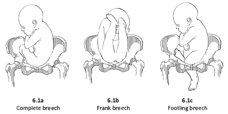
- In a complete breech presentation, the legs are tucked, and the foetus is in a crouching position (Figure 6.1a).
- In a frank breech presentation, the legs are extended, raised in front of the torso, with the feet near the head (Figure 6.1b).
- In a footling breech presentation (rare), one or both feet present first, with the buttocks higher up and the lower limbs extended or half-bent (Figure 6.1c).
6.1.2 Diagnosis
- The cephalic pole is palpable in the uterine fundus; round, hard, and mobile; the indentation of the neck can be felt.
- The inferior pole is voluminous, irregular, less hard, and less mobile than the head.
- During labour, vaginal examination reveals a “soft mass” divided by the cleft between the buttocks, with a hard projection at end of the cleft (the coccyx and sacrum).
- After rupture of the membranes: the anus can be felt in the middle of the cleft; a foot may also be felt.
- The clinical diagnosis may be difficult: a hand may be mistaken for a foot, a face for a breech.
6.1.3 Management
Route of delivery.
Before labour, external version (Chapter 7, Section 7.7 ) may be attempted to avoid breech delivery.
If external version is contra-indicated or unsuccessful, the breech position alone – in the absence of any other anomaly – is not, strictly speaking, a dystocic presentation, and does not automatically require a caesarean section. Deliver vaginally, if possible – even if the woman is primiparous.
Breech deliveries must be done in a CEmONC facility, especially for primiparous women.
Favourable factors for vaginal delivery are:
- Frank breech presentation;
- A history of vaginal delivery (whatever the presentation);
- Normally progressing dilation during labour.
The footling breech presentation is a very unfavourable position for vaginal delivery (risk of foot or cord prolapse). In this situation, the route of delivery depends on the number of previous births, the state of the membranes and how far advanced the labour is.
During labour
- Monitor dilation every 2 to 4 hours.
- If contractions are of good quality, dilation is progressing, and the foetal heart rate is regular, an expectant approach is best. Do not rupture the membranes unless dilation stops.
- If the uterine contractions are inadequate, labour can be actively managed with oxytocin.
Note : if the dilation stales, transfer the mother to a CEmONC facility unless already done, to ensure access to surgical facility for potential caesarean section.
At delivery
- Insert an IV line before expulsion starts.
- Consider episiotomy at expulsion. Episiotomy is performed when the perineum is sufficiently distended by the foetus's buttocks.
- Presence of meconium or meconium-stained amniotic fluid is common during breech delivery and is not necessarily a sign of foetal distress.
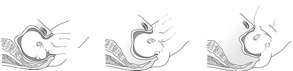
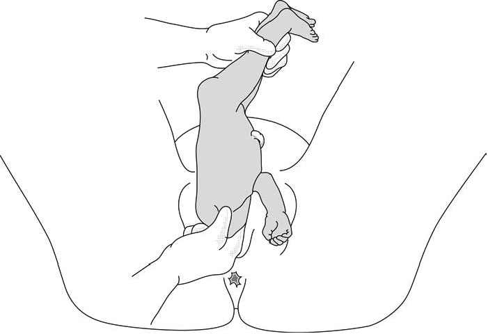
- The infant delivers unaided , as a result of the mother's pushing, simply supported by the birth attendant who gently holds the infant by the bony parts (hips and sacrum), with no traction. Do not pull on the legs.
Once the umbilicus is out, the rest of the delivery must be completed within 3 minutes, otherwise compression of the cord will deprive the infant of oxygen. Do not touch the infant until the shoulder blades appear to avoid triggering the respiratory reflex before the head is delivered.
- Monitor the position of the infant's back; impede rotation into posterior position.
Figures 6.2 - Breech delivery
6.1.4 Breech delivery problems
Posterior orientation.
If the infant’s back is posterior during expulsion, take hold of the hips and turn into an anterior position (this is a rare occurrence).
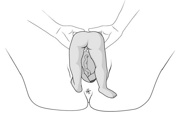
Obstructed shoulders
The shoulders can become stuck and hold back the infant's upper chest and head. This can occur when the arms are raised as the shoulders pass through the mother's pelvis. There are 2 methods for lowering the arms so that the shoulders can descend:
1 - Lovset's manoeuvre
- With thumbs on the infant's sacrum, take hold of the hips and pelvis with the other fingers.
- Turn the infant 90° (back to the left or to the right), to bring the anterior shoulder underneath the symphysis and engage the arm. Deliver the anterior arm.
- Then do a 180° counter-rotation (back to the right or to the left); this engages the posterior arm, which is then delivered.
Figures 6.3 - Lovset's manoeuvre
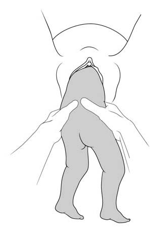
6.3c - Delivering the anterior arm and shoulder
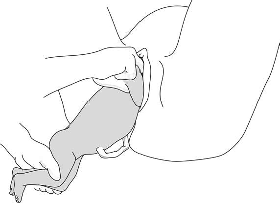
2 - Suzor’s manoeuvre
In case the previous method fails:
- Turn the infant 90° (its back to the right or to the left).
- Pull the infant downward: insert one hand along the back to look for the anterior arm. With the operator thumb in the infant armpit and middle finger along the arm, bring down the arm (Figure 6.4a).
- Lift infant upward by the feet in order to deliver the posterior shoulder (Figure 6.4b).
Figures 6.4 - Suzor's manoeuvre
6.4b - Delivering the posterior shoulder
Head entrapment
The infant's head is bulkier than the body, and can get trapped in the mother's pelvis or soft tissue.
There are various manoeuvres for delivering the head by flexing it, so that it descends properly, and then pivoting it up and around the mother's symphysis. These manoeuvres must be done without delay, since the infant must be allowed to breathe as soon as possible. All these manoeuvres must be performed smoothly, without traction on the infant.
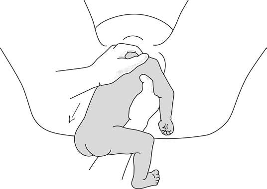
1 - Bracht's manoeuvre
- After the arms are delivered, the infant is grasped by the hips and lifted with two hands toward the mother's stomach, without any traction, the neck pivoting around the symphysis.
- Having an assistant apply suprapubic pressure facilitates delivery of the aftercoming head.
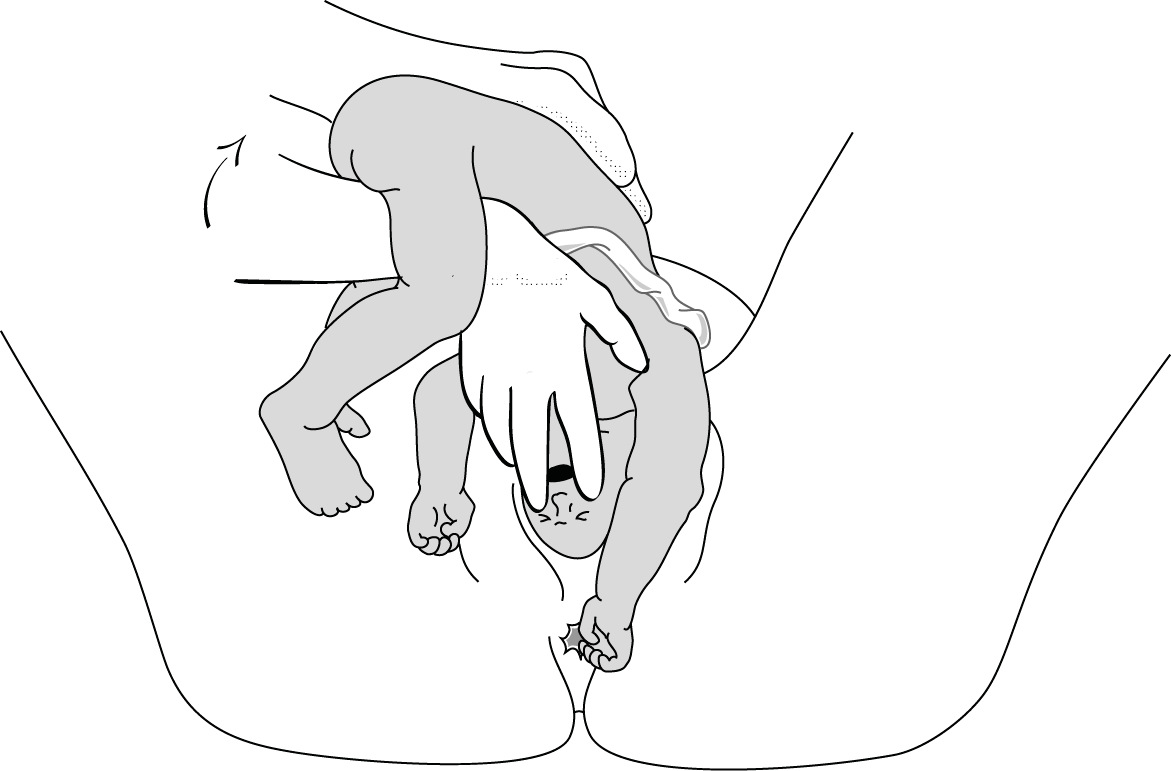
2 - Modified Mauriceau manoeuvre
- Infant's head occiput anterior.
- Kneel to get a good traction angle: 45° downward.
- Support the infant on the hand and forearm, then insert the index and middle fingers, placing them on the infant’s maxilla. Placing the index and middle fingers into the infant’s mouth is not recommended, as this can fracture the mandible.
- Place the index and middle fingers of the other hand on either side of the infant's neck and lower the infant's head to bring the sub-occiput under the symphysis (Figure 6.6a).
- Tip the infant’s head and with a sweeping motion bring the back up toward the mother's abdomen, pivoting the occiput around her symphysis pubis (Figure 6.6b).
- Suprapubic pressure on the infant's head along the pelvic axis helps delivery of the head.
- As a last resort, symphysiotomy (Chapter 5, Section 5.7 ) can be combined with this manoeuvre.
Figures 6.6 - Modified Mauriceau manoeuvre
6.6a - Step 1 Infant straddles the birth attendant's forearm; the head, occiput anterior, is lowered to bring the occiput in contact with the symphysis.
6.6b - Step 2 The infant's back is tipped up toward the mother's abdomen.
3 - Forceps on aftercoming head
This procedure can only be performed by an operator experienced in using forceps.
Breech Presentation
- 📖 Geeky Medics OSCE Book
- ⚡ Geeky Medics Bundles
- ✨ 1300+ OSCE Stations
- ✅ OSCE Checklist PDF Booklet
- 🧠 UKMLA AKT Question Bank
- 💊 PSA Question Bank
- 💉 Clinical Skills App
- 🗂️ Flashcard Collections | OSCE , Medicine , Surgery , Anatomy
- 💬 SCA Cases for MRCGP
To be the first to know about our latest videos subscribe to our YouTube channel 🙌
Table of Contents
Suggest an improvement
- Hidden Post Title
- Hidden Post URL
- Hidden Post ID
- Type of issue * N/A Fix spelling/grammar issue Add or fix a link Add or fix an image Add more detail Improve the quality of the writing Fix a factual error
- Please provide as much detail as possible * You don't need to tell us which article this feedback relates to, as we automatically capture that information for you.
- Your Email (optional) This allows us to get in touch for more details if required.
- Which organ is responsible for pumping blood around the body? * Enter a five letter word in lowercase
- Email This field is for validation purposes and should be left unchanged.
Introduction
Breech presentation is a type of malpresentation and occurs when the fetal head lies over the uterine fundus and fetal buttocks or feet present over the maternal pelvis (instead of cephalic/head presentation).
The incidence in the United Kingdom of breech presentation is 3-4% of all fetuses. 1
Breech presentation is most commonly idiopathic .
Types of breech presentation
The three types of breech presentation are:
- Complete (flexed) breech : one or both knees are flexed (Figure 1)
- Footling (incomplete) breech : one or both feet present below the fetal buttocks, with hips and knees extended (Figure 2)
- Frank (extended) breech : both hips flexed and both knees extended. Babies born in frank breech are more likely to have developmental dysplasia of the hip (Figure 3)

Risk factors
Risk factors for breech presentation can be divided into maternal , fetal and placental risk factors:
- Maternal : multiparity, fibroids, previous breech presentation, Mullerian duct abnormalities
- Fetal : preterm, macrosomia, fetal abnormalities (anencephaly, hydrocephalus, cystic hygroma), multiple pregnancy
- Placental : placenta praevia , polyhydramnios, oligohydramnios , amniotic bands
Clinical features
Before 36 weeks , breech presentation is not significant, as the fetus is likely to revert to a cephalic presentation. The mother will often be asymptomatic with the diagnosis being incidental.
The incidence of breech presentation is approximately 20% at 28 weeks gestation, 16% at 32 weeks gestation and 3-4% at term . Therefore, breech presentation is more common in preterm labour . Most fetuses with breech presentation in the early third trimester will turn spontaneously and be cephalic at term.
However, spontaneous version rates for nulliparous women with breech presentation at 36 weeks of gestation are less than 10% .
Clinical examination
Typical clinical findings of a breech presentation include:
- Longitudinal lie
- Head palpated at the fundus
- Irregular mass over pelvis (feet, legs and buttocks)
- Fetal heart auscultated higher on the maternal abdomen
- Palpation of feet or sacrum at the cervical os during vaginal examination
For more information, see the Geeky Medics guide to obstetric abdominal examination .
Positions in breech presentation
There are multiple fetal positions in breech presentation which are described according to the relation of the fetal sacrum to the maternal pelvis .
These are: direct sacroanterior, left sacroanterior, right sacroanterior, direct sacroposterior, right sacroposterior, left sacroposterior, left sacrotransverse and right sacrotranverse. 5
Investigations
An ultrasound scan is diagnostic for breech presentation. Growth, amniotic fluid volume and anatomy should be assessed to check for abnormalities.
There are three management options for breech presentation at term, with consideration of maternal choice: external cephalic version , vaginal delivery and Caesarean section .
External cephalic version
External cephalic version (ECV) involves manual rotation of the fetus into a cephalic presentation by applying pressure to the maternal abdomen under ultrasound guidance. Entonox and subcutaneous terbutaline are used to relax the uterus.
ECV has a 40% success rate in primiparous women and 60% in multiparous women . It should be offered to nulliparous women at 36 weeks and multiparous women at 37 weeks gestation.
If ECV is unsuccessful, then delivery options include elective caesarean section or vaginal delivery.
Contraindications for undertaking external cephalic version include:
- Antepartum haemorrhage
- Ruptured membranes
- Previous caesarean section
- Major uterine abnormality
- Multiple pregnancy
- Abnormal cardiotocography (CTG)
Vaginal delivery
Vaginal delivery is an option but carries risks including head entrapment, birth asphyxia, intracranial haemorrhage, perinatal mortality, cord prolapse and fetal and/or maternal trauma.
The preference is to deliver the baby without traction and with an anterior sacrum during delivery to decrease the risk of fetal head entrapment .
The mother may be offered an epidural , as vaginal breech delivery can be very painful. 6
Contraindications for vaginal delivery in a breech presentation include:
- Footling breech: the baby’s head and trunk are more likely to be trapped if the feet pass through the dilated cervix too soon
- Macrosomia: usually defined as larger than 3800g
- Growth restricted baby: usually defined as smaller than 2000g
- Other complications of vaginal birth: for example, placenta praevia and fetal compromise
- Lack of clinical staff trained in vaginal breech delivery
Caesarean section
A caesarian section booked as an elective procedure at term is the most common management for breech presentation.
Caesarean section is preferred for preterm babies (due to an increased head to abdominal circumference ratio in preterm babies) and is used if the external cephalic version is unsuccessful or as a maternal preference. This option has fewer risks than a vaginal delivery.
Complications
Fetal complications of breech presentation include:
- Developmental dysplasia of the hip (DDH)
- Cord prolapse
- Fetal head entrapment
- Birth asphyxia
- Intracranial haemorrhage
- Perinatal mortality
Complications of external cephalic version include:
- Transient fetal heart abnormalities (common)
- Fetomaternal haemorrhage
- Placental abruption (rare)
- There are three types of breech presentation: complete, incomplete and frank breech
- The most common clinical findings include: longitudinal lie, smooth fetal head-shape at the fundus, irregular masses over the pelvis and abnormal placement being required for fetal hear auscultation
- The diagnostic investigation is an ultrasound scan
- Breech presentation can be managed in three ways: external cephalic version , vaginal delivery or elective caesarean section
- Complications are more common in vaginal delivery , such as cord prolapse, fetal head entrapment, intracranial haemorrhage and birth asphyxia
Miss Saba Al Juboori
Consultant in Obstetrics and Gynaecology
Miss Neeraja Kuruba
Dr chris jefferies.
- Oxford Handbook of Obstetrics and Gynaecology. Breech Presentation: Overview. Published in 2011.
- Jemimah Thomas. Image: Complete breech.
- Bonnie Urquhart Gruenberg. Footling breech. Licence: [ CC BY-SA ]
- Bonnie Urquhart Gruenberg. Frank breech . Licence: [ CC BY-SA ]
- A Comprehensive Textbook of Obstetrics and Gynaecology. Chapter 50: Malpresentation and Malposition: Breech Presentation. Published in 2011.
- Diana Hamilton Fairley. Lecture Notes: Obstetrics and Gynaecology, Malpresentation, Breech Presentation. Published in 2009.

Other pages
- Product Bundles 🎉
- Join the Team 🙌
- Institutional Licence 📚
- OSCE Station Creator Tool 🩺
- Create and Share Flashcards 🗂️
- OSCE Group Chat 💬
- Newsletter 📰
- Advertise With Us
Join the community
Management of Breech Presentation (Green-top Guideline No. 20b)
Summary: The aim of this guideline is to aid decision making regarding the route of delivery and choice of various techniques used during delivery. It does not include antenatal or postnatal care. Information regarding external cephalic version is the topic of the separate Royal College of Obstetricians and Gynaecologists Green-top Guideline No. 20a, External Cephalic Version and Reducing the Incidence of Term Breech Presentation .
Breech presentation occurs in 3–4% of term deliveries and is more common in preterm deliveries and nulliparous women. Breech presentation is associated with uterine and congenital abnormalities, and has a significant recurrence risk. Term babies presenting by the breech have worse outcomes than cephalic presenting babies, irrespective of the mode of delivery.
A large reduction in the incidence of planned vaginal breech birth followed publication of the Term Breech Trial. Nevertheless, due to various circumstances vaginal breech births will continue. Lack of experience has led to a loss of skills essential for these deliveries. Conversely, caesarean section can has serious long-term consequences.
COVID disclaimer: This guideline was developed as part of the regular updates to programme of Green-top Guidelines, as outlined in our document Developing a Green-top Guideline: Guidance for developers , and prior to the emergence of COVID-19.
Version history: This is the fourth edition of this guideline.
Please note that the RCOG Guidelines Committee regularly assesses the need to update the information provided in this publication. Further information on this review is available on request.
Developer declaration of interests:
Mr M Griffiths is a member of Doctors for a Woman's right to Choose on Abortion. He is an unpaid member of a Quality Standards Advisory Committee at NICE, for which he does receive expenses for related travel, accommodation and meals.
Mr LWM Impey is Director of Oxford Fetal Medicine Ltd. and a member of the International Society of Ultrasound in Obstetrics and Gynecology. He also holds patents related to ultrasound processing, which are of no relevance to the Breech guidelines.
Professor DJ Murphy provides medicolegal expert opinions in Scotland and Ireland for which she is remunerated.
Dr LK Penna: None declared.
- Access the PDF version of this guideline on Wiley
- Access the web version of this guideline on Wiley
This page was last reviewed 16 March 2017.
Better Birth Blog
Information & Inspiration for Pregnancy, Birth & Parenting
What is a footling breech baby?
January 17, 2018 By Lauren McClain

Babies in utero spend most of their gestation head-up. As they get heavier and prepare for delivery, they turn head-down. This usually happens around 32 weeks. When it doesn’t, and baby is still head-up at term, we call this baby breech.
When the baby’s head is up by the mother’s heart, the baby may be curled up in any number of positions—all of which are breech.
A footling breech baby is presenting feet or foot first . If we could look through the cervix, the first thing we’d see would be a foot. If labor started and the baby was pushed out this way, the first thing to emerge would be a foot.
A single footling has one knee drawn up so that only one foot is down and a double footling breech has both her feet together over the cervix.
Most breech babies come butt-first–Frank breech or complete breech. In this case, when the mother pushes her baby out, it’s a butt that is crowning, not a head (often called ‘rumping’).
Only about 20% of breech babies are footling breeches.
Footling breeches are trickier vaginal births. For one thing, there isn’t anything nice and solid and heavy pressing on the cervix to help it dilate. With a butt or a head over the cervix, it’s likely to dilate quicker and more efficiently.
Another risk of footling breech birth is cord prolapse . If the water is broken and a part of the umbilical cord falls through the cervix, it can create a dangerous situation for the baby due to cord compression and congealing.
Extremely rare in head-down birth, the incidence of cord prolapse rises considerably with breeches. Footling breeches have the highest risk (10-25%) because there is no butt or head blocking the cervix.
For many providers who are comfortable delivering a breech baby normally, footling breech position is a contraindication . For vaginal breech birth to be considered safe, a number of conditions must be met. The baby’s position is high on the list of questions.
Frank breech babies are generally seen as the safest because they can be delivered like a baby tube, neatly packaged with bum over cervix. These babies tend to do well in labor.
Part of the reason they do well is that the baby’s compacted body ‘opens the door’ for the head to pass through easily. With a footling, the baby’s feet can come down anytime and the birth canal may not be stretched as fully. Sometimes this makes birthing the head more difficult or dangerous.
Sometimes it does no such thing.
Check out some of the great footling breech birth stories and videos here.
Sometimes a footling will convert to a complete breech and come down butt-first once contractions begin. Keep this in mind when thinking about and discussing your options.
Finding a provider to attend and assist at your vaginal breech birth is already hard. Finding one who feels comfortable with a footling breech is even more difficult.
Some people do think it is easier to turn a footling . If you’re interested in ways to help a breech baby flip , it may just work. An ECV is safe and does reduce the likelihood a mother will end up with a cesarean.
Only you can make the decision, and I hope that if you feel comfortable with it you will convince your doctor to get some training or even consider traveling to avoid a cesarean.


An official website of the United States government
The .gov means it’s official. Federal government websites often end in .gov or .mil. Before sharing sensitive information, make sure you’re on a federal government site.
The site is secure. The https:// ensures that you are connecting to the official website and that any information you provide is encrypted and transmitted securely.
- Publications
- Account settings
Preview improvements coming to the PMC website in October 2024. Learn More or Try it out now .
- Advanced Search
- Journal List
- PMC11262641
Maternal and neonatal outcomes associated with breech presentation in planned community (home and birth center) births in the United States: A prospective observational cohort study
Robyn schafer.
1 Division of Advanced Nursing Practice, School of Nursing, Rutgers University, Newark, NJ, United States of America
2 Department of Obstetrics, Gynecology, and Reproductive Sciences, Robert Wood Johnson Medical School, Rutgers University, New Brunswick, NJ, United States of America
Marit L. Bovbjerg
3 Epidemiology Program, College of Public Health and Human Sciences, Oregon State University, Corvallis, OR, United States of America
Melissa Cheyney
4 Department of Anthropology, Oregon State University, Corvallis, OR, United States of America
Julia C. Phillippi
5 School of Nursing, Vanderbilt University, Nashville, TN, United States of America
Associated Data
MANA Stats data are available to researchers with an approved data use agreement. Researchers can apply for access to use MANA Stats data by emailing gro.statsanam@snoitacilppahcraeser .
Investigate maternal and neonatal outcomes associated with breech presentation in planned community births in the United States, including outcomes associated with types of breech presentation (i.e., frank, complete, footling/kneeling)
Secondary analysis of prospective cohort data from a national perinatal data registry (MANA Stats)
Planned community birth (homes and birth centers), United States
Individuals with a term, singleton gestation (N = 71,943) planning community birth at labor onset
Descriptive statistics to calculate associations between types of breech presentation and maternal and neonatal outcomes
Main outcome measures
Maternal : intrapartum/postpartum transfer, hospitalization, cesarean, hemorrhage, severe perineal laceration, duration of labor stages and membrane rupture
Neonatal : transfer, hospitalization, NICU admission, congenital anomalies, umbilical cord prolapse, birth injury, intrapartum/neonatal death
One percent (n = 695) of individuals experienced breech birth (n = 401, 57.6% vaginally). Most fetuses presented frank breech (57%), with 19% complete, 18% footling/kneeling, and 5% unknown type of breech presentation. Among all breech labors, there were high rates of intrapartum transfer and cesarean birth compared to cephalic presentation (OR 9.0, 95% CI 7.7–10.4 and OR 18.6, 95% CI 15.9–21.7, respectively), with no substantive difference based on parity, planned site of birth, or level of care integration into the health system. For all types of breech presentations, there was increased risk for nearly all assessed neonatal outcomes including hospital transfer, NICU admission, birth injury, and umbilical cord prolapse. Breech presentation was also associated with increased risk of intrapartum/neonatal death (OR 8.5, 95% CI 4.4–16.3), even after congenital anomalies were excluded.
Conclusions
All types of breech presentations in community birth settings are associated with increased risk of adverse neonatal outcomes. These research findings contribute to informed decision-making and reinforce the need for breech training and research and an increase in accessible, high-quality care for planned vaginal breech birth in US hospitals.
Introduction
There has been a recent increase in breech birth in community settings (homes and birth centers) in the United States [ 1 ]. This is despite research demonstrating increased risk of intrapartum or neonatal death (16.8/1000 adjusted odds ratio [aOR] 8.2, 95% CI, 3.7–18.4) [ 2 ] in breech community births and consensus obstetric and midwifery recommendations that classify breech presentation as a contraindication to home birth [ 3 , 4 ]. Since 2000, planned cesarean has been the standard of care for breech presentation, following a landmark large-scale, randomized controlled trial (the Term Breech Trial) [ 5 ] and subsequent American College of Obstetricians and Gynecologists (ACOG) committee opinion [ 6 ] recommending planned cesarean delivery for all singleton term breech fetuses. However, more recent research has called those recommendations into question [ 7 – 12 ], concluding that although risk of adverse outcomes is higher in planned vaginal breech birth than planned cesarean, the absolute risk is quite low [ 13 – 16 ]. Internationally, support for vaginal breech birth is increasing [ 17 – 20 ], but nearly all breech fetuses (95.5%) in the US are born via cesarean [ 1 , 13 , 14 , 21 ]. ACOG committee opinion now recommends that for a term, singleton fetus, planned vaginal breech birth “may be reasonable under hospital-specific protocol guidelines for eligibility and labor management” [ 22 ]. However, hospital-based care for planned vaginal breech birth in the US is very difficult to obtain, in part due to a lack of skilled providers and medicolegal concerns [ 22 – 24 ], leading some individuals to seek care in community-based settings (homes and birth centers) [ 25 – 27 ].
Breech presentation affects approximately 3–4% of term pregnancies, and community births currently comprise about 2% of US births [ 28 , 29 ]. Based on birth certificate data from the National Center for Health Statistics, rates of US community births rose 33.2% from 2019 to 2022, including a 61.7% increase in breech births (n = 423 in 2019, n = 684 in 2022), in tandem with a decrease in hospital births [ 1 ]. In 2022, 12.5% (n = 488) of all reported singleton, term (greater than or equal to 37 + 0/7 weeks’ gestation) vaginal breech births in the US occurred in a community birth setting [ 1 ]. Research has established that intrapartum and neonatal death rates are higher in breech birth than cephalic births [ 2 ], but little is known about neonatal and maternal outcomes associated with breech presentation managed in community birth settings.
Data is also limited about maternal and neonatal outcomes based on type of breech presentation. Breech presentation is classified based on the position of the lower fetal extremities (see Table 1 ). Breech presentation nomenclature has been applied inconsistently in research and clinical practice recommendations, and there is ambiguity about variations of presentation types (such as partial flexion, location of feet alongside or just below the buttocks, or dynamic presentations that change during labor) [ 5 , 15 , 18 , 30 – 32 ]. Alternative nomenclatures have been proposed, but none have gained widespread acceptance [ 33 , 34 ]. Footling or kneeling breech presentation is generally considered a contraindication to vaginal birth due to increased risk of perinatal morbidity from umbilical cord prolapse or head entrapment leading to hypoxic injury [ 17 – 19 , 22 ]. However, there is limited evidence to support this recommendation since, with rare exceptions [ 30 , 35 ], vaginal breech trials historically have excluded (or not reported data regarding) footling or kneeling presentations [ 5 , 15 , 16 , 36 , 37 ]. Research that examines potential differences in community birth outcomes associated with type of breech presentation is needed to guide informed decision-making and optimize perinatal outcomes [ 2 ]. The purpose of this study was to analyze associations between breech birth and maternal and neonatal outcomes compared to cephalic presentations in planned community births and assess differences in outcomes associated with type of breech presentation.
| Type | Attitude at hip | Attitude at knee | Position of feet |
|---|---|---|---|
| Frank (or “extended”) | Flexed (both) | Extended (both) | Proximal to the fetal head |
| Complete (or “flexed”) | Flexed (both) | Flexed (both) | Lack of consensus |
| Incomplete | Lack of consensus | Lack of consensus | Lack of consensus |
| Footling (single or double footling) | Extended (partially or fully, one or both) | Flexed or extended | Presenting below the level of the buttocks |
| Kneeling (single or double kneeling) | Extended (one or both) | Flexed (one or both) | Below the level of the buttocks and above the level of the knee(s), with one or both knees presenting |
* There is not a consensus definition for position of the fetal feet in a complete presentation, which either (a) cannot be below the fetal buttocks [ 5 ] or (b) may be palpable at or just below the buttocks [ 16 , 18 ].
† The term “incomplete” is inconsistently defined in the literature as either (a) both hips flexed with one knee flexed and one knee extended [ 18 , 30 ] or (b) one or both hips not completely flexed, regardless of attitude at the knee (in essence, an umbrella term for footling and kneeling presentations) [ 38 , 39 ].
Materials and methods
This cohort study used registry data (birth years 2012–2018) from the Midwives Alliance of North America Statistics Project (MANA Stats). MANA Stats includes extensive prenatal, birth, and postpartum data from individuals who received care from midwives in community birth settings in the United States. Individuals are prospectively enrolled in the registry at the onset of care in pregnancy with informed consent, and midwives enter data throughout perinatal care. MANA Stats development, data collection protocols, and evidence of reliability and validity are described elsewhere [ 40 , 41 ]. Ethical approval was received from Oregon State University’s IRB. All pregnant persons and midwives gave informed consent for research participation.
MANA data were accessed July 1, 2019. The study sample (N = 71,943) included all singleton, term births for individuals who planned community birth at the onset of labor and had a documented fetal presentation at birth ( Fig 1 ). Pregnancies missing information on fetal presentation at birth were excluded, as were persons who changed their intended site of birth to a hospital setting prior to onset of labor. Both vaginal and cesarean births were included. The main exposure of interest was breech presentation at birth (n = 695) in comparison to cephalic presentation, subdivided by type of breech presentation as defined by the data set variable “breech presentation at birth” as frank, complete, footling, kneeling, or unknown. No formal definitions of breech types were provided to midwives entering data into the registry; those who were uncertain could contact MANA Stats support staff for assistance.

We explored associations between breech presentations at time of birth and multiple perinatal outcomes including durations of labor stages and membrane rupture. Labor stages were defined in the MANA Stats system as follows: first stage as the interval between frequent, intense contractions and onset of pushing; second stage as the start of active pushing efforts until birth of the neonate; and third stage as time from birth of the neonate until placental expulsion, as described in prior publications [ 42 ]. The management of impossible or improbable duration values are described in supplemental materials ( S1 Table ). Because this was a cohort of planned community births, intrapartum or postpartum transfer to hospital within six hours after birth was assessed, along with the reason(s) for transfer and urgency. Determination of indication(s) for transfer and associated urgency were based on assessment of the transferring midwife. We also analyzed maternal hospitalization in the first six weeks postpartum, including new admissions following community birth and postpartum readmissions. Finally, we evaluated adverse maternal outcomes, including severe (i.e., third- or fourth-degree) perineal laceration, retained placenta, and obstetric hemorrhage (defined as ≥1000 mL and/or diagnosed hemorrhage regardless of estimated blood loss) [ 43 ].
Neonatal outcomes included transfer to hospital in the first six hours of life (including indications and urgency), hospitalization (any) and/or NICU admission in the first six weeks of life (whether primary or readmission), umbilical cord prolapse, birth injury (defined as “skeletal fracture, peripheral nerve injury, and soft tissue or solid organ hemorrhage requiring intervention”), and intrapartum or neonatal death up to six weeks. Because term breech presentation is associated with congenital anomalies [ 44 – 46 ], we also assessed the presence of congenital anomalies (diagnosed antenatally or in the first six weeks of life) and explored deaths associated with anomalies separately. For every intrapartum or neonatal death, we explored free-text data entered by the community birth midwives describing the clinical course and circumstances surrounding care and provided brief case summaries.
Statistical analyses were performed using SPSS V 24.0.0.0 (IBM Corporation, Armonk, NY, USA) and R version 3.3.2 (R Foundation for Statistical Computing, Vienna, Austria). Initial analysis compared all types of breech presentation, collectively, to cephalic presentation. Analyses were then repeated to compare outcomes by presentation type. Medians and interquartile range are reported for labor durations and frequencies for all other outcomes. Because multivariable models were not possible due to low event counts for adverse outcomes, bivariable analyses were performed. We reported counts and proportions, including odds ratios (ORs) and confidence intervals (CI) for outcomes with five or more events in both comparison groups. Standard bivariable statistics were used to explore associations. We used unadjusted logistic regression models to calculate ORs and 95% CIs for categorical outcomes and the Kruskal-Wallis test to assess associations between breech presentation and labor duration, stratified by parity.
To contextualize our study sample, we compared the overall proportion of breech presentation to the expected proportion in the general US childbearing population based on vital statistics data (2016–2021) [ 47 ]. With the understanding that maternity care policies related to breech birth care may affect access to care and health outcomes [ 48 ], we also explored the two most frequent outcomes (cesarean and intrapartum transfer) for both cephalic and breech presentation stratified by covariables of planned site of community birth (i.e., home or birth center) and region of the country. Finally, since there is evidence that the level of integration of community birth providers into regional health systems affects maternal and neonatal birth outcomes [ 49 ], we explored associations state-level midwifery care integration scores (defined by Vedam et al., 2018) as an additional covariable in this analysis.
In this sample of 71,943 individuals, 1% (n = 695) gave birth to a term, singleton, breech neonate. Incidence of breech births in this low-risk sample of planned community births was, predictably, lower than the rate of 2.8% found the general US childbearing population (based on term, singleton births with known presentation from 2016–2021). As shown in Table 2 , demographic characteristics of individuals in this sample who experienced breech birth were generally similar to those with a cephalic birth, except for increased likelihood of being nulliparous (48.7% breech, 32.6% cephalic) and not eligible for low-income public health insurance (19.5% breech, 23.2% cephalic). Of the 695 breech neonates in this sample, the majority presented frank breech at birth (57.0%, n = 396), followed by complete (19.3%, n = 134), footling (17.7%, n = 123), and kneeling (0.7%, n = 5) presentations. Type of breech presentation was unknown in 5.3% (n = 37) of births.
| Comparison variable | total n (%) | breech n (%) | cephalic n (%) | p-value (chi-square test) |
|---|---|---|---|---|
| total | 71,943 | 695 (1.0%) | 71,248 (99.0%) | |
| Age, | 30.6 (5.0) | 31.2 (5.0) | 30.6 (5.0) | 0.004 |
| Race identified as White | 66,883 (93.2%) | 660 (95.5%) | 66,223 (93.2%) | 0.02 |
| Married or partnered | 68,293 (94.9%) | 667 (96.0%) | 67,626 (94.9) | 0.26 |
| Level of education bachelor’s degree or higher | 35,804 (50.3%) | 349 (50.7%) | 35,455 (50.3%) | 0.85 |
| Eligible for Medicaid (public health insurance) based on income | 16,646 (23.2%) | 135 (19.5%) | 16,511 (23.2%) | 0.02 |
| Pre-gravid BMI <18.5 18.5–24.9 25–29.9 30–34.9 ≥ 35 Missing | 2803 (3.9%) 42,753 (59.4%) 13,977 (19.4%) 5141 (7.1%) 2868 (4.0%) 4401 (6.1%) | 30 (4.3%) 412 (59.3%) 136 (19.6%) 49 (7.1%) 25 (3.6%) 43 (6.2%) | 2773 (3.9%) 42,341 (59.4%) 13,841 (19.4%) 5092 (7.1%) 2843 (4.0%) 4358 (6.1%) | 0.99 |
| Nulliparous | 23,457 (32.6%) | 338 (48.7%) | 23,119 (32.5%) | <0.001 |
| Parous, with: History of cesarean with prior vaginal birth | 2072 (4.3%) | 14 (3.9%) | 2058 (4.3%) | 0.07 |
| History of cesarean only | 1756 (3.6%) | 21 (5.9%) | 1735 (3.6%) | |
| Gestational age at birth, mean (SD) | 281.5 (7.7) | 279.5 (8.6) | 281.5 (7.7) | <0.001 |
| Post-dates gestation | 2808 (3.9%) | 19 (2.7%) | 2789 (3.9%) | 0.12 |
| Planned place of birth home birth center | 50,324 (69.9%) 21,619 (30.1%) | 531 (76.4%) 164 (23.6%) | 49,793 (69.9%) 21,455 (30.1%) | <0.001 |
| Primary provider credential Certified professional midwife (CPM) Certified nurse-midwife (CNM) Dually certified midwife (CPM/CNM) Other type of provider | 52,077 (72.4%) 8462 (11.8%) 2368 (3.3%) 9019 (12.5%) | 524 (75.4%) 72 (10.4%) 19 (2.7%) 80 (11.5%) | 51,553 (72.4%) 8390 (11.8%) 2349 (3.3%) 8939 (12.5%) | 0.48 |
a For maternal age, the p-value is from a t-test assuming equal variances
b Denominator is multiparas
c Other types of providers included student midwives under supervision, clinicians with other credentials (e.g., ND, DO, lay midwives), and unknown or missing provider credential information.
Notes: Data come from the Midwives Alliance of North America Statistics Project (MANA Stats), birth years 2012–2018. Comparison of demographic and pregnancy risk factor variables between births including a breech fetus, compared to births with a cephalic fetus. Sample was limited to singleton, not preterm, and not missing information on presentation.
Associations between breech presentation and maternal and neonatal outcomes are presented in Table 3 , with reasons for transfer detailed and compared in Table 4 . Nearly half (42.4%) of all breech neonates in planned community births were born via cesarean (versus 3.8% for cephalic), and, relatedly, more individuals with a breech fetus transferred from community birth settings to the hospital in the intrapartum period (OR 9.0, 95% CI 7.7–10.4). Midwives classified more breech intrapartum transfers as urgent (46% v. 17%, p < 0.001), with malpresentation/malposition (85%) being the most common reason for intrapartum transfer. Multiple indications for transfer were commonly cited. Other than cord prolapse and fetal malpresentation, all other reasons for transfer were more common among cephalic labors. After intrapartum transfer (n = 344), 50 breech neonates were born vaginally (14.5%, vs. 61.4% of cephalic intrapartum transfers) in hospital settings. Vaginal hospital births included 30 frank breech, 7 complete, 12 footling, and 1 unknown breech type.
| Outcome | Cephalic n (%) N = 71,248 | Breech n (%) N = 695 | OR (95% CI) |
|---|---|---|---|
| Intrapartum transfer (any) | 7030 (9.9%) | 344 (49.5%) | 9.0 (7.7–10.4) |
| Intrapartum transfer (urgent) | 1171 (1.6%) | 159 (22.9%) | 17.7 (14.7–23.4) |
| Cesarean | 2713 (3.8%) | 294 (42.4%) | 18.6 (15.9–21.7) |
| Postpartum transfer (any) | 1699 (2.6%) | 22 (6.3%) | 2.5 (1.6–3.8) |
| Postpartum transfer (urgent) | 912 (1.4%) | 13 (3.7%) | 2.7 (1.5–4.6) |
| Severe perineal laceration | 948 (1.4%) | 11 (2.8%) | 2.0 (1.1–3.7) |
| Hemorrhage (any) | 3836 (5.4%) | 33 (4.7%) | 0.88 (0.62–1.2) |
| Hemorrhage ≥1000 mL | 1594 (2.4%) | 9 (2.0%) | 0.82 (0.42–1.6) |
| Hospitalization | 1681 (2.4%) | 21 (3.1%) | 1.3 (0.86–2.1) |
| Neonatal transfer (any) | 1126 (1.8%) | 27 (7.7%) | 4.7 (3.1–7.0) |
| Neonatal transfer (urgent) | 727 (1.1%) | 22 (6.3%) | 5.8 (3.8–9.1) |
| Umbilical cord prolapse | 50 (0.1%) | 15 (2.2%) | 32.2 (18.0–57.7) |
| Congenital anomaly (any) | 627 (0.9%) | 14 (2.0%) | 2.3 (1.4–4.0) |
| Birth injury | 212 (0.3%) | 16 (2.3%) | 7.9 (4.7–13.2) |
| Hospitalization (any) | 2576 (3.6%) | 30 (4.5%) | 1.2 (0.86–1.8) |
| NICU admission | 1868 (2.6%) | 44 (6.6%) | 2.6 (1.9–3.5) |
| Intrapartum or neonatal death (any) | 122/71,248 (1.7/1000) | 10/695 (14.4/1000) | 8.5 (4.4–16.3) |
| Intrapartum or neonatal death (not attributed to congenital anomaly) | 100/71,215 (1.4/1000) | 8/693 (11.5/1000) | 8.3 (4.0–17.1) |
a limited to those who completed community birth
b limited to vaginal births; includes third- and fourth-degree lacerations
Notes: Odds Ratios are breech vs. cephalic, so OR > 1 means the outcome is more common in breech labors, and OR < 1 means outcome is less common in breech labors. All ORs are unadjusted because of small sample sizes.
| Reason for transfer | Cephalic | Breech | Chi-square p-value |
|---|---|---|---|
| N = 7027 | N = 344 | ||
| Arrest of labor/failure to progress, first stage of labor | 2810 ( | 27 (7.8%) | <0.001 |
| Arrest of labor/failure to progress, second stage of labor | 1154 ( | 9 (2.6%) | <0.001 |
| Prolonged labor | 617 ( ) | 4 (1.2%) | --- |
| Prolonged rupture of membranes | 1048 ( ) | 18 (5.2%) | <0.001 |
| Maternal dehydration | 182 ( | 0 | --- |
| Hypertensive disorders of pregnancy | 213 ( ) | 2 (0.6%) | --- |
| Maternal exhaustion | 1799 ( | 8 (2.3%) | <0.001 |
| Maternal request for additional pain relief | 2387 ( ) | 15 (4.4%) | <0.001 |
| Signs or symptoms of infection | 124 ( ) | 0 | --- |
| Uterine rupture | 6 ( ) | 0 | --- |
| Umbilical cord prolapse | 24 (0.3%) | 7 ( | <0.001 |
| Malposition or malpresentation | 1273 (18.1%) | 293 ( ) | <0.001 |
| Light/thin meconium | 408 ( ) | 10 (2.9%) | <0.02 |
| Heavy/thick meconium | 501 (7.1%) | 23 (6.7%) | 0.83 |
| Non-reassuring fetal heart tones | 1101 ( ) | 10 (2.9%) | <0.001 |
| Placental abruption | 70 ( | 2 (0.6%) | --- |
| Other | 506 (7.2%) | 25 (7.3%) | 0.92 |
| N = 1707 | N = 22 | ||
| Cervical or uterine prolapse | 5 (0.3%) | 0 | --- |
| Hemorrhage | 677 ( | 5 (22.7%) | 0.13 |
| Laceration repair | 602 ( ) | 6 (27.3%) | 0.51 |
| Hypertension | 14 (0.8%) | 0 | --- |
| Retained placenta | 510 ( ) | 6 (27.3%) | 1.0 |
| Signs/symptoms of infection | 9 (0.5%) | 0 | --- |
| Other reason | 241 (14.1%) | 9 ( ) | 0.002 |
| N = 1132 | N = 27 | ||
| Birth trauma/injury | 40 (3.5%) | 5 ( ) | 0.003 |
| Suspected congenital anomaly | 75 (6.6%) | 0 | --- |
| Meconium aspiration syndrome | 87 (7.7%) | 0 | --- |
| Signs of prematurity | 6 (0.5%) | 1 (3.7%) | |
| Respiratory distress syndrome | 687 ( | 11 (40.7%) | 0.05 |
| Neonatal seizures | 16 (1.4%) | 0 | --- |
| Symptoms of infection | 77 (36.8%) | 1 (3.7%) | --- |
| Other reason | 350 (30.9%) | 15 ( | 0.01 |
a Multiple item selection permissible on data entry
b P-values are suppressed unless there were at least 5 events in both groups.
c Other reasons were reported as
Medical complications: abnormal vital signs, seizure, stroke, active herpes simplex infection, cardiac condition, excessive nausea, and vomiting
Obstetric complications: prolonged rupture of membranes, precipitous labor (unattended), hypertensive disorders of pregnancy, oligohydramnios, postterm gestation, cervical edema, urinary retention
Situational or environmental factors: poor weather conditions, independent maternal decision to transfer, state regulations, lack of availability of birth attendant
d Postpartum and neonatal transfer include transfers within the first 6 hours after birth.
e Other reasons were reported as
Medical complications: abnormal vital signs, postpartum psychosis, syncope
Obstetric complications: precipitous labor
Situational or environmental factors: maternal intuition (“didn’t feel right”), desire to remain with neonate requiring transfer
f Other reasons were reported as
Neonatal complications: lethargy, cardiac arrythmia, unspecified (baby “didn’t look right”), protocol following resuscitation (not meeting criteria for respiratory distress syndrome)
Situational or environmental factors: precipitous and/or unattended birth, desire for neonate to remain with postpartum person requiring transfer
Maternal postpartum transfers were also more likely to be considered urgent in breech births (OR 2.7, 95% CI 1.5–4.6), even though prevailing maternal indications for transfer (including hemorrhage, laceration repair, and retained placenta) were more common in the cephalic group. Neither postpartum hemorrhage nor maternal hospitalization increased significantly with breech presentation compared to cephalic. There were insufficient events of operative births (i.e., forceps) (n = 4) or retained placenta (n = 7) for analysis.
Distributions of labor duration variables are shown in Fig 2 , stratified by presentation and parity. Median active labor for breech fetuses among nulliparas was shorter than cephalic fetuses (406 vs. 480 minutes), but the opposite was true for multiparous individuals (228 breech vs. 207 cephalic). There were no significant differences in duration of second or third stages based on fetal presentation, although breech labors were associated with significantly longer durations of membrane rupture for both nulliparas (median 336 minutes for breech vs. 268 cephalic) and multiparas (84 breech vs. 31 cephalic).

For neonates, breech presentation was associated with increased odds of neonatal transfer, NICU admission, and birth injury (OR 4.7, 95% CI 3.1–7.0; OR 2.6, 95% CI 1.9–3.5; and OR 7.9, 95% CI 4.7–13.2, respectively) ( Table 3 ). There was no association between presentation at birth and neonatal hospitalization. Regarding indications for neonatal transfer ( Table 4 ), breech neonates were more likely to transfer for birth injury (18.5% vs. 3.5%) and “other” (not listed) reasons (55.6% vs. 30.9%) and less likely to transfer for respiratory distress (40.7% vs. 60.7%). Breech births were also more likely to experience umbilical cord prolapse (2.2% v. 0.1%, OR 32.2, 95% CI 18.0–57.7).
There was also a substantive increase in odds of intrapartum or neonatal death for the breech fetus (OR 8.5, 95% CI 4.4–16.3). Although based on only ten perinatal deaths (five intrapartum and five neonatal), this association persisted even when deaths related to congenital anomalies were excluded (OR 8.3, 95% CI 4.0–17.1). Deaths (described in S2 Table ) were attributed to congenital anomalies (n = 4), head entrapment (n = 3), cord prolapse (n = 2), and unknown causes (interoperative death, suspected placental abruption) (n = 1). Several intrapartum/neonatal deaths were complicated by late diagnosis of breech presentation and inefficient transfer of care including medical errors by emergency medical services (EMS), delays in hospital assessment and treatment, and conflicts with EMS or hospital staff. It is also worth noting that intrapartum/neonatal deaths included several instances of late onset of community-based care, with the midwives describing assuming responsibility for antepartum care only after hospital providers declined care for planned vaginal birth due to breech presentation in the absence of other risk factors.
Maternal and neonatal outcomes stratified by type of breech presentation are shown in Table 5 . For many outcomes, the small sample size of breech births and correspondingly low event counts preclude firm conclusions; however, a few patterns do emerge from the limited data. Rates of intrapartum transfer and cesarean birth are similar across all breech types, and postpartum hemorrhage was less common with frank breech (3.3% frank vs. 6.0% complete, 7.0% footling/kneeling). Neonatal transfers, hospitalization, and NICU admissions were twice as common in footling/kneeling presentations. Umbilical cord prolapse was also significantly more common, occurring in 7.3% of footling/kneeling breech births (0.8% frank, 2.3% complete); however, perinatal death was half as likely (7.8/1000 footling/kneeling vs. 20/1000 frank, 22/1000 complete)—a finding that should be interpreted with caution given the low incidence of death (n = 1) in the footling/kneeling group.
| Frank breech N = 396 | Complete breech N = 134 | Footling/kneeling breech N = 128 | |||
|---|---|---|---|---|---|
| Outcome | n (%) | n (%) | OR (95% CI) | n (%) | OR (95% CI) |
| Intrapartum transfer | 189 (47.7%) | 65 (48.5%) | 1.03 (0.70–1.5) | 65 (50.8%) | 1.1 (0.76–1.7) |
| Cesarean | 159 (40.3%) | 58 (43.3%) | 1.1 (0.76–1.7) | 53 (41.4%) | 1.0 (0.70–1.6) |
| Postpartum transfer | 11 (5.3%) | 4 (5.8%) | --- | 5 (7.9%) | 1.5 (0.52–4.6) |
| Severe perineal laceration | 9 (2.3%) | 1 (0.7%) | --- | 2 (1.6%) | --- |
| Hemorrhage (any) | 13 (3.3%) | 8 (6.0%) | 1.9 (0.76–4.6) | 9 (7.0%) | 2.2 (0.93–5.3) |
| Hemorrhage ≥1000 mL | 4 (1.4%) | 2 (2.4%) | --- | 2 (2.5%) | --- |
| Hospitalization | 10 (2.6%) | 2 (1.5%) | --- | 6 (4.8%) | 1.9 (0.68–5.3) |
| Neonatal transfer | 15 (7.2%) | 3 (4.4%) | --- | 8 (12.9%) | 1.9 (0.77–4.7) |
| Umbilical cord prolapse | 3 (0.8%) | 3 (2.3%) | --- | 9 (7.3%) | --- |
| Congenital anomaly, any | 8 (2.0%) | 3 (2.2%) | --- | 3 (2.3%) | --- |
| Birth injury | 8 (2.0%) | 4 (3.0%) | --- | 4 (3.1%) | --- |
| Hospitalization | 14 (3.7%) | 5 (3.8%) | 1.0 (0.40–3.0) | 9 (7.3%) | 2.0 (0.86–4.8) |
| NICU admission | 23 (6.1%) | 8 (6.2%) | 1.0 (0.44–2.3) | 12 (9.6%) | 1.6 (0.80–3.4) |
| Intrapartum or neonatal death (any) | 6/396 (15.2/1000) | 3/134 (22.4/1000) | --- | 1/128 (7.8/1000) | --- |
| Intrapartum or neonatal death (not attributed to congenital anomaly) | 4/394 (10.1/1000) | 3/134 (22.4/1000) | --- | 1/128 (7.8/1000) | --- |
a limited to those who completed community birth: 64,176 cephalic, 208 frank, 68 complete, 62 footling or kneeling presentations
Data are from planned community births in the USA, 2012–2018, limited to singleton term labors for which fetal presentation at birth was identified. Odds ratios use frank breech as the reference group (i.e., complete vs. frank; footling/kneeling vs. frank). Breech presentations of unknown type (N = 37) were excluded from this analysis.
Odds ratios have been suppressed for any category for which there were <5 events in either the numerator or denominator.
Finally, analysis of contextual variables ( S3 Table ) found higher rates of cesarean and intrapartum transfer for breech labors in the New England region (OR 17.6, 95% CI 7.6–40.9 and OR 47.2, 95% CI 20.1–110.7, respectively) compared to other regions of the country. There were no substantive differences in outcomes based on planned site of community birth (i.e., home or birth center) or level of integration of community birth midwifery services into the healthcare system, as defined by Vedam et al.[ 49 ]
Among this sample of planned community births, breech presentation was associated with high rates of intrapartum transfer and cesarean birth (OR 9.0 and 18.6, respectively) and no increased risk of maternal hospitalization or postpartum hemorrhage. Associations with nearly all assessed adverse neonatal outcomes were increased in breech births, including transfer, NICU admission, and birth injury. Umbilical cord prolapse occurred in 2.2% of breech births (OR 32.2, 95% CI 18.0–57.7). There was a high rate of intrapartum and neonatal death (14.4/1000, OR 8.5, 95% CI 4.4–16.3), which persisted even after excluding congenital anomalies.
All types of breech presentation carry additional risk for adverse neonatal outcomes. Although sample sizes precluded meaningful analysis of perinatal outcomes associated with type of breech presentation, our findings support existing research that increased incidence of umbilical cord prolapse in footling/kneeling breech presentations may not be associated with increased risk of severe complications [ 50 ], though this result should be interpreted with caution. Labor duration was not affected by type of breech presentation, as consistent with prior findings [ 51 ]. Although there was some regional variation in rates of maternal transfer and cesarean, there were no substantive differences in outcomes based on parity, planned site of birth, or level of care integration of community-based midwifery services.
Due to logistical and ethical concerns about randomizing individuals to site or mode of birth [ 10 , 52 , 53 ], assessment of outcomes associated with breech presentation relies primarily on observational evidence. This descriptive analysis is useful for guiding decision-making for breech labor and birth. The size and scope of this dataset are a strength of this study, with a large sample of individuals across community birth settings throughout the United States and high rates of participation in data collection from community midwives (>95%) [ 40 ]. Prospective enrollment in pregnancy ensured that all birth outcomes were included, thereby minimizing selection bias and potential underreporting of adverse outcomes [ 40 ]. Additionally, this dataset includes vaginal breech births and footling/kneeling presentations, which are often excluded from research.
Despite these strengths, there are also several limitations to the research based on this dataset. First, because participation in data collection is voluntary, outcomes may differ between providers who participate in data collection and those who do not. Second, as with any dataset, research findings are limited by the existing variables and their definitions. For example, because community birth providers avoid frequent or unnecessary cervical examinations, the dataset defined onset of second stage by initiation of pushing (rather than with onset of full cervical dilation as it is commonly defined). Although these definitions are used elsewhere in the literature [ 42 ], these findings may not correlate exactly to other studies exploring labor durations. Similarly, the lack of variables regarding comprehensive clinical and environmental factors prohibited investigation of predictive factors associated with breech birth outcomes. For example, we could not distinguish between planned and unplanned breech births, assess relationships with external cephalic version, determine when breech presentation was identified or whether a skilled breech attendant was present, or correlate outcomes with regulatory scope of practice restrictions, such as state regulations that limit community birth providers’ care for breech labors.
One additional limitation of this study is the possibility that not all presentation types were classified accurately. In community birth settings, there is rarely access ultrasound technology to confirm presentation, and evidence has demonstrated poor reliability in determining presentation by physical examination alone [ 54 ]. Due to constraints of existing breech nomenclature, there was also potential for unreliable classifications of presentation variants (such as when the hips and knees are incompletely flexed or feet are located alongside or just below the buttocks) or those that changed during labor (such as a complete breech fetus who extends a leg). Finally, because community birth care utilizes low levels of intervention, findings from breech community birth may not be generalizable to high-resource hospital settings [ 14 ].
Interpretation and implications
Findings from this study reinforce existing evidence of increased risk of adverse neonatal outcomes in breech community birth [ 2 , 55 , 56 ]. Although many emergent interventions and technologies are not readily accessible in community births, the physiologic approach exemplified in these settings is widely considered by expert breech clinicians to be optimal for perinatal outcomes [ 57 , 58 ]. However, even physiologic management in a low-risk population does not appear to circumvent risks to the breech neonate.
This research has implications for clinical practice, health care policy, and future research. Pregnant people should be counselled about the increased risk of adverse neonatal outcomes for breech fetuses in planned community births. These risks should be considered in context of the risks and benefits associated with sites and modes of birth, including risks to future pregnancies and individuals’ unique needs, preferences, values, and risk tolerance [ 13 , 17 ]. Care providers in all settings should take steps to identify breech presentation at term and provide evidence-based information about breech birth outcomes to ensure informed choice. Skills in breech assessment and management should be incorporated into midwifery and obstetric training to optimize outcomes. Recognizing that breech community births will inevitably occur, both accidentally and intentionally, community and hospital birth providers should develop guidelines to identify and manage complications and provide timely and efficient transfer when needed [ 59 ].
Community birth is not well integrated into the health care system throughout the United States [ 49 , 60 ], and this lack of coordination of care across birth settings was evident in several intrapartum and neonatal deaths in this sample. In addition, it was noted in a few cases that individuals were late to community-based care after they were declined care for planned hospital vaginal birth due to breech presentation in the absence of other risk factors. Community birth in the presence of high-risk conditions often indicates a failure of the medical system to meet patient’s needs for less interventive care and autonomy in decision-making [ 27 , 61 – 64 ]. Restrictive policies preventing hospital providers from offering care for planned vaginal breech birth to appropriate candidates should be eliminated as they impede patient autonomy and access to care and inadvertently push more medically complex births into community settings [ 24 , 26 , 57 , 59 , 65 , 66 ].
In prior published analyses using this data set, members of this research team recommended that, due to increased risk of adverse outcomes in community birth, breech presentations were better managed in birth settings with immediate access to hospital staff and facilities [ 2 ]. However, despite US recommendations supporting care for planned vaginal breech birth for appropriately screened candidates in hospitals [ 17 , 22 ], access to vaginal breech birth and skilled breech providers in hospitals remains limited [ 25 , 26 ]. Findings from this study, along with the recent increase in US breech community births, reinforce consensus recommendations that US hospitals have a “clear and urgent responsibility” [ 25 ] to increase access to care for planned vaginal breech given the increased risk of adverse perinatal outcomes associated with breech community birth compared to cephalic presentations. Policies and medicolegal reforms that incorporate best available evidence and center the birthing person and their rights to autonomy are necessary to improve maternal and neonatal outcomes and support informed choice for breech pregnancy and birth.
Breech presentation in all birth settings is associated with increased risk of adverse outcomes compared to cephalic presentation, and further research is needed to explore maternal and neonatal outcomes in matched cohorts of breech births in different settings with skilled breech providers. There is a need for development and adoption of a consistent and well-defined breech nomenclature to minimize ambiguity between presentation types and facilitate evidence synthesis. Future studies should explore outcomes based on type of breech presentation using this standardized nomenclature and report outcomes according to a standardized core outcome set (e.g., Breech-COS, currently in development) [ 67 ]. Research on breech labor outcomes is needed to guide decision-making, given that comparisons of prelabor cesarean to planned vaginal birth are not generalizable to laboring persons facing either emergent cesarean or unplanned vaginal breech birth. Researchers should assess the proportion of breech presentations correlated with underlying conditions (i.e., fetal growth restriction, congenital anomalies, oligohydramnios, placenta previa, maternal gestational diabetes mellitus or hypertensive disorders, uterine malformation, or history of cesarean) [ 45 , 68 ] and investigate how these conditions affect morbidity and mortality, regardless of mode or site of birth. Finally, research is needed to explore the barriers and facilitators of breech birth care in the United States to guide recommendations to improve access to quality care [ 26 ].
In planned community births, all types of breech presentation pose substantial risk of adverse outcomes, including high rates of intrapartum and neonatal death. This research provides evidence about breech labor in community birth settings and adverse maternal and neonatal outcomes associated with breech birth to inform decision-making. There is a need for increased training and research on vaginal breech birth. Reforms are needed to ensure accessible, high-quality care for planned vaginal breech birth in US hospitals.
Supporting information
S1 appendix, acknowledgments.
The authors gratefully acknowledge the midwives who contributed data to the MANA Stats Project.
Funding Statement
Marit L. Bovbjerg and Melissa Cheyney received funding from the United States National Institute of Health (Grant R03HD096094) towards this research effort. The funders played no role in the study design, data collection and analysis, decision to publish, or preparation of the manuscript.
Data Availability
Anesthesia for Multiparity, Multiple Gestation, and Breech Presentation
- First Online: 04 September 2024
Cite this chapter

- Jessica Chan 3 &
- Joseph Villaluz 4
Risk factors associated with a breech presentation include uterine distension (e.g., multiple gestation, polyhydramnios, multiparity), maternal pelvic abnormalities (e.g., pelvic tumors, uterine anomalies), and fetal abnormalities (e.g., anencephaly, hydrocephalus). Fetal head entrapment from breech presentation is more likely to occur before 32-week gestation. The fetal head may be entrapped due to suboptimal cervical dilatation, a relatively large fetal head, or an underdeveloped fetal skull that may not be able to mold to the maternal pelvis. External cephalic version (ECV) is the first-line approach to a full-term pregnancy in breech presentation. Patients with multiple gestation are at a high risk for preterm labor. Preterm labor occurs in women with spontaneous twins at a rate of 22%, while preterm labor in women with twins from in vitro fertilization occurs at a rate of 52%.
This is a preview of subscription content, log in via an institution to check access.
Access this chapter
Subscribe and save.
- Get 10 units per month
- Download Article/Chapter or eBook
- 1 Unit = 1 Article or 1 Chapter
- Cancel anytime
- Available as PDF
- Read on any device
- Instant download
- Own it forever
- Available as EPUB and PDF
- Durable hardcover edition
- Dispatched in 3 to 5 business days
- Free shipping worldwide - see info
Tax calculation will be finalised at checkout
Purchases are for personal use only
Institutional subscriptions
Orbach-Zinger S, Ashwal E, Hazan L, Bracco D, Ioscovich A, Hiersch L, Khinchuck A, Aviram A, Eidelman LA. Risk factors for unintended Dural puncture in obstetric patients: a retrospective cohort study. Anesth Analg. 2016;123(4):972–6. https://doi.org/10.1213/ANE.0000000000001510 .
Article PubMed Google Scholar
Alsammani MA, Jafer AM, Khieri SA, Ali AO, Shaaeldin MA. Effect of grand multiparity on pregnancy outcomes in women under 35 years of age: a comparative study. Med Arch. 2019;73(2):92–6. https://doi.org/10.5455/medarh.2019.73.92-96 .
Article PubMed PubMed Central Google Scholar
Liu B, Beral V. Balkwill a; million women study collaborators. Childbearing, breastfeeding, other reproductive factors and the subsequent risk of hospitalization for gallbladder disease. Int J Epidemiol. 2009;38(1):312–8. https://doi.org/10.1093/ije/dyn174 . Epub 2008 Sep 4.
Chestnut DH. Chestnut’s obstetric anesthesia: principles and practice. Elsevier; 2020.
Google Scholar
Al JF. Grandmultiparity: a potential risk factor for adverse pregnancy outcomes. J Reprod Med. 2012;57(1–2):53–7.
PubMed Google Scholar
Nassar AH, Fayyumy R, Saab W, Mehio G, Usta IM. Grandmultiparas in modern obstetrics. Am J Perinatol. 2006;23(6):345–9. https://doi.org/10.1055/s-2006-947158 . Epub 2006 Jul 13.
Kao DP, Hsich E, Lindenfeld J. Characteristics, adverse events, and racial differences among delivering mothers with peripartum cardiomyopathy. JACC Heart Fail. 2013;1(5):409–16. https://doi.org/10.1016/j.jchf.2013.04.011 .
Mant J, Painter R, Vessey M. Epidemiology of genital prolapse: observations from the Oxford Family Planning Association Study. Br J Obstet Gynaecol. 1997;104(5):579–85. https://doi.org/10.1111/j.1471-0528.1997.tb11536.x .
Article CAS PubMed Google Scholar
Högnäs E, Kauppila A, Pukkala E, Tapanainen JS. Cancer risk in women with 10 or more deliveries. Obstet Gynecol. 2014;123(4):811–6. https://doi.org/10.1097/AOG.0000000000000182 .
Peker N, Tosun ÖÇ. Is grand multiparity a risk factor for the development of postmenopausal osteoporosis? Clin Interv Aging. 2018;13:505–8. https://doi.org/10.2147/CIA.S155793 .
Article CAS PubMed PubMed Central Google Scholar
Turan V. Grand-grand multiparity (more than 10 deliveries) does not convey a risk for osteoporosis. Acta Obstet Gynecol Scand. 2011;90(12):1440–2. https://doi.org/10.1111/j.1600-0412.2011.01250.x . Epub 2011 Oct 10.
Boyle A, Reddy UM, Landy HJ, Huang CC, Driggers RW, Laughon SK. Primary cesarean delivery in the United States. Obstet Gynecol. 2013;122(1):33–40. https://doi.org/10.1097/AOG.0b013e3182952242 .
American College of Obstetricians and Gynecologists. Practice Bulletin No. 169: Multifetal gestations: twin, triplet, and higher-order multifetal pregnancies. Obstet Gynecol. 2016;128:e131–46.
Pratt SD. Anesthesia for breech presentation and multiple gestation. Clin Obstet Gynecol. 2003;46(3):711–29.
Shanahan MM, Gray CJ. External cephalic version. [updated 2022 Jul 4]. In: StatPearls [internet]. Treasure Island (FL): StatPearls Publishing; 2023. https://www.ncbi.nlm.nih.gov/books/NBK482475/
Pollack KL, Chestnut DH. Anesthesia for complicated vaginal deliveries. Anesthesiol Clin North Am. 1990;8(1):115–29. https://doi.org/10.1016/s0889-8537(21)00157-7 .
Article Google Scholar
Boushra M, Stone A, Rathbun KM. Umbilical Cord Prolapse. [Updated 2022 Jun 5]. In: StatPearls [Internet]. Treasure Island (FL): StatPearls Publishing; 2023 Jan. https://www.ncbi.nlm.nih.gov/books/NBK542241/
Suggested Reading
Chestnut DH, Wong CA, Tsen LC, et al. Chestnut’s obstetric anesthesia: principles and practice. 6th ed. Elsevier; 2020. Kacmar RM, Gaiser R (eds).
Download references
Author information
Authors and affiliations.
Kaweah Health Medical Center, Visalia, CA, USA
Jessica Chan
University of California Irvine, Irvine, CA, USA
Joseph Villaluz
You can also search for this author in PubMed Google Scholar
Corresponding author
Correspondence to Jessica Chan .
Editor information
Editors and affiliations.
Department of Anesthesiology, Loma Linda University, Loma Linda, CA, USA
Ashish C. Sinha
Department of Anesthesiology, Critical Care, and Neurocritical Care, Loma Linda University at Riverside University Health System, Loma Linda, CA, USA
Ioana F. Pasca
Rights and permissions
Reprints and permissions
Copyright information
© 2024 The Author(s), under exclusive license to Springer Nature Switzerland AG
About this chapter
Chan, J., Villaluz, J. (2024). Anesthesia for Multiparity, Multiple Gestation, and Breech Presentation. In: Sinha, A.C., Pasca, I.F. (eds) Peripartum Care of the Pregnant Patient. Springer, Cham. https://doi.org/10.1007/978-3-031-62756-9_19
Download citation
DOI : https://doi.org/10.1007/978-3-031-62756-9_19
Published : 04 September 2024
Publisher Name : Springer, Cham
Print ISBN : 978-3-031-62755-2
Online ISBN : 978-3-031-62756-9
eBook Packages : Medicine Medicine (R0)
Share this chapter
Anyone you share the following link with will be able to read this content:
Sorry, a shareable link is not currently available for this article.
Provided by the Springer Nature SharedIt content-sharing initiative
- Publish with us
Policies and ethics
- Find a journal
- Track your research
Extended Breech, Flexed Breech and Footling Breech
- Download PDF Copy
.jpg "breech presentation with footling")
The position of the baby inside the uterus keeps changing during pregnancy. At the time of delivery, the best position for the baby to be in is the head-down position. This position allows safer and easier passage of the baby via the birth canal and minimizes complications during child birth. However, this is not always the case and other fetal positions are common.
What is breech?
A fetus is said to be in a breech position if the baby is in a bottom-down position instead of head down. In breech presentation, the buttocks or foot / feet of the baby presents at the bottom of the uterus instead of the baby’s head.
During the last few weeks of pregnancy, health care providers check the position of the fetus with the help of a clinical examination and an ultrasound. In case breech presentation is confirmed, doctors discuss the matter with the woman and her partner and decide on options that will ensure a safe delivery.
Types of breech
Depending on which part is presenting as the lowest part of the fetus during a clinical examination, breech is classified into 3 types.
Extended breech – In this position, the hips of the baby are flexed and its legs are extended at the knee joint. The baby appears bottom first with its feet seen near its head.
Complete or flexed breech – Here, both the hips and knees of the fetus are flexed, the feet are stretched up and not seen below the level of the buttocks.
Footling breech – In this type of breech, one or both feet of the fetus extends downwards and lowers over the cervix, presenting as the lowest part of the fetus.

Risk factors contributing to breech presentation
Some risk factors at the maternal and fetal level can increase the chances of breech presentation. They are as follows:
Related Stories
- Study reveals high failure rate for tubal sterilization as birth control
- Mother's gut bacteria during pregnancy promote healthy brain development in the fetus
- Smoking during pregnancy affects children’s academic achievement
Maternal factors
- Previous breech birth
- First pregnancy
- Early labor
- Abnormal shape of the uterus
- Presence of fibroids in the uterus
- Excess or low levels of or amniotic fluid
- More than one baby in the womb
- Placental abnormalities such as placenta previa in which placenta is on the lower part of the uterine wall and blocks the cervix
Fetal factors
- Short umbilical cord
- Extended fetal legs
- Abnormal fetus
- Poor growth of the fetus
Clinical examination
In late pregnancy, a clinical examination is carried out to assess the presentation of the fetus.
Breech presentation is suspected in the following cases:
- During abdominal examination, the lowest part of the fetus or the presenting part feels irregular and a hard round part is found in the fundus, which is the upper part of the uterus.
- Heart sound of the fetus comes from the upper portion of the abdomen, typically above the navel or the umbilicus.
- During pelvic examination, the hard round head cannot be felt in the pelvis and instead the soft buttocks and / or the feet of the fetus are felt. Once the membrane breaks, thick, dark green feces called meconium may be present
When a breech presentation is suspected by 37 weeks of pregnancy, an ultrasound may be done to confirm the type of breech - extended, flexed, or footling breech - and exclude fetal head hyperextension and placenta previa.
In case of fetal head hyperextension beyond 90 degrees, there is a high risk of damage to the spinal cord during vaginal delivery and hence delivery via caesarean is recommended. The chance of a breech baby spontaneously turning into a cephalic position declines as pregnancy advances, but it is still possible in nearly 25% of women after 36 weeks.
- https://www.nlm.nih.gov/medlineplus/ency/patientinstructions/000623.htm
- https://thewomens.r.worldssl.net/
- http://apps.who.int/iris/bitstream/10665/43972/1/9241545879_eng.pdf
Further Reading
- All Pregnancy Content
- Early Signs of Pregnancy
- Is it Safe to Exercise During Pregnancy?
- Pregnancy: 0-8 weeks
- Pregnancy: 9 - 12 weeks
Last Updated: Dec 29, 2022
Susha Cheriyedath
Susha is a scientific communication professional holding a Master's degree in Biochemistry, with expertise in Microbiology, Physiology, Biotechnology, and Nutrition. After a two-year tenure as a lecturer from 2000 to 2002, where she mentored undergraduates studying Biochemistry, she transitioned into editorial roles within scientific publishing. She has accumulated nearly two decades of experience in medical communication, assuming diverse roles in research, writing, editing, and editorial management.
Please use one of the following formats to cite this article in your essay, paper or report:
Cheriyedath, Susha. (2022, December 29). Extended Breech, Flexed Breech and Footling Breech. News-Medical. Retrieved on September 08, 2024 from https://www.news-medical.net/health/Extended-Breech-Flexed-Breech-and-Footling-Breech.aspx.
Cheriyedath, Susha. "Extended Breech, Flexed Breech and Footling Breech". News-Medical . 08 September 2024. <https://www.news-medical.net/health/Extended-Breech-Flexed-Breech-and-Footling-Breech.aspx>.
Cheriyedath, Susha. "Extended Breech, Flexed Breech and Footling Breech". News-Medical. https://www.news-medical.net/health/Extended-Breech-Flexed-Breech-and-Footling-Breech.aspx. (accessed September 08, 2024).
Cheriyedath, Susha. 2022. Extended Breech, Flexed Breech and Footling Breech . News-Medical, viewed 08 September 2024, https://www.news-medical.net/health/Extended-Breech-Flexed-Breech-and-Footling-Breech.aspx.
Suggested Reading
_which_the_pregnant_woman_in_a_coral_dress_embraces_-_ARegina_be8fb2211c1b4cca93378f2b8fa975cb-125x110.jpg "breech presentation with footling")
Cancel reply to comment
- Trending Stories
- Latest Interviews
- Top Health Articles

How can microdialysis benefit drug development
Ilona Vuist
In this interview, discover how Charles River uses the power of microdialysis for drug development as well as CNS therapeutics.

Global and Local Efforts to Take Action Against Hepatitis
Lindsey Hiebert and James Amugsi
In this interview, we explore global and local efforts to combat viral hepatitis with Lindsey Hiebert, Deputy Director of the Coalition for Global Hepatitis Elimination (CGHE), and James Amugsi, a Mandela Washington Fellow and Physician Assistant at Sandema Hospital in Ghana. Together, they provide valuable insights into the challenges, successes, and the importance of partnerships in the fight against hepatitis.

Addressing Important Cardiac Biology Questions with Shotgun Top-Down Proteomics
In this interview conducted at Pittcon 2024, we spoke to Professor John Yates about capturing cardiomyocyte cell-to-cell heterogeneity via shotgun top-down proteomics.

Latest News

Newsletters you may be interested in
Your AI Powered Scientific Assistant
Hi, I'm Azthena, you can trust me to find commercial scientific answers from News-Medical.net.
A few things you need to know before we start. Please read and accept to continue.
- Use of “Azthena” is subject to the terms and conditions of use as set out by OpenAI .
- Content provided on any AZoNetwork sites are subject to the site Terms & Conditions and Privacy Policy .
- Large Language Models can make mistakes. Consider checking important information.
Great. Ask your question.
Azthena may occasionally provide inaccurate responses. Read the full terms .
While we only use edited and approved content for Azthena answers, it may on occasions provide incorrect responses. Please confirm any data provided with the related suppliers or authors. We do not provide medical advice, if you search for medical information you must always consult a medical professional before acting on any information provided.
Your questions, but not your email details will be shared with OpenAI and retained for 30 days in accordance with their privacy principles.
Please do not ask questions that use sensitive or confidential information.
Read the full Terms & Conditions .
Provide Feedback

Breech Presentation
- Author: Richard Fischer, MD; Chief Editor: Ronald M Ramus, MD more...
- Sections Breech Presentation
- Vaginal Breech Delivery
- Cesarean Delivery
- Comparative Studies
- External Cephalic Version
- Conclusions
- Media Gallery
Breech presentation is defined as a fetus in a longitudinal lie with the buttocks or feet closest to the cervix. This occurs in 3-4% of all deliveries. The percentage of breech deliveries decreases with advancing gestational age from 22-25% of births prior to 28 weeks' gestation to 7-15% of births at 32 weeks' gestation to 3-4% of births at term. [ 1 ]
Predisposing factors for breech presentation include prematurity , uterine malformations or fibroids, polyhydramnios , placenta previa , fetal abnormalities (eg, CNS malformations, neck masses, aneuploidy), and multiple gestations . Fetal abnormalities are observed in 17% of preterm breech deliveries and in 9% of term breech deliveries.
Perinatal mortality is increased 2- to 4-fold with breech presentation, regardless of the mode of delivery. Deaths are most often associated with malformations, prematurity, and intrauterine fetal demise .
Types of breeches
The types of breeches are as follows:
Frank breech (50-70%) - Hips flexed, knees extended (pike position)
Complete breech (5-10%) - Hips flexed, knees flexed (cannonball position)
Footling or incomplete (10-30%) - One or both hips extended, foot presenting
Historical considerations
Vaginal breech deliveries were previously the norm until 1959 when it was proposed that all breech presentations should be delivered abdominally to reduce perinatal morbidity and mortality. [ 2 ]
Vaginal breech delivery
Three types of vaginal breech deliveries are described, as follows:
Spontaneous breech delivery: No traction or manipulation of the infant is used. This occurs predominantly in very preterm, often previable, deliveries.
Assisted breech delivery: This is the most common type of vaginal breech delivery. The infant is allowed to spontaneously deliver up to the umbilicus, and then maneuvers are initiated to assist in the delivery of the remainder of the body, arms, and head.
Total breech extraction: The fetal feet are grasped, and the entire fetus is extracted. Total breech extraction should be used only for a noncephalic second twin; it should not be used for a singleton fetus because the cervix may not be adequately dilated to allow passage of the fetal head. Total breech extraction for the singleton breech is associated with a birth injury rate of 25% and a mortality rate of approximately 10%. Total breech extractions are sometimes performed by less experienced accoucheurs when a foot unexpectedly prolapses through the vagina. As long as the fetal heart rate is stable in this situation, it is permissible to manage expectantly to allow the cervix to completely dilate around the breech (see the image below).

Technique and tips for assisted vaginal breech delivery
The fetal membranes should be left intact as long as possible to act as a dilating wedge and to prevent overt cord prolapse .
Oxytocin induction and augmentation are controversial. In many previous studies, oxytocin was used for induction and augmentation, especially for hypotonic uterine dysfunction. However, others are concerned that nonphysiologic forceful contractions could result in an incompletely dilated cervix and an entrapped head.
An anesthesiologist and a pediatrician should be immediately available for all vaginal breech deliveries. A pediatrician is needed because of the higher prevalence of neonatal depression and the increased risk for unrecognized fetal anomalies. An anesthesiologist may be needed if intrapartum complications develop and the patient requires general anesthesia .
Some clinicians perform an episiotomy when the breech delivery is imminent, even in multiparas, as it may help prevent soft tissue dystocia for the aftercoming head (see the images below).

The Pinard maneuver may be needed with a frank breech to facilitate delivery of the legs but only after the fetal umbilicus has been reached. Pressure is exerted in the popliteal space of the knee. Flexion of the knee follows, and the lower leg is swept medially and out of the vagina.
No traction should be exerted on the infant until the fetal umbilicus is past the perineum, after which time maternal expulsive efforts should be used along with gentle downward and outward traction of the infant until the scapula and axilla are visible (see the image below).

Use a dry towel to wrap around the hips (not the abdomen) to help with gentle traction of the infant (see the image below).

An assistant should exert transfundal pressure from above to keep the fetal head flexed.
Once the scapula is visible, rotate the infant 90° and gently sweep the anterior arm out of the vagina by pressing on the inner aspect of the arm or elbow (see the image below).

Rotate the infant 180° in the reverse direction, and sweep the other arm out of the vagina. Once the arms are delivered, rotate the infant back 90° so that the back is anterior (see the image below).

The fetal head should be maintained in a flexed position during delivery to allow passage of the smallest diameter of the head. The flexed position can be accomplished by using the Mauriceau Smellie Veit maneuver, in which the operator's index and middle fingers lift up on the fetal maxillary prominences, while the assistant applies suprapubic pressure (see the image below).

Alternatively, Piper forceps can be used to maintain the head in a flexed position (see the image below).

In many early studies, routine use of Piper forceps was recommended to protect the head and to minimize traction on the fetal neck. Piper forceps are specialized forceps that are placed from below the infant and, unlike conventional forceps, are not tailored to the position of the fetal head (ie, it is a pelvic, not cephalic, application). The forceps are applied while the assistant supports the fetal body in a horizontal plane.
During delivery of the head, avoid extreme elevation of the body, which may result in hyperextension of the cervical spine and potential neurologic injury (see the images below).

Lower Apgar scores, especially at 1 minute, are more common with vaginal breech deliveries. Many advocate obtaining an umbilical cord artery and venous pH for all vaginal breech deliveries to document that neonatal depression is not due to perinatal acidosis.
Fetal head entrapment may result from an incompletely dilated cervix and a head that lacks time to mold to the maternal pelvis. This occurs in 0-8.5% of vaginal breech deliveries. [ 3 ] This percentage is higher with preterm fetuses (< 32 wk), when the head is larger than the body. Dührssen incisions (ie, 1-3 cervical incisions made to facilitate delivery of the head) may be necessary to relieve cervical entrapment. However, extension of the incision can occur into the lower segment of the uterus, and the operator must be equipped to deal with this complication. The Zavanelli maneuver has been described, which involves replacement of the fetus into the abdominal cavity followed by cesarean delivery. While success has been reported with this maneuver, fetal injury and even fetal death have occurred.
Nuchal arms, in which one or both arms are wrapped around the back of the neck, are present in 0-5% of vaginal breech deliveries and in 9% of breech extractions. [ 3 ] Nuchal arms may result in neonatal trauma (including brachial plexus injuries) in 25% of cases. Risks may be reduced by avoiding rapid extraction of the infant during delivery of the body. To relieve nuchal arms when it is encountered, rotate the infant so that the fetal face turns toward the maternal symphysis pubis (in the direction of the impacted arm); this reduces the tension holding the arm around the back of the fetal head, allowing for delivery of the arm.
Cervical spine injury is predominantly observed when the fetus has a hyperextended head prior to delivery. Ballas and Toaff (1976) reported 20 cases of hyperextended necks, defined as an angle of extension greater than 90° ("star-gazing"), discovered on antepartum radiographs. [ 4 ] Of the 11 fetuses delivered vaginally, 8 (73%) sustained complete cervical spinal cord lesions, defined as either transection or nonfunction.
Cord prolapse may occur in 7.4% of all breech labors. This incidence varies with the type of breech: 0-2% with frank breech, 5-10% with complete breech, and 10-25% with footling breech. [ 3 ] Cord prolapse occurs twice as often in multiparas (6%) than in primigravidas (3%). Cord prolapse may not always result in severe fetal heart rate decelerations because of the lack of presenting parts to compress the umbilical cord (ie, that which predisposes also protects).
Prior to the 2001 recommendations by the American College of Obstetricians and Gynecologists (ACOG), approximately 50% of breech presentations were considered candidates for vaginal delivery. Of these candidates, 60-82% were successfully delivered vaginally.
Candidates can be classified based on gestational age. For pregnancies prior to 26 weeks' gestation, prematurity, not mode of delivery, is the greatest risk factor. Unfortunately, no randomized clinical trials to help guide clinical management have been reported. Vaginal delivery can be considered, but a detailed discussion of the risks from prematurity and the lack of data regarding the ideal mode of delivery should take place with the parent(s). For example, intraventricular hemorrhage, which can occur in an infant of extremely low birth weight, should not be misinterpreted as proof of a traumatic vaginal breech delivery.
For pregnancies between 26 and 32 weeks, retrospective studies suggest an improved outcome with cesarean delivery, although these reports are subject to selection bias. In contrast, between 32 and 36 weeks' gestation, vaginal breech delivery may be considered after a discussion of risks and benefits with the parent(s).
After 37 weeks' gestation, parents should be informed of the results of a multicenter randomized clinical trial that demonstrated significantly increased perinatal mortality and short-term neonatal morbidity associated with vaginal breech delivery (see Comparative Studies). For those attempting vaginal delivery, if estimated fetal weight (EFW) is more than 4000 g, some recommend cesarean delivery because of concern for entrapment of the unmolded head in the maternal pelvis, although data to support this practice are limited.
A frank breech presentation is preferred when vaginal delivery is attempted. Complete breeches and footling breeches are still candidates, as long as the presenting part is well applied to the cervix and both obstetrical and anesthesia services are readily available in the event of a cord prolapse.
The fetus should show no neck hyperextension on antepartum ultrasound imaging (see the image below). Flexed or military position is acceptable.
Regarding prior cesarean delivery, a retrospective study by Ophir et al of 71 women with one prior low transverse cesarean delivery who subsequently delivered a breech fetus found that 24 women had an elective repeat cesarean and 47 women had a trial of labor. [ 5 ] In the 47 women with a trial of labor, 37 (78.7%) resulted in a vaginal delivery. Two infants in the trial of labor group had nuchal arms (1 with a transient brachial plexus injury) and 1 woman required a hysterectomy for hemorrhage due to a uterine dehiscence discovered after vaginal delivery. Vaginal breech delivery after one prior cesarean delivery is not contraindicated, though larger studies are needed.
Primigravida versus multiparous
It had been commonly believed that primigravidas with a breech presentation should have a cesarean delivery, although no data (prospective or retrospective) support this view. The only documented risk related to parity is cord prolapse, which is 2-fold higher in parous women than in primigravid women.
Radiographic and CT pelvimetry
Historically, radiograph pelvimetry was believed to be useful to quantitatively assess the inlet and mid pelvis. Recommended pelvimetry criteria included a transverse inlet diameter larger than 11.5 cm, anteroposterior inlet diameter larger than 10.5 cm, transverse midpelvic diameter (between the ischial spines) larger than 10 cm, and anteroposterior midpelvic diameter larger than 11.5 cm. However, radiographic pelvimetry is rarely, if ever, used in the United States.
CT pelvimetry , which is associated with less fetal radiation exposure than conventional radiographic pelvimetry, was more recently advocated by some investigators. It, too, is rarely used today.
Ultimately, if the obstetrical operator is not experienced or comfortable with vaginal breech deliveries, cesarean delivery may be the best choice. Unfortunately, with the dwindling number of experienced obstetricians who still perform vaginal breech deliveries and who can teach future generations of obstetricians, this technique may soon be lost due to attrition.
In 1970, approximately 14% of breeches were delivered by cesarean delivery. By 1986, that rate had increased to 86%. In 2003, based on data from the National Center for Health Statistics, the rate of cesarean delivery for all breech presentations was 87.2%. Most of the remaining breeches delivered vaginally were likely second twins, fetal demises, and precipitous deliveries. However, the rise in cesarean deliveries for breeches has not necessarily equated with an improvement in perinatal outcome. Green et al compared the outcome for term breeches prior to 1975 (595 infants, 22% cesarean delivery rate for breeches) with those from 1978-1979 (164 infants, 94% cesarean delivery rate for breeches). [ 6 ] Despite the increase in rates of cesarean delivery, the differences in rates of asphyxia, birth injury, and perinatal deaths were not significant.
Maneuvers for cesarean delivery are similar to those for vaginal breech delivery, including the Pinard maneuver, wrapping the hips with a towel for traction, head flexion during traction, rotation and sweeping out of the fetal arms, and the Mauriceau Smellie Veit maneuver.
An entrapped head can still occur during cesarean delivery as the uterus contracts after delivery of the body, even with a lower uterine segment that misleadingly appears adequate prior to uterine incision. Entrapped heads occur more commonly with preterm breeches, especially with a low transverse uterine incision. As a result, some practitioners opt to perform low vertical uterine incisions for preterm breeches prior to 32 weeks' gestation to avoid head entrapment and the kind of difficult delivery that cesarean delivery was meant to avoid. Low vertical incisions usually require extension into the corpus, resulting in cesarean delivery for all future deliveries.
If a low transverse incision is performed, the physician should move quickly once the breech is extracted in order to deliver the head before the uterus begins to contract. If any difficulty is encountered with delivery of the fetal head, the transverse incision can be extended vertically upward (T incision). Alternatively, the transverse incision can be extended laterally and upward, taking great care to avoid trauma to the uterine arteries. A third option is the use of a short-acting uterine relaxant (eg, nitroglycerin) in an attempt to facilitate delivery.
Only 3 randomized studies have evaluated the mode of delivery of the term breech. All other studies were nonrandomized or retrospective, which may be subject to selection bias.
In 1980, Collea et al randomized 208 women in labor with term frank breech presentations to either elective cesarean delivery or attempted vaginal delivery after radiographic pelvimetry. [ 7 ] Oxytocin was allowed for dysfunctional labor. Of the 60 women with adequate pelves, 49 delivered vaginally. Two neonates had transient brachial plexus injuries. Women randomized to elective cesarean delivery had higher postpartum morbidity rates (49.3% vs 6.7%).
In 1983, Gimovsky et al randomized 105 women in labor with term nonfrank breech presentations to a trial of labor versus elective cesarean delivery. [ 8 ] In this group of women, 47 had complete breech presentations, 16 had incomplete breech presentations (hips flexed, 1 knee extended/1 knee flexed), 32 had double-footling presentations, and 10 had single-footling presentations. Oxytocin was allowed for dysfunctional labor. Of the labor group, 44% had successful vaginal delivery. Most cesarean deliveries were performed for inadequate pelvic dimensions on radiographic pelvimetry. The rate of neonatal morbidity did not differ between neonates delivered vaginally and those delivered by cesarean delivery, although a higher maternal morbidity rate was noted in the cesarean delivery group.
In 2000, Hannah and colleagues completed a large, multicenter, randomized clinical trial involving 2088 term singleton fetuses in frank or complete breech presentations at 121 institutions in 26 countries. [ 9 ] In this study, popularly known as the Term Breech Trial, subjects were randomized into a planned cesarean delivery group or a planned vaginal birth group. Exclusion criteria were estimated fetal weight (EFW) more than 4000 g, hyperextension of the fetal head, lethal fetal anomaly or anomaly that might result in difficulty with delivery, or contraindication to labor or vaginal delivery (eg, placenta previa ).
Subjects randomized to cesarean delivery were scheduled to deliver after 38 weeks' gestation unless conversion to cephalic presentation had occurred. Subjects randomized to vaginal delivery were treated expectantly until labor ensued. Electronic fetal monitoring was either continuous or intermittent. Inductions were allowed for standard obstetrical indications, such as postterm gestations. Augmentation with oxytocin was allowed in the absence of apparent fetopelvic disproportion, and epidural analgesia was permitted.
Adequate labor was defined as a cervical dilation rate of 0.5 cm/h in the active phase of labor and the descent of the breech fetus to the pelvic floor within 2 hours of achieving full dilation. Vaginal delivery was spontaneous or assisted and was attended by an experienced obstetrician. Cesarean deliveries were performed for inadequate progress of labor, nonreassuring fetal heart rate, or conversion to footling breech. Results were analyzed by intent-to-treat (ie, subjects were analyzed by randomization group, not by ultimate mode of delivery).
Of 1041 subjects in the planned cesarean delivery group, 941 (90.4%) had cesarean deliveries. Of 1042 subjects in the planned vaginal delivery group, 591 (56.7%) had vaginal deliveries. Indications for cesarean delivery included: fetopelvic disproportion or failure to progress in labor (226), nonreassuring fetal heart rate tracing (129), footling breech (69), request for cesarean delivery (61), obstetrical or medical indications (45), or cord prolapse (12).
The composite measurement of either perinatal mortality or serious neonatal morbidity by 6 weeks of life was significantly lower in the planned cesarean group than in the planned vaginal group (5% vs 1.6%, P < .0001). Six of 16 neonatal deaths were associated with difficult vaginal deliveries, and 4 deaths were associated with fetal heart rate abnormalities. The reduction in risk in the cesarean group was even greater in participating countries with overall low perinatal mortality rates as reported by the World Health Organization. The difference in perinatal outcome held after controlling for the experience level of the obstetrician. No significant difference was noted in maternal mortality or serious maternal morbidity between the 2 groups within the first 6 weeks of delivery (3.9% vs 3.2%, P = .35).
A separate analysis showed no difference in breastfeeding, sexual relations, or depression at 3 months postpartum, though the reported rate of urinary incontinence was higher in the planned vaginal group (7.3% vs 4.5%).
Based on the multicenter trial, the ACOG published a Committee Opinion in 2001 that stated "planned vaginal delivery of a singleton term breech may no longer be appropriate." This did not apply to those gravidas presenting in advanced labor with a term breech and imminent delivery or to a nonvertex second twin.
A follow-up study by Whyte et al was conducted in 2004 on 923 children who were part of the initial multicenter study. [ 10 ] The authors found no differences between the planned cesarean delivery and planned vaginal breech delivery groups with regards to infant death rates or neurodevelopmental delay by age 2 years. Similarly, among 917 participating mothers from the original trial, no substantive differences were apparent in maternal outcome between the 2 groups. [ 11 ] No longer-term maternal effects, such as the impact of a uterine scar on future pregnancies, have yet been reported.
A meta-analysis of the 3 above mentioned randomized trials was published in 2015. The findings included a reduction in perinatal/neonatal death, reduced composite short-term outcome of perinatal/neonatal death or serious neonatal morbidity with planned cesarean delivery versus planned vaginal delivery. [ 12 ] However, at 2 years of age, there was no significant difference in death or neurodevelopmental delay between the two groups. Maternal outcomes assessed at 2 years after delivery were not significantly different.
With regard to preterm breech deliveries, only one prospective randomized study has been performed, which included only 38 subjects (28-36 wk) with preterm labor and breech presentation. [ 13 ] Of these subjects, 20 were randomized to attempted vaginal delivery and 18 were randomized to immediate cesarean delivery. Of the attempted vaginal delivery group, 25% underwent cesarean delivery for nonreassuring fetal heart rate tracings. Five neonatal deaths occurred in the vaginal delivery group, and 1 neonatal death occurred in the cesarean delivery group. Two neonates died from fetal anomalies, 3 from respiratory distress, and 1 from sepsis.
Nonanomalous infants who died were not acidotic at delivery and did not have birth trauma. Differences in Apgar scores were not significant, although the vaginal delivery group had lower scores. The small number of enrolled subjects precluded any definitive conclusions regarding the safety of vaginal breech delivery for a preterm breech.
Retrospective analyses showed a higher mortality rate in vaginal breech neonates weighing 750-1500 g (26-32 wk), but less certain benefit was shown with cesarean delivery if the fetal weight was more than 1500 g (approximately 32 wk). Therefore, this subgroup of very preterm infants (26-32 wk) may benefit from cesarean delivery, although this recommendation is based on potentially biased retrospective data.
A large cohort study was published in 2015 from the Netherlands Perinatal Registry, which included 8356 women with a preterm (26-36 6/7 weeks) breech from 2000 to 2011, over three quarters of whom intended to deliver vaginally. In this overall cohort, there was no significant difference in perinatal mortality between the planned vaginal delivery and planned cesarean delivery groups (adjusted odds ratio 0.97, 95% confidence interval 0.60 – 1.57). However, the subgroup delivering at 28 to 32 weeks had a lower perinatal mortality with planned cesarean section (aOR 0.27, 95% CI 0.10 – 0.77). After adding a composite of perinatal morbidity, planned cesarean delivery was associated with a better outcome than a planned vaginal delivery (aOR 0.77, 95% CI 0.63 – 0.93. [ 14 ]
A Danish study found that nulliparous women with a singleton breech presentation who had a planned vaginal delivery were at significantly higher risk for postoperative complications, such as infection, compared with women who had a planned cesarean delivery. This increased risk was due to the likelihood of conversion to an emergency cesarean section, which occurred in over 69% of the planned vaginal deliveries in the study. [ 15 ]
The Maternal-Fetal Medicine Units Network of the US National Institute of Child Health and Human Development considered a multicenter randomized clinical trial of attempted vaginal delivery versus elective cesarean delivery for 24- to 28-week breech fetuses. [ 16 ] However, it was not initiated because of anticipated difficulty with recruitment, inadequate numbers to show statistically significant differences, and medicolegal concerns. Therefore, this study is not likely to be performed.
External cephalic version (ECV) is the transabdominal manual rotation of the fetus into a cephalic presentation.
Initially popular in the 1960s and 1970s, ECV virtually disappeared after reports of fetal deaths following the procedure. Reintroduced to the United States in the 1980s, it became increasingly popular in the 1990s.
Improved outcome may be related to the use of nonstress tests both before and after ECV, improved selection of low-risk fetuses, and Rh immune globulin to prevent isoimmunization.
Prepare for the possibility of cesarean delivery. Obtain a type and screen as well as an anesthesia consult. The patient should have nothing by mouth for at least 8 hours prior to the procedure. Recent ultrasonography should have been performed for fetal position, to check growth and amniotic fluid volume, to rule out a placenta previa, and to rule out anomalies associated with breech. Another sonogram should be performed on the day of the procedure to confirm that the fetus is still breech.
A nonstress test (biophysical profile as backup) should be performed prior to ECV to confirm fetal well-being.
Perform ECV in or near a delivery suite in the unlikely event of fetal compromise during or following the procedure, which may require emergent delivery.
ECV can be performed with 1 or 2 operators. Some prefer to have an assistant to help turn the fetus, elevate the breech out of the pelvis, or to monitor the position of the baby with ultrasonography. Others prefer a single operator approach, as there may be better coordination between the forces that are raising the breech and moving the head.
ECV is accomplished by judicious manipulation of the fetal head toward the pelvis while the breech is brought up toward the fundus. Attempt a forward roll first and then a backward roll if the initial attempts are unsuccessful. No consensus has been reached regarding how many ECV attempts are appropriate at one time. Excessive force should not be used at any time, as this may increase the risk of fetal trauma.
Following an ECV attempt, whether successful or not, repeat the nonstress test (biophysical profile if needed) prior to discharge. Also, administer Rh immune globulin to women who are Rh negative. Some physicians traditionally induce labor following successful ECV. However, as virtually all of these recently converted fetuses are unengaged, many practitioners will discharge the patient and wait for spontaneous labor to ensue, thereby avoiding the risk of a failed induction of labor. Additionally, as most ECV’s are attempted prior to 39 weeks, as long as there are no obstetrical or medical indications for induction, discharging the patient to await spontaneous labor would seem most prudent.
In those with an unsuccessful ECV, the practitioner has the option of sending the patient home or proceeding with a cesarean delivery. Expectant management allows for the possibility of spontaneous version. Alternatively, cesarean delivery may be performed at the time of the failed ECV, especially if regional anesthesia is used and the patient is already in the delivery room (see Regional anesthesia). This would minimize the risk of a second regional analgesia.
In those with an unsuccessful ECV, the practitioner may send the patient home, if less than 39 weeks, with plans for either a vaginal breech delivery or scheduled cesarean after 39 weeks. Expectant management allows for the possibility of a spontaneous version. Alternatively, if ECV is attempted after 39 weeks, cesarean delivery may be performed at the time of the failed ECV, especially if regional anesthesia is used and the patient is already in the delivery room (see Regional anesthesia). This would minimize the risk of a second regional analgesia.
Success rate
Success rates vary widely but range from 35% to 86% (average success rate in the 2004 National Vital Statistics was 58%). Improved success rates occur with multiparity, earlier gestational age, frank (versus complete or footling) breech presentation, transverse lie, and in African American patients.
Opinions differ regarding the influence of maternal weight, placental position, and amniotic fluid volume. Some practitioners find that thinner patients, posterior placentas, and adequate fluid volumes facilitate successful ECV. However, both patients and physicians need to be prepared for an unsuccessful ECV; version failure is not necessarily a reflection of the skill of the practitioner.
Zhang et al reviewed 25 studies of ECV in the United States, Europe, Africa, and Israel. [ 17 ] The average success rate in the United States was 65%. Of successful ECVs, 2.5% reverted back to breech presentation (other estimates range from 3% to 5%), while 2% of unsuccessful ECVs had spontaneous version to cephalic presentation prior to labor (other estimates range from 12% to 26%). Spontaneous version rates depend on the gestational age when the breech is discovered, with earlier breeches more likely to undergo spontaneous version.
A prospective study conducted in Germany by Zielbauer et al demonstrated an overall success rate of 22.4% for ECV among 353 patients with a singleton fetus in breech presentation. ECV was performed at 38 weeks of gestation. Factors found to increase the likelihood of success were a later week of gestation, abundant amniotic fluid, fundal and anterior placental location, and an oblique lie. [ 18 ]
A systematic review in 2015 looked at the effectiveness of ECV with eight randomized trials of ECV at term. Compared to women with no attempt at ECV, ECV reduced non-cephalic presentation at birth by 60% and reduced cesarean sections by 40% in the same group. [ 19 ] Although the rate of cesarean section is lower when ECV is performed than if not, the overall rate of cesarean section remains nearly twice as high after successful ECV due to both dystocia and non-reassuring fetal heart rate patterns. [ 20 ] Nulliparity was the only factor shown in follow-up to increase the risk of instrumental delivery following successful ECV. [ 21 ]
While most studies of ECV have been performed in university hospitals, Cook showed that ECV has also been effective in the private practice setting. [ 22 ] Of 65 patients with term breeches, 60 were offered ECV. ECV was successful in 32 (53%) of the 60 patients, with vaginal delivery in 23 (72%) of the 32 patients. Of the remaining breech fetuses believed to be candidates for vaginal delivery, 8 (80%) had successful vaginal delivery. The overall vaginal delivery rate was 48% (31 of 65 patients), with no significant morbidity.
Cost analysis
In 1995, Gifford et al performed a cost analysis of 4 options for breech presentations at term: (1) ECV attempt on all breeches, with attempted vaginal breech delivery for selected persistent breeches; (2) ECV on all breeches, with cesarean delivery for persistent breeches; (3) trial of labor for selected breeches, with scheduled cesarean delivery for all others; and (4) scheduled cesarean delivery for all breeches prior to labor. [ 23 ]
ECV attempt on all breeches with attempted vaginal breech delivery on selected persistent breeches was associated with the lowest cesarean delivery rate and was the most cost-effective approach. The second most cost-effective approach was ECV attempt on all breeches, with cesarean delivery for persistent breeches.
Uncommon risks of ECV include fractured fetal bones, precipitation of labor or premature rupture of membranes , abruptio placentae , fetomaternal hemorrhage (0-5%), and cord entanglement (< 1.5%). A more common risk of ECV is transient slowing of the fetal heart rate (in as many as 40% of cases). This risk is believed to be a vagal response to head compression with ECV. It usually resolves within a few minutes after cessation of the ECV attempt and is not usually associated with adverse sequelae for the fetus.
Trials have not been large enough to determine whether the overall risk of perinatal mortality is increased with ECV. The Cochrane review from 2015 reported perinatal death in 2 of 644 in ECV and 6 of 661 in the group that did not attempt ECV. [ 19 ]
A 2016 Practice Bulletin by ACOG recommended that all women who are near term with breech presentations should be offered an ECV attempt if there are no contraindications (see Contraindications below). [ 24 ] ACOG guidelines issued in 2020 recommend that ECV should be performed starting at 37+0 weeks, in order to reduce the likelihood of reversion and to increase the rate of spontaneous version. [ 25 ]
ACOG recommends that ECV be offered as an alternative to a planned cesarean section for a patient who has a term singleton breech fetus, wishes to have a planned vaginal delivery of a vertex-presenting fetus, and has no contraindications. ACOG also advises that ECV be attempted only in settings where cesarean delivery services are available. [ 26 ]
ECV is usually not performed on preterm breeches because they are more likely to undergo spontaneous version to cephalic presentation and are more likely to revert to breech after successful ECV (approximately 50%). Earlier studies of preterm ECV did not show a difference in the rates of breech presentations at term or overall rates of cesarean delivery. Additionally, if complications of ECV were to arise that warranted emergent delivery, it would result in a preterm neonate with its inherent risks. The Early External Cephalic Version (ECV) 2 trial was an international, multicentered, randomized clinical trial that compared ECV performed at 34-35 weeks’ gestation compared with 37 weeks’ gestation or more. [ 27 ] Early ECV increased the chance of cephalic presentation at birth; however, no difference in cesarean delivery rates was noted, along with a nonstatistical increase in preterm births.
A systematic review looked at 5 studies of ECV completed prior to 37 weeks and concluded that compared with no ECV attempt, ECV commenced before term reduces the non-cephalic presentation at birth, however early ECV may increase the risk of late preterm birth. [ 28 ]
Given the increasing awareness of the risks of late preterm birth and early term deliveries, the higher success of earlier ECV should be weighed against the risks of iatrogenic prematurity should a complication arise necessitating delivery.
Contraindications
Absolute contraindications for ECV include multiple gestations with a breech presenting fetus, contraindications to vaginal delivery (eg, herpes simplex virus infection, placenta previa), and nonreassuring fetal heart rate tracing.
Relative contraindications include polyhydramnios or oligohydramnios , fetal growth restriction , uterine malformation , and major fetal anomaly.
Controversial candidates
Women with prior uterine incisions may be candidates for ECV, but data are scant. In 1991, Flamm et al attempted ECV on 56 women with one or more prior low transverse cesarean deliveries. [ 29 ] The success rate of ECV was 82%, with successful vaginal births in 65% of patients with successful ECVs. No uterine ruptures occurred during attempted ECV or subsequent labor, and no significant fetal complications occurred.
In 2010 ACOG acknowledged that although there is limited data in both the above study and one more recently, [ 30 ] no serious adverse events occurred in these series. A larger prospective cohort study that was published in 2014 reported similar success rates of ECV among women with and without prior cesarean section, although lower vaginal birth rates. There were, however, no cases of uterine rupture or other adverse outcomes. [ 31 ]
Another controversial area is performing ECV on a woman in active labor. In 1985, Ferguson and Dyson reported on 15 women in labor with term breeches and intact membranes. [ 32 ] Four patients were dilated greater than 5 cm (2 women were dilated 8 cm). Tocolysis was administered, and intrapartum ECV was attempted. ECV was successful in 11 of 15 patients, with successful vaginal births in 10 patients. No adverse effects were noted. Further studies are needed to evaluate the safety and efficacy of intrapartum ECV.
Data regarding the benefit of intravenous or subcutaneous beta-mimetics in improving ECV rates are conflicting.
In 1996, Marquette et al performed a prospective, randomized, double-blinded study on 283 subjects with breech presentations between 36 and 41 weeks' gestation. [ 33 ] Subjects received either intravenous ritodrine or placebo. The success rate of ECV was 52% in the ritodrine group versus 42% in the placebo group ( P = .35). When only nulliparous subjects were analyzed, significant differences were observed in the success of ECV (43% vs 25%, P < .03). ECV success rates were significantly higher in parous versus nulliparous subjects (61% vs 34%, P < .0001), with no additional improvement with ritodrine.
A systematic review published in 2015 of six randomized controlled trials of ECV that compared the use of parenteral beta-mimetic tocolysis during ECV concluded that tocolysis was effective in increasing the rate of cephalic presentation in labor and reducing the cesarean delivery rate by almost 25% in both nulliparous and multiparous women. [ 34 ] Data on adverse effects and other tocolytics was insufficient. A review published in 2011 on Nifedipine did not show an improvement in ECV success. [ 35 ]
Regional anesthesia
Regional analgesia, either epidural or spinal, may be used to facilitate external cephalic version (ECV) success. When analgesia levels similar to that for cesarean delivery are given, it allows relaxation of the anterior abdominal wall, making palpation and manipulation of the fetal head easier. Epidural or spinal analgesia also eliminates maternal pain that may cause bearing down and tensing of the abdominal muscles. If ECV is successful, the epidural can be removed and the patient sent home to await spontaneous labor. If ECV is unsuccessful, a patient can proceed to cesarean delivery under her current anesthesia, if the gestational age is more than 39 weeks.
The main disadvantage is the inherent risk of regional analgesia, which is considered small. Additionally, lack of maternal pain could potentially result in excessive force being applied to the fetus without the knowledge of the operator.
In 1994, Carlan et al retrospectively analyzed 61 women who were at more than 36 weeks' gestation and had ECV with or without epidural. [ 36 ] The success rate of ECV was 59% in the epidural group and 24% in the nonepidural group ( P < .05). In 7 of 8 women with unsuccessful ECV without epidural, a repeat ECV attempt after epidural was successful. No adverse effects on maternal or perinatal morbidity or mortality occurred.
In 1997, Schorr et al randomized 69 subjects who were at least 37 weeks' gestation to either epidural or control groups prior to attempted ECV. [ 37 ] Those in whom ECV failed underwent cesarean delivery. The success rate of ECV was 69% in the epidural group and 32% in the control group (RR, 2.12; 95% CI, 1.24-3.62). The cesarean delivery rate was 79% in the control group and 34% in the epidural group ( P = .001). No complications of epidural anesthesia and no adverse fetal effects occurred.
In 1999, Dugoff et al randomized 102 subjects who were at more than 36 weeks' gestation with breech presentations to either spinal anesthesia or a control group. [ 38 ] All subjects received 0.25 mg terbutaline subcutaneously. The success rate of ECV was 44% in the spinal group and 42% in the nonspinal group, which was not statistically significant.
In contrast, a 2007 randomized clinical trial of spinal analgesia versus no analgesia in 74 women showed a significant improvement in ECV success (66.7% vs 32.4%, p = .004), with a significantly lower pain score by the patient. [ 39 ]
The 2015 systematic review asserted that regional analgesia in combination with a tocolytic was more effective than the tocolytic alone for increasing ECV success; however there was no difference in cephalic presentation in labor. Data from the same review was insufficient to assess regional analgesia without tocolysis [ 34 ]
Acoustic stimulation
Johnson and Elliott performed a randomized, blinded trial on 23 subjects to compare acoustic stimulation prior to ECV with a control group when the fetal spine was in the midline (directly back up or back down). [ 40 ] Of those who received acoustic stimulation, 12 of 12 fetuses shifted to a spine-lateral position after acoustic stimulation, and 11 (91%) underwent successful ECV. In the control group, 0 of 11 shifts and 1 (9%) successful ECV ( P < .0001) occurred. Additional studies are needed.
Amnioinfusion
Although an earlier study reported on the utility of amnioinfusion to successfully turn 6 fetuses who initially failed ECV, [ 41 ] a subsequent study was published of 7 women with failed ECV who underwent amniocentesis and amnioinfusion of up to 1 liter of crystalloid. [ 42 ] Repeat attempts of ECV were unsuccessful in all 7 cases. Amnioinfusion to facilitate ECV cannot be recommended at this time.
Vaginal delivery rates after successful version
The rate of cesarean delivery ranges from 0-31% after successful external cephalic version (ECV). Controversy has existed on whether there is a higher rate of cesarean delivery for labor dystocia following ECV. In 1994, a retrospective study by Egge et al of 76 successful ECVs matched with cephalic controls by delivery date, parity, and gestational age failed to note any significant difference in the cesarean delivery rate (8% in ECV group, 6% in control group). [ 43 ]
However, in 1997, Lau et al compared 154 successful ECVs to 308 spontaneously occurring cephalic controls (matched for age, parity, and type of labor onset) with regard to the cesarean delivery rate. [ 44 ] Cesarean delivery rates were higher after ECV (16.9% vs 7.5%, P < .005) because of higher rates of cephalopelvic disproportion and nonreassuring fetal heart rate tracings. This may be related to an increased frequency of compound presentations after ECV. Immediate induction of labor after successful ECV may also contribute to an increase in the cesarean delivery rate due to failed induction in women with unripe cervices and unengaged fetal heads.
Further, in another cohort study from 2015, factors were described which decreased the vaginal delivery rate after successful ECV including labor induction, less than 2 weeks between ECV and delivery, high body mass index, and previous cesarean. [ 45 ] The overall caesarean delivery rate in this cohort was 15%.
Vaginal breech delivery requires an experienced obstetrician and careful counseling of the parents. Although studies on the delivery of the preterm breech are limited, the multicenter Term Breech Trial found an increased rate of perinatal mortality and serious immediate perinatal morbidity, though no differences were seen in infant outcome at 2 years of age.
Parents must be informed about potential risks and benefits to the mother and neonate for both vaginal breech delivery and cesarean delivery. Discussion of risks should not be limited only to the current pregnancy. The risks of a cesarean on subsequent pregnancies, including uterine rupture and placental attachment abnormalities ( placenta previa , abruption , accreta), as well as maternal and perinatal sequelae from these complications, should be reviewed as well.
It remains concerning that the dearth of experienced physicians to teach younger practitioners will lead to the abandonment of vaginal breeches altogether. For those wishing to learn the art of vaginal breech deliveries, simulation training with pelvic models has been advocated to familiarize trainees with the procedure in a nonthreatening environment. [ 46 ] Once comfortable with the appropriate maneuvers, vaginal delivery of the second, noncephalic twin, may be attempted under close supervision by an experienced physician. The cervix will already be fully dilated, and, assuming the second twin is not significantly larger, the successful vaginal delivery rate has been quoted to be as high as 96%.
External cephalic version (ECV) is a safe alternative to vaginal breech delivery or cesarean delivery, reducing the cesarean delivery rate for breech by 50%. ACOG recommends offering ECV to all women with a breech fetus near term. [ 24 ] Adjuncts such as tocolysis, regional anesthesia, and acoustic stimulation when appropriate may improve ECV success rates.
Hickok DE, Gordon DC, Milberg JA, Williams MA, Daling JR. The frequency of breech presentation by gestational age at birth: a large population-based study. Am J Obstet Gynecol . 1992 Mar. 166(3):851-2. [QxMD MEDLINE Link] .
Wright RC. Reduction of perinatal mortality and morbidity in breech delivery through routine use of cesarean section. Obstet Gynecol . 1959. 14:758-63.
Cheng M, Hannah M. Breech delivery at term: a critical review of the literature. Obstet Gynecol . 1993 Oct. 82(4 Pt 1):605-18. [QxMD MEDLINE Link] .
Ballas S, Toaff R. Hyperextension of the fetal head in breech presentation: radiological evaluation and significance. Br J Obstet Gynaecol . 1976 Mar. 83(3):201-4. [QxMD MEDLINE Link] .
Ophir E, Oettinger M, Yagoda A, Markovits Y, Rojansky N, Shapiro H. Breech presentation after cesarean section: always a section?. Am J Obstet Gynecol . 1989 Jul. 161(1):25-8. [QxMD MEDLINE Link] .
Green JE, McLean F, Smith LP, Usher R. Has an increased cesarean section rate for term breech delivery reduced in incidence of birth asphyxia, trauma, and death?. Am J Obstet Gynecol . 1982 Mar 15. 142(6 Pt 1):643-8. [QxMD MEDLINE Link] .
Collea JV, Chein C, Quilligan EJ. The randomized management of term frank breech presentation: a study of 208 cases. Am J Obstet Gynecol . 1980 May 15. 137(2):235-44. [QxMD MEDLINE Link] .
Gimovsky ML, Wallace RL, Schifrin BS, Paul RH. Randomized management of the nonfrank breech presentation at term: a preliminary report. Am J Obstet Gynecol . 1983 May 1. 146(1):34-40. [QxMD MEDLINE Link] .
Hannah ME, Hannah WJ, Hewson SA, Hodnett ED, Saigal S, Willan AR. Planned caesarean section versus planned vaginal birth for breech presentation at term: a randomised multicentre trial. Term Breech Trial Collaborative Group. Lancet . 2000 Oct 21. 356(9239):1375-83. [QxMD MEDLINE Link] .
Whyte H, Hannah ME, Saigal S, et al. Outcomes of children at 2 years after planned cesarean birth versus planned vaginal birth for breech presentation at term: the International Randomized Term Breech Trial. Am J Obstet Gynecol . 2004 Sep. 191(3):864-71. [QxMD MEDLINE Link] .
Hannah ME, Whyte H, Hannah WJ, Hewson S, Amankwah K, Cheng M. Maternal outcomes at 2 years after planned cesarean section versus planned vaginal birth for breech presentation at term: the international randomized Term Breech Trial. Am J Obstet Gynecol . 2004 Sep. 191(3):917-27. [QxMD MEDLINE Link] .
Hofmeyr GJ, Hannah M, Lawrie TA. Planned caesarean section for term breech delivery. Cochrane Database Syst Rev . 2015 Jul 21. 7:CD000166. [QxMD MEDLINE Link] .
Zlatnik FJ. The Iowa premature breech trial. Am J Perinatol . 1993 Jan. 10(1):60-3. [QxMD MEDLINE Link] .
Bergenhenegouwen L, Vlemmix F, Ensing S, Schaaf J, van der Post J, Abu-Hanna A, et al. Preterm Breech Presentation: A Comparison of Intended Vaginal and Intended Cesarean Delivery. Obstet Gynecol . 2015 Dec. 126 (6):1223-30. [QxMD MEDLINE Link] .
Caning MM, Rasmussen SC, Krebs L. Maternal outcomes of planned mode of delivery for term breech in nulliparous women. PLoS One . 2024. 19 (4):e0297971. [QxMD MEDLINE Link] . [Full Text] .
Eller DP, VanDorsten JP. Route of delivery for the breech presentation: a conundrum. Am J Obstet Gynecol . 1995 Aug. 173(2):393-6; discussion 396-8. [QxMD MEDLINE Link] .
Zhang J, Bowes WA Jr, Fortney JA. Efficacy of external cephalic version: a review. Obstet Gynecol . 1993 Aug. 82(2):306-12. [QxMD MEDLINE Link] .
Zielbauer AS, Louwen F, Jennewein L. External cephalic version at 38 weeks' gestation at a specialized German single center. PLoS One . 2021. 16 (8):e0252702. [QxMD MEDLINE Link] . [Full Text] .
Hofmeyr GJ, Kulier R, West HM. External cephalic version for breech presentation at term. Cochrane Database Syst Rev . 2015 Apr 1. 4:CD000083. [QxMD MEDLINE Link] .
de Hundt M, Velzel J, de Groot CJ, Mol BW, Kok M. Mode of delivery after successful external cephalic version: a systematic review and meta-analysis. Obstet Gynecol . 2014 Jun. 123 (6):1327-34. [QxMD MEDLINE Link] .
de Hundt M, Vlemmix F, Bais JM, de Groot CJ, Mol BW, Kok M. Risk factors for cesarean section and instrumental vaginal delivery after successful external cephalic version. J Matern Fetal Neonatal Med . 2016 Jun. 29 (12):2005-7. [QxMD MEDLINE Link] .
Cook HA. Experience with external cephalic version and selective vaginal breech delivery in private practice. Am J Obstet Gynecol . 1993 Jun. 168(6 Pt 1):1886-9; discussion 1889-90. [QxMD MEDLINE Link] .
Gifford DS, Keeler E, Kahn KL. Reductions in cost and cesarean rate by routine use of external cephalic version: a decision analysis. Obstet Gynecol . 1995 Jun. 85(6):930-6. [QxMD MEDLINE Link] .
Practice Bulletin No. 161 Summary: External Cephalic Version. Obstet Gynecol . 2016 Feb. 127 (2):412-3. [QxMD MEDLINE Link] .
[Guideline] External Cephalic Version: ACOG Practice Bulletin, Number 221. Obstet Gynecol . 2020 May. 135 (5):e203-e212. [QxMD MEDLINE Link] .
[Guideline] ACOG Committee Opinion No. 745: Mode of Term Singleton Breech Delivery. Obstet Gynecol . 2018 Aug; reaffirmed 2023. 132 (2):e60-e63. [QxMD MEDLINE Link] . [Full Text] .
Hutton E, Hannah M, Ross S, Delisle MF, Carson G, Windrim R, et al. The Early External Cephalic Version (ECV) 2 Trial: an international multicentre randomised controlled trial of timing of ECV for breech pregnancies. BJOG . 2011 Apr. 118(5):564-577. [QxMD MEDLINE Link] .
Hutton EK, Hofmeyr GJ, Dowswell T. External cephalic version for breech presentation before term. Cochrane Database Syst Rev . 2015 Jul 29. 7:CD000084. [QxMD MEDLINE Link] .
Flamm BL, Fried MW, Lonky NM, Giles WS. External cephalic version after previous cesarean section. Am J Obstet Gynecol . 1991 Aug. 165(2):370-2. [QxMD MEDLINE Link] .
de Meeus JB, Ellia F, Magnin G. External cephalic version after previous cesarean section: a series of 38 cases. Eur J Obstet Gynecol Reprod Biol . 1998 Oct. 81 (1):65-8. [QxMD MEDLINE Link] .
Burgos J, Cobos P, Rodríguez L, Osuna C, Centeno MM, Martínez-Astorquiza T, et al. Is external cephalic version at term contraindicated in previous caesarean section? A prospective comparative cohort study. BJOG . 2014 Jan. 121 (2):230-5; discussion 235. [QxMD MEDLINE Link] .
Ferguson JE 2nd, Dyson DC. Intrapartum external cephalic version. Am J Obstet Gynecol . 1985 Jun 1. 152(3):297-8. [QxMD MEDLINE Link] .
Marquette GP, Boucher M, Theriault D, Rinfret D. Does the use of a tocolytic agent affect the success rate of external cephalic version?. Am J Obstet Gynecol . 1996 Oct. 175(4 Pt 1):859-61. [QxMD MEDLINE Link] .
Cluver C, Gyte GM, Sinclair M, Dowswell T, Hofmeyr GJ. Interventions for helping to turn term breech babies to head first presentation when using external cephalic version. Cochrane Database Syst Rev . 2015 Feb 9. 2:CD000184. [QxMD MEDLINE Link] .
Wilcox CB, Nassar N, Roberts CL. Effectiveness of nifedipine tocolysis to facilitate external cephalic version: a systematic review. BJOG . 2011 Mar. 118 (4):423-8. [QxMD MEDLINE Link] .
Carlan SJ, Dent JM, Huckaby T, Whittington EC, Shaefer D. The effect of epidural anesthesia on safety and success of external cephalic version at term. Anesth Analg . 1994 Sep. 79(3):525-8. [QxMD MEDLINE Link] .
Schorr SJ, Speights SE, Ross EL, et al. A randomized trial of epidural anesthesia to improve external cephalic version success. Am J Obstet Gynecol . 1997 Nov. 177(5):1133-7. [QxMD MEDLINE Link] .
Dugoff L, Stamm CA, Jones OW 3rd, Mohling SI, Hawkins JL. The effect of spinal anesthesia on the success rate of external cephalic version: a randomized trial. Obstet Gynecol . 1999 Mar. 93(3):345-9. [QxMD MEDLINE Link] .
Weiniger CF, Ginosar Y, Elchalal U, Sharon E, Nokrian M, Ezra Y. External cephalic version for breech presentation with or without spinal analgesia in nulliparous women at term: a randomized controlled trial. Obstet Gynecol . 2007 Dec. 110(6):1343-50. [QxMD MEDLINE Link] .
Johnson RL, Elliott JP. Fetal acoustic stimulation, an adjunct to external cephalic version: a blinded, randomized crossover study. Am J Obstet Gynecol . 1995 Nov. 173(5):1369-72. [QxMD MEDLINE Link] .
Benifla JL, Goffinet F, Darai E, Madelenat P. Antepartum transabdominal amnioinfusion to facilitate external cephalic version after initial failure. Obstet Gynecol . 1994 Dec. 84(6):1041-2. [QxMD MEDLINE Link] .
Adama van Scheltema PN, Feitsma AH, Middeldorp JM, Vandenbussche FP, Oepkes D. Amnioinfusion to facilitate external cephalic version after initial failure. Obstet Gynecol . 2006 Sep. 108(3 Pt 1):591-2. [QxMD MEDLINE Link] .
Egge T, Schauberger C, Schaper A. Dysfunctional labor after external cephalic version. Obstet Gynecol . 1994 May. 83(5 Pt 1):771-3. [QxMD MEDLINE Link] .
Lau TK, Lo KW, Rogers M. Pregnancy outcome after successful external cephalic version for breech presentation at term. Am J Obstet Gynecol . 1997 Jan. 176(1 Pt 1):218-23. [QxMD MEDLINE Link] .
Burgos J, Iglesias M, Pijoan JI, Rodriguez L, Fernández-Llebrez L, Martínez-Astorquiza T. Probability of cesarean delivery after successful external cephalic version. Int J Gynaecol Obstet . 2015 Nov. 131 (2):192-5. [QxMD MEDLINE Link] .
Deering S, Brown J, Hodor J, Satin AJ. Simulation training and resident performance of singleton vaginal breech delivery. Obstet Gynecol . 2006 Jan. 107(1):86-9. [QxMD MEDLINE Link] .
- Footling breech presentation. Once the feet have delivered, one may be tempted to pull on the feet. However, a singleton gestation should not be pulled by the feet because this action may precipitate head entrapment in an incompletely dilated cervix or may precipitate nuchal arms. As long as the fetal heart rate is stable and no physical evidence of a prolapsed cord is evident, management may be expectant while awaiting full cervical dilation.
- Assisted vaginal breech delivery. Thick meconium passage is common as the breech is squeezed through the birth canal. This is usually not associated with meconium aspiration because the meconium passes out of the vagina and does not mix with the amniotic fluid.
- Assisted vaginal breech delivery. The Ritgen maneuver is applied to take pressure off the perineum during vaginal delivery. Episiotomies are often performed for assisted vaginal breech deliveries, even in multiparous women, to prevent soft tissue dystocia.
- Assisted vaginal breech delivery. No downward or outward traction is applied to the fetus until the umbilicus has been reached.
- Assisted vaginal breech delivery. With a towel wrapped around the fetal hips, gentle downward and outward traction is applied in conjunction with maternal expulsive efforts until the scapula is reached. An assistant should be applying gentle fundal pressure to keep the fetal head flexed.
- Assisted vaginal breech delivery. After the scapula is reached, the fetus should be rotated 90° in order to deliver the anterior arm.
- Assisted vaginal breech delivery. The anterior arm is followed to the elbow, and the arm is swept out of the vagina.
- Assisted vaginal breech delivery. The fetus is rotated 180°, and the contralateral arm is delivered in a similar manner as the first. The infant is then rotated 90° to the backup position in preparation for delivery of the head.
- Assisted vaginal breech delivery. The fetal head is maintained in a flexed position by using the Mauriceau maneuver, which is performed by placing the index and middle fingers over the maxillary prominence on either side of the nose. The fetal body is supported in a neutral position, with care to not overextend the neck.
- Piper forceps application. Piper forceps are specialized forceps used only for the after-coming head of a breech presentation. They are used to keep the fetal head flexed during extraction of the head. An assistant is needed to hold the infant while the operator gets on one knee to apply the forceps from below.
- Assisted vaginal breech delivery. Low 1-minute Apgar scores are not uncommon after a vaginal breech delivery. A pediatrician should be present for the delivery in the event that neonatal resuscitation is needed.
- Assisted vaginal breech delivery. The neonate after birth.
- Ultrasound demonstrating a fetus in breech presentation with a hyperextended head (ie, "star gazing").

Contributor Information and Disclosures
Richard Fischer, MD Professor, Division Head, Department of Obstetrics and Gynecology, Division of Maternal-Fetal Medicine, Cooper University Hospital Richard Fischer, MD is a member of the following medical societies: American College of Obstetricians and Gynecologists , American Institute of Ultrasound in Medicine , Association of Professors of Gynecology and Obstetrics , Society for Maternal-Fetal Medicine Disclosure: Stock ownership for: Pfizer Pharmaceuticals (< 5% of portfolio); Johnson & Johnson (< 5% of portfolio).
Alisa B Modena, MD, FACOG Assistant Professor, Cooper Medical School of Rowan University; Attending Physician, Division of Maternal-Fetal Medicine, Cooper University Hospital Alisa B Modena, MD, FACOG is a member of the following medical societies: American College of Obstetricians and Gynecologists , American Institute of Ultrasound in Medicine , Philadelphia Perinatal Society, Society for Maternal-Fetal Medicine Disclosure: Nothing to disclose.
Francisco Talavera, PharmD, PhD Adjunct Assistant Professor, University of Nebraska Medical Center College of Pharmacy; Editor-in-Chief, Medscape Drug Reference Disclosure: Received salary from Medscape for employment. for: Medscape.
Richard S Legro, MD Professor, Department of Obstetrics and Gynecology, Division of Reproductive Endocrinology, Pennsylvania State University College of Medicine; Consulting Staff, Milton S Hershey Medical Center Richard S Legro, MD is a member of the following medical societies: American College of Obstetricians and Gynecologists , Society of Reproductive Surgeons , American Society for Reproductive Medicine , Endocrine Society , Phi Beta Kappa Disclosure: Received honoraria from Korea National Institute of Health and National Institute of Health (Bethesda, MD) for speaking and teaching; Received honoraria from Greater Toronto Area Reproductive Medicine Society (Toronto, ON, CA) for speaking and teaching; Received honoraria from American College of Obstetrics and Gynecologists (Washington, DC) for speaking and teaching; Received honoraria from National Institute of Child Health and Human Development Pediatric and Adolescent Gynecology Research Thi.
Ronald M Ramus, MD Professor of Obstetrics and Gynecology, Director, Division of Maternal-Fetal Medicine, Virginia Commonwealth University School of Medicine Ronald M Ramus, MD is a member of the following medical societies: American College of Obstetricians and Gynecologists , American Institute of Ultrasound in Medicine , Medical Society of Virginia , Society for Maternal-Fetal Medicine Disclosure: Nothing to disclose.
What would you like to print?
- Print this section
- Print the entire contents of
- Print the entire contents of article
- HIV in Pregnancy
- Pulmonary Disease and Pregnancy
- Kidney Disease and Pregnancy
- Vaccinations/Immunizations During Pregnancy
- Common Pregnancy Complaints and Questions
- Shock and Pregnancy
- Anemia and Thrombocytopenia in Pregnancy
- Is immunotherapy for cancer safe in pregnancy?
- The Pregnancy Challenges of Women With Chronic Conditions
- Labetalol, Nifedipine: Outcome on Pregnancy Hypertension

- Drug Interaction Checker
- Pill Identifier
- Calculators

- 2020/viewarticle/immunotherapy-cancer-safe-pregnancy-2024a100083dnews news Is immunotherapy for cancer safe in pregnancy?

- 2002261369-overviewDiseases & Conditions Diseases & Conditions Postterm Pregnancy


An official website of the United States government
Here’s how you know
Official websites use .gov A .gov website belongs to an official government organization in the United States.
Secure .gov websites use HTTPS A lock ( Lock Locked padlock icon ) or https:// means you’ve safely connected to the .gov website. Share sensitive information only on official, secure websites.

- Health Topics
- Drugs & Supplements
- Medical Tests
- Medical Encyclopedia
- About MedlinePlus
- Customer Support
Breech - series—Types of breech presentation
- Go to slide 1 out of 7
- Go to slide 2 out of 7
- Go to slide 3 out of 7
- Go to slide 4 out of 7
- Go to slide 5 out of 7
- Go to slide 6 out of 7
- Go to slide 7 out of 7

There are three types of breech presentation: complete, incomplete, and frank.
Complete breech is when both of the baby's knees are bent and his feet and bottom are closest to the birth canal.
Incomplete breech is when one of the baby's knees is bent and his foot and bottom are closest to the birth canal.
Frank breech is when the baby's legs are folded flat up against his head and his bottom is closest to the birth canal.
There is also footling breech where one or both feet are presenting.
Review Date 11/21/2022
Updated by: LaQuita Martinez, MD, Department of Obstetrics and Gynecology, Emory Johns Creek Hospital, Alpharetta, GA. Also reviewed by David C. Dugdale, MD, Medical Director, Brenda Conaway, Editorial Director, and the A.D.A.M. Editorial team.
Related MedlinePlus Health Topics
- Childbirth Problems
- Search Please fill out this field.
- Newsletters
- Sweepstakes
- Labor & Delivery
What Causes Breech Presentation?
Learn more about the types, causes, and risks of breech presentation, along with how breech babies are typically delivered.
What Is Breech Presentation?
Types of breech presentation, what causes a breech baby, can you turn a breech baby, how are breech babies delivered.
FatCamera/Getty Images
Toward the end of pregnancy, your baby will start to get into position for delivery, with their head pointed down toward the vagina. This is otherwise known as vertex presentation. However, some babies turn inside the womb so that their feet or buttocks are poised to be delivered first, which is commonly referred to as breech presentation, or a breech baby.
As you near the end of your pregnancy journey, an OB-GYN or health care provider will check your baby's positioning. You might find yourself wondering: What causes breech presentation? Are there risks involved? And how are breech babies delivered? We turned to experts and research to answer some of the most common questions surrounding breech presentation, along with what causes this positioning in the first place.
During your pregnancy, your baby constantly moves around the uterus. Indeed, most babies do somersaults up until the 36th week of pregnancy , when they pick their final position in the womb, says Laura Riley , MD, an OB-GYN in New York City. Approximately 3-4% of babies end up “upside-down” in breech presentation, with their feet or buttocks near the cervix.
Breech presentation is typically diagnosed during a visit to an OB-GYN, midwife, or health care provider. Your physician can feel the position of your baby's head through your abdominal wall—or they can conduct a vaginal exam if your cervix is open. A suspected breech presentation should ultimately be confirmed via an ultrasound, after which you and your provider would have a discussion about delivery options, potential issues, and risks.
There are three types of breech babies: frank, footling, and complete. Learn about the differences between these breech presentations.
Frank Breech
With frank breech presentation, your baby’s bottom faces the cervix and their legs are straight up. This is the most common type of breech presentation.
Footling Breech
Like its name suggests, a footling breech is when one (single footling) or both (double footling) of the baby's feet are in the birth canal, where they’re positioned to be delivered first .
Complete Breech
In a complete breech presentation, baby’s bottom faces the cervix. Their legs are bent at the knees, and their feet are near their bottom. A complete breech is the least common type of breech presentation.
Other Types of Mal Presentations
The baby can also be in a transverse position, meaning that they're sideways in the uterus. Another type is called oblique presentation, which means they're pointing toward one of the pregnant person’s hips.
Typically, your baby's positioning is determined by the fetus itself and the shape of your uterus. Because you can't can’t control either of these factors, breech presentation typically isn’t considered preventable. And while the cause often isn't known, there are certain risk factors that may increase your risk of a breech baby, including the following:
- The fetus may have abnormalities involving the muscular or central nervous system
- The uterus may have abnormal growths or fibroids
- There might be insufficient amniotic fluid in the uterus (too much or too little)
- This isn’t your first pregnancy
- You have a history of premature delivery
- You have placenta previa (the placenta partially or fully covers the cervix)
- You’re pregnant with multiples
- You’ve had a previous breech baby
In some cases, your health care provider may attempt to help turn a baby in breech presentation through a procedure known as external cephalic version (ECV). This is when a health care professional applies gentle pressure on your lower abdomen to try and coax your baby into a head-down position. During the entire procedure, the fetus's health will be monitored, and an ECV is often performed near a delivery room, in the event of any potential issues or complications.
However, it's important to note that ECVs aren't for everyone. If you're carrying multiples, there's health concerns about you or the baby, or you've experienced certain complications with your placenta or based on placental location, a health care provider will not attempt an ECV.
The majority of breech babies are born through C-sections . These are usually scheduled between 38 and 39 weeks of pregnancy, before labor can begin naturally. However, with a health care provider experienced in delivering breech babies vaginally, a natural delivery might be a safe option for some people. In fact, a 2017 study showed similar complication and success rates with vaginal and C-section deliveries of breech babies.
That said, there are certain known risks and complications that can arise with an attempt to deliver a breech baby vaginally, many of which relate to problems with the umbilical cord. If you and your medical team decide on a vaginal delivery, your baby will be monitored closely for any potential signs of distress.
Ultimately, it's important to know that most breech babies are born healthy. Your provider will consider your specific medical condition and the position of your baby to determine which type of delivery will be the safest option for a healthy and successful birth.
ACOG. If Your Baby Is Breech .
American Pregnancy Association. Breech Presentation .
Gray CJ, Shanahan MM. Breech Presentation . [Updated 2022 Nov 6]. In: StatPearls [Internet]. Treasure Island (FL): StatPearls Publishing; 2023 Jan-.
Mount Sinai. Breech Babies .
Takeda J, Ishikawa G, Takeda S. Clinical Tips of Cesarean Section in Case of Breech, Transverse Presentation, and Incarcerated Uterus . Surg J (N Y). 2020 Mar 18;6(Suppl 2):S81-S91. doi: 10.1055/s-0040-1702985. PMID: 32760790; PMCID: PMC7396468.
Shanahan MM, Gray CJ. External Cephalic Version . [Updated 2022 Nov 6]. In: StatPearls [Internet]. Treasure Island (FL): StatPearls Publishing; 2023 Jan-.
Fonseca A, Silva R, Rato I, Neves AR, Peixoto C, Ferraz Z, Ramalho I, Carocha A, Félix N, Valdoleiros S, Galvão A, Gonçalves D, Curado J, Palma MJ, Antunes IL, Clode N, Graça LM. Breech Presentation: Vaginal Versus Cesarean Delivery, Which Intervention Leads to the Best Outcomes? Acta Med Port. 2017 Jun 30;30(6):479-484. doi: 10.20344/amp.7920. Epub 2017 Jun 30. PMID: 28898615.
Related Articles
Breech Delivery Treatment & Management
- Author: Philippe H Girerd, MD; Chief Editor: Ronald M Ramus, MD more...
- Sections Breech Delivery
- Practice Essentials
- Pathophysiology
- Epidemiology
- Patient Education
- Physical Examination
- Prehospital Care
- Emergency Department Care
- Consultations
- Further Inpatient Care
- Media Gallery
If a vaginal delivery is planned, or the fetus has an underlying concern leading to a breech presentation transport the mother to the nearest facility with neonatal intensive care. If the mother is in the second-stage of labor or if amniotic membranes have ruptured, take the mother to the nearest hospital or urgent care center for emergency delivery.
Administer supportive oxygen and IV fluids. Transport the mother in a comfortable position or in the left lateral decubitus position.
Inform the hospital of an impending arrival and of the clinical situation.
Note the following:
Provide supportive care, including IV, oxygen, monitor, complete blood cell (CBC) count, and blood type and screen.
Consult an obstetrician and neonatologist.
Alert labor & delivery.
Three types of vaginal breech delivery exist:
Spontaneous breech (rare): No manipulation of the infant is necessary, other than supporting the infant.
Partial breech extraction (most common): Fetus descends spontaneously to the point where the umbilicus is at the vaginal introitus; then, the fetus is further extracted.
Total breech extraction: The entire body is extracted. This is typically only done for a second twin delivery, and with a singleton is indicated only if there is evidence of fetal distress unresponsive to routine maneuvers and a cesarean delivery is not possible. As mentioned earlier, it is imperative that the cervix be fully dilated and effaced before the infant is delivered past its umbilicus. Note: The presence of the feet at the vulva is not an indication to the physician to proceed with active extraction.
Technique for footling extraction (see image below)

Advance the hand into the vagina and grasp the feet. How do you know the extremity is a foot? Feel for the heel. Place a finger between the legs and apply gentle traction (see image below).

After the feet are pulled through the vulva, an episiotomy can be made, if necessary (see image below).

Wrap the legs with a towel to aid in grasping the fetus (see image below).

Perform gentle downward traction to deliver the hips, and, then, the buttocks. At this point, the fetus's back should rotate anteriorly.
Adjust grip so that the thumbs overlay the sacrum. With the fingers over the hips, continue gentle downward traction with a left and right rotation (to reduce any nuchal arms).
As the scapulae are delivered, the fetus's back rotates laterally. If this does not occur spontaneously, gently rotate the fetus.
Once the lower halves of the scapula have passed the vulva and the axillae are identified, deliver the shoulders by 1 of 2 maneuvers:
In the first method, rotate the trunk posteriorly until the anterior arm and shoulder are delivered; then, rotate the body in the reverse direction to deliver the other shoulder and arm beneath the symphysis pubis (see images below).

If the rotation and counter-rotation method is unsuccessful, deliver the posterior shoulder first. Grasp the feet of the fetus in one hand and, with upward traction, pull the fetus over the mother's groin. The posterior shoulder and extremity slide out above the perineum. Afterward, deliver the anterior shoulder and upper extremity with downward traction.
If the arm does not pass with the shoulder, deliver the upper extremity manually. Slide two fingers along the humerus until the elbow is reached. Use fingers to splint the humerus, and sweep the forearm of the fetus across the chest and out of the vagina.
The last part to pass is the head. Typically, the fetal chin is posterior. The head is extracted using the Mauriceau maneuver, as follows (see image below):

With the fetus resting on your hand and forearm, insert index and middle fingers into the vagina to rest upon the fetal maxilla.
This maneuver accomplishes flexion of the head. Use caution to avoid placing fingers into the mouth or pushing hard on the neck, as tears may occur.
Hook 2 fingers from the other hand on either side of the fetus's neck. Grasp the shoulders and apply downward traction until the fetal subocciput appears beneath the symphysis pubis.
The fetus subsequently is elevated toward the maternal abdomen with delivery of the mouth, nose, brow, and occiput beyond the perineum.
An assistant may apply suprapubic pressure during the Mauriceau maneuver to aid in delivery of the head.
As an alternative, Piper forceps may be used to deliver the aftercoming head. These forceps are designed to prevent hyperextension of the fetal neck with delivery.
• Technique for frank delivery
After episiotomy, allow breech birth to proceed spontaneously as far as possible. Then, apply posterior traction with a finger from each hand placed around the hips of the fetus and into each inguinal region.
Once the knees appear, flex the legs gently to assist in delivery.
Inform an obstetrician skilled in breech delivery of its possibility. Their presence at the bedside is imperative.
As most infants delivered breech are premature, notify a neonatologist or a pediatric intensivist.
Premature infants do not have great pulmonary reserve. Thus, airway support and intubation may be necessary. [ 15 ]
Warm and dry the infant. Place him or her in an infant incubator. If the infant is younger than 37 weeks' gestation, the lungs may be premature. Consider endotracheal intubation with mechanical ventilation. Even infants older than 37 weeks' gestation still should be placed in a hospital with a nursery.
Inspect the maternal birth canal, and repair lacerations of the cervix and vagina, as required. Administer 300 mcg RhoGAM IM if the mother is Rh negative.
When the infant is stable, transfer him or her to the nearest hospital with pediatric intensive care. Otherwise, transfer the infant and mother to a hospital with newborn facilities.
ACOG Committee on Obstetric Practice. ACOG Committee Opinion No. 745: Mode of Term Singleton Breech Delivery. Obstet Gynecol . 2018 Aug; reaffirmed 2023. 132 (2):e60-e63. [QxMD MEDLINE Link] . [Full Text] .
Hannah ME, Hannah WJ, Hewson SA, Hodnett ED, Saigal S, Willan AR. Planned caesarean section versus planned vaginal birth for breech presentation at term: a randomised multicentre trial. Term Breech Trial Collaborative Group. Lancet . 2000 Oct 21. 356(9239):1375-83. [QxMD MEDLINE Link] .
Committee on Obstetric Practice. ACOG committee opinion. Mode of term singleton breech delivery. Number 265, December 2001. American College of Obstetricians and Gynecologists. Int J Gynaecol Obstet . 2002 Apr. 77 (1):65-6. [QxMD MEDLINE Link] .
Whyte H, Hannah ME, Saigal S, Hannah WJ, Hewson S, Amankwah K, et al. Outcomes of children at 2 years after planned cesarean birth versus planned vaginal birth for breech presentation at term: the International Randomized Term Breech Trial. Am J Obstet Gynecol . 2004 Sep. 191 (3):864-71. [QxMD MEDLINE Link] .
External Cephalic Version: ACOG Practice Bulletin Summary, Number 221. Obstet Gynecol . 2020 May. 135 (5):1239-41. [QxMD MEDLINE Link] .
Hofmeyr GJ, Barrett JF, Crowther CA. Planned caesarean section for women with a twin pregnancy. Cochrane Database Syst Rev . 2015 Dec 19. 12:CD006553. [QxMD MEDLINE Link] .
Aviram A, Barrett JFR, Melamed N, Mei-Dan E. Mode of delivery in multiple pregnancies. Am J Obstet Gynecol MFM . 2022 Mar. 4 (2S):100470. [QxMD MEDLINE Link] .
Roecker CB. Breech repositioning unresponsive to Webster technique: coexistence of oligohydramnios. J Chiropr Med . 2013 Jun. 12(2):74-8. [QxMD MEDLINE Link] . [Full Text] .
Tunde-Byass MO, Hannah ME. Breech vaginal delivery at or near term. Semin Perinatol . 2003 Feb. 27(1):34-45. [QxMD MEDLINE Link] .
Rayl J, Gibson PJ, Hickok DE. A population-based case-control study of risk factors for breech presentation. Am J Obstet Gynecol . 1996 Jan. 174(1 Pt 1):28-32. [QxMD MEDLINE Link] .
Toijonen AE, Heinonen ST, Gissler MVM, Macharey G. A comparison of risk factors for breech presentation in preterm and term labor: a nationwide, population-based case-control study. Arch Gynecol Obstet . 2020 Feb. 301 (2):393-403. [QxMD MEDLINE Link] .
Caning MM, Rasmussen SC, Krebs L. Maternal outcomes of planned mode of delivery for term breech in nulliparous women. PLoS One . 2024. 19 (4):e0297971. [QxMD MEDLINE Link] . [Full Text] .
Bergenhenegouwen LA, Meertens LJ, Schaaf J, Nijhuis JG, Mol BW, Kok M, et al. Vaginal delivery versus caesarean section in preterm breech delivery: a systematic review. Eur J Obstet Gynecol Reprod Biol . 2013 Oct 16. [QxMD MEDLINE Link] .
Knights S, Prasad S, Kalafat E, et al. Impact of point-of-care ultrasound and routine third trimester ultrasound on undiagnosed breech presentation and perinatal outcomes: An observational multicentre cohort study. PLoS Med . 2023 Apr. 20 (4):e1004192. [QxMD MEDLINE Link] . [Full Text] .
Miwa I, Sase M, Nakamura Y, Hasegawa K, Kawasaki M, Ueda K. Congenital high airway obstruction syndrome in the breech presentation managed by ex utero intrapartum treatment procedure after intraoperative external cephalic version. J Obstet Gynaecol Res . 2012 Mar 22. [QxMD MEDLINE Link] .
- Footling breech presentation. Once the feet have delivered, one may be tempted to pull on the feet. However, a singleton gestation should not be pulled by the feet because this action may precipitate head entrapment in an incompletely dilated cervix or may precipitate nuchal arms. As long as the fetal heart rate is stable and no physical evidence of a prolapsed cord is evident, management may be expectant while awaiting full cervical dilation.
- Assisted vaginal breech delivery. Thick meconium passage is common as the breech is squeezed through the birth canal. This is usually not associated with meconium aspiration because the meconium passes out of the vagina and does not mix with the amniotic fluid.
- Assisted vaginal breech delivery. The Ritgen maneuver is applied to take pressure off the perineum during vaginal delivery. Episiotomies are often performed for assisted vaginal breech deliveries, even in multiparous women, to prevent soft tissue dystocia.
- Assisted vaginal breech delivery. No downward or outward traction is applied to the fetus until the umbilicus has been reached.
- Assisted vaginal breech delivery. With a towel wrapped around the fetal hips, gentle downward and outward traction is applied in conjunction with maternal expulsive efforts until the scapula is reached. An assistant should be applying gentle fundal pressure to keep the fetal head flexed.
- Assisted vaginal breech delivery. After the scapula is reached, the fetus should be rotated 90° in order to deliver the anterior arm.
- Assisted vaginal breech delivery. The anterior arm is followed to the elbow, and the arm is swept out of the vagina.
- Assisted vaginal breech delivery. The fetus is rotated 180°, and the contralateral arm is delivered in a similar manner as the first. The infant is then rotated 90° to the backup position in preparation for delivery of the head.
- Assisted vaginal breech delivery. The fetal head is maintained in a flexed position by using the Mauriceau maneuver, which is performed by placing the index and middle fingers over the maxillary prominence on either side of the nose. The fetal body is supported in a neutral position, with care to not overextend the neck.
- Piper forceps application. Piper forceps are specialized forceps used only for the after-coming head of a breech presentation. They are used to keep the fetal head flexed during extraction of the head. An assistant is needed to hold the infant while the operator gets on one knee to apply the forceps from below.
- Assisted vaginal breech delivery. Low 1-minute Apgar scores are not uncommon after a vaginal breech delivery. A pediatrician should be present for the delivery in the event that neonatal resuscitation is needed.
- Assisted vaginal breech delivery. The neonate after birth.
- Ultrasound demonstrating a fetus in breech presentation with a hyperextended head (ie, "star gazing").
- Table. Gestational age and frequency of breech birth
|
|
21-24 | 33 |
25-28 | 28 |
29-32 | 14 |
33-36 | 9 |
37-40 | 3-4 |
Contributor Information and Disclosures
Philippe H Girerd, MD Associate Professor, Department of Obstetrics and Gynecology, Virginia Commonwealth University, Medical College of Virginia Philippe H Girerd, MD is a member of the following medical societies: American College of Obstetricians and Gynecologists , Association of Professors of Gynecology and Obstetrics , Medical Society of Virginia , AAGL Disclosure: Nothing to disclose.
Francisco Talavera, PharmD, PhD Adjunct Assistant Professor, University of Nebraska Medical Center College of Pharmacy; Editor-in-Chief, Medscape Drug Reference Disclosure: Received salary from Medscape for employment. for: Medscape.
John G Pierce, Jr, MD Chairman of Women’s Health and Medical Specialties, Liberty University College of Osteopathic Medicine; Obstetrician/Gynecologist, Women’s Health of Central Virginia John G Pierce, Jr, MD is a member of the following medical societies: American College of Obstetricians and Gynecologists , Association of Professors of Gynecology and Obstetrics , Christian Medical and Dental Associations , Medical Society of Virginia , Society of Laparoscopic and Robotic Surgeons Disclosure: Nothing to disclose.
Ronald M Ramus, MD Professor of Obstetrics and Gynecology, Director, Division of Maternal-Fetal Medicine, Virginia Commonwealth University School of Medicine Ronald M Ramus, MD is a member of the following medical societies: American College of Obstetricians and Gynecologists , American Institute of Ultrasound in Medicine , Medical Society of Virginia , Society for Maternal-Fetal Medicine Disclosure: Nothing to disclose.
Assaad J Sayah, MD, FACEP President and Chief Executive Officer, Cambridge Health Alliance Assaad J Sayah, MD, FACEP is a member of the following medical societies: American College of Emergency Physicians , Massachusetts Medical Society Disclosure: Nothing to disclose.
Andrew D Jenis, MD Chair, Department of Emergency Medicine, Memorial Hospital, York, PA
Andrew D Jenis, MD is a member of the following medical societies: American College of Emergency Physicians and Medical Society of the State of New York
Disclosure: Nothing to disclose.
What would you like to print?
- Print this section
- Print the entire contents of
- Print the entire contents of article
- HIV in Pregnancy
- Pulmonary Disease and Pregnancy
- Kidney Disease and Pregnancy
- Vaccinations/Immunizations During Pregnancy
- Common Pregnancy Complaints and Questions
- Shock and Pregnancy
- Anemia and Thrombocytopenia in Pregnancy
- Is immunotherapy for cancer safe in pregnancy?
- The Pregnancy Challenges of Women With Chronic Conditions
- Labetalol, Nifedipine: Outcome on Pregnancy Hypertension
- Drug Interaction Checker
- Pill Identifier
- Calculators
- 2020/viewarticle/immunotherapy-cancer-safe-pregnancy-2024a100083dnews news Is immunotherapy for cancer safe in pregnancy?
- 2002261369-overviewDiseases & Conditions Diseases & Conditions Postterm Pregnancy
Maternal and neonatal outcomes associated with breech presentation in planned community (home and birth center) births in the United States: A prospective observational cohort study
- Schafer, Robyn
- Bovbjerg, Marit L.
- Cheyney, Melissa
- Phillippi, Julia C.
Objective Investigate maternal and neonatal outcomes associated with breech presentation in planned community births in the United States, including outcomes associated with types of breech presentation (i.e., frank, complete, footling/kneeling) Design Secondary analysis of prospective cohort data from a national perinatal data registry (MANA Stats) Setting Planned community birth (homes and birth centers), United States Sample Individuals with a term, singleton gestation (N = 71,943) planning community birth at labor onset Methods Descriptive statistics to calculate associations between types of breech presentation and maternal and neonatal outcomes Main outcome measures Maternal: intrapartum/postpartum transfer, hospitalization, cesarean, hemorrhage, severe perineal laceration, duration of labor stages and membrane rupture Neonatal: transfer, hospitalization, NICU admission, congenital anomalies, umbilical cord prolapse, birth injury, intrapartum/neonatal death Results One percent (n = 695) of individuals experienced breech birth (n = 401, 57.6% vaginally). Most fetuses presented frank breech (57%), with 19% complete, 18% footling/kneeling, and 5% unknown type of breech presentation. Among all breech labors, there were high rates of intrapartum transfer and cesarean birth compared to cephalic presentation (OR 9.0, 95% CI 7.7–10.4 and OR 18.6, 95% CI 15.9–21.7, respectively), with no substantive difference based on parity, planned site of birth, or level of care integration into the health system. For all types of breech presentations, there was increased risk for nearly all assessed neonatal outcomes including hospital transfer, NICU admission, birth injury, and umbilical cord prolapse. Breech presentation was also associated with increased risk of intrapartum/neonatal death (OR 8.5, 95% CI 4.4–16.3), even after congenital anomalies were excluded. Conclusions All types of breech presentations in community birth settings are associated with increased risk of adverse neonatal outcomes. These research findings contribute to informed decision-making and reinforce the need for breech training and research and an increase in accessible, high-quality care for planned vaginal breech birth in US hospitals.
IMAGES
VIDEO
COMMENTS
At full term, around 3%-4% of births are breech. The different types of breech presentations include: Complete: The fetus's knees are bent, and the buttocks are presenting first. Frank: The fetus's legs are stretched upward toward the head, and the buttocks are presenting first. Footling: The fetus's foot is showing first.
Breech Presentation - StatPearls
This is called a vertex presentation. A breech presentation occurs when the baby's buttocks, feet, or both are positioned to come out first during birth. This happens in 3-4% of full-term births. ... Footling breech: In this position, one or both of the baby's feet point downward and will deliver before the rest of the body.
Single or double footling presentation: One or both legs are completely extended and present before the buttocks. Types of breech presentations. Breech presentation makes delivery difficult ,primarily because the presenting part is a poor dilating wedge. Having a poor dilating wedge can lead to incomplete cervical dilation, because the ...
Breech delivery is the single most common abnormal presentation. The incidence is highly dependent on the gestational age. At 20 weeks, about one in four pregnancies are breech presentation. By full term, the incidence is about 4%. Other contributing factors include: Abnormal shape of the pelvis, uterus, or abdominal wall,
6.1 Breech presentation
A footling breech is often listed as a contraindication to a VBB [13,15,17]. The SOGC guidelines defines a footling breech presentation as a fetus where "…one or both hips are extended" and provides an explanation for the recommendation of a C/S, that being a ten-fold risk of cord prolapse (10% versus 1%) compared to frank breech .
Aetiology. Breech presentation is most commonly idiopathic.. Types of breech presentation. The three types of breech presentation are: Complete (flexed) breech: one or both knees are flexed (Figure 1); Footling (incomplete) breech: one or both feet present below the fetal buttocks, with hips and knees extended (Figure 2); Frank (extended) breech: both hips flexed and both knees extended.
Footling breech presentation. Once the feet have delivered, one may be tempted to pull on the feet. However, a singleton gestation should not be pulled by the feet because this action may precipitate head entrapment in an incompletely dilated cervix or may precipitate nuchal arms. As long as the fetal heart rate is stable and no physical ...
Breech presentation occurs in 3-4% of term deliveries and is more common in preterm deliveries and nulliparous women. Breech presentation is associated with uterine and congenital abnormalities, and has a significant recurrence risk. Term babies presenting by the breech have worse outcomes than cephalic presenting babies, irrespective of the ...
A footling breech baby is presenting feet or foot first. If we could look through the cervix, the first thing we'd see would be a foot. If labor started and the baby was pushed out this way, the first thing to emerge would be a foot. A single footling has one knee drawn up so that only one foot is down and a double footling breech has both ...
Footling or kneeling breech presentation is generally considered a contraindication to vaginal birth due to increased risk of perinatal morbidity from umbilical cord prolapse or head entrapment leading to hypoxic injury [17-19, 22].
Even though frank breech makes up approximately half of all the breech presentations, incomplete breech, also known as footling, is associated with a higher risk for umbilical cord prolapse with risk of 15-18%. Umbilical cord prolapse occurs when the umbilical cord exits the cervical os before the fetal presenting part.
When a breech presentation is suspected by 37 weeks of pregnancy, an ultrasound may be done to confirm the type of breech - extended, flexed, or footling breech - and exclude fetal head ...
Breech Presentation: Overview, Vaginal Breech Delivery, ...
A footling breech is often listed as a contraindication to a VBB [13, 15, 17]. The SOGC guidelines defines a footling breech presentation as a fetus where "…one or both hips are extended" and provides an explanation for the recommendation of a C/S, that being a ten-fold risk of cord prolapse (10% versus 1%) compared to frank breech [15].
Overview. There are three types of breech presentation: complete, incomplete, and frank. Complete breech is when both of the baby's knees are bent and his feet and bottom are closest to the birth canal. Incomplete breech is when one of the baby's knees is bent and his foot and bottom are closest to the birth canal.
Breech Presentation: Types, Causes, Risks
Footling breech presentation. Once the feet have delivered, one may be tempted to pull on the feet. However, a singleton gestation should not be pulled by the feet because this action may precipitate head entrapment in an incompletely dilated cervix or may precipitate nuchal arms. As long as the fetal heart rate is stable and no physical ...
Breech birth - Wikipedia ... Breech birth
Objective Investigate maternal and neonatal outcomes associated with breech presentation in planned community births in the United States, including outcomes associated with types of breech presentation (i.e., frank, complete, footling/kneeling) Design Secondary analysis of prospective cohort data from a national perinatal data registry (MANA Stats) Setting Planned community birth (homes and ...