
- Copy/Paste Link Link Copied

What are some different areas of neuroscience?
There are many different branches of neuroscience. Each focuses on a specific topic, body system, or function:
- Developmental neuroscience describes how the brain forms, grows, and changes.
- Cognitive neuroscience is about how the brain creates and controls thought, language, problem-solving, and memory.
- Molecular and cellular neuroscience explores the genes, proteins, and other molecules that guide how neurons function.
- Neurogenetics focuses on inherited changes to neurons, including studies of certain genetic diseases, such as Huntington’s disease and Duchenne muscular dystrophy.
- Behavioral neuroscience examines the brain areas and processes underlying how animals and humans act.
- Clinical neuroscience explores how to treat and prevent neurological disorders and how to rehabilitate patients whose nervous system has been injured.
- Neurophysiology describes the study of the nervous system itself and how it functions.
- Sensory neuroscience examines features of the body’s sensory systems and how the nervous system interprets and processes sensory information.
Fields of Study in Neuroscience
Reviewed by Psychology Today Staff
Neuroscience is a vast field of study containing a range of narrower subfields. Each involves a spotlight on the brain and other parts of the nervous system, connecting them to one or more zones of psychology and behavior—from thought processes to social interactions to mental illness.
Given how enmeshed the different aspects of mental life are, there is plenty of overlap between the different domains of neuroscience. Different branches can blend and feed into one another: Scientific research on cognition or emotions can be of value to neuroscientists who study psychiatric disorders, for instance.
Commonly recognized categories such as the ones below offer a sense of the breadth and diversity of neuroscience as an endeavor. Among the other fields of neuroscience are neuroanatomy, cellular and molecular neuroscience, and neurogenetics (the study of the nervous system’s genetic basis). Neuroscientists in each field are typically researchers with a doctoral-level degree (such as a Ph.D. or MD).
On This Page
- Cognitive Neuroscience
- Social Neuroscience
- Clinical Neuroscience
- Developmental Neuroscience
- Affective Neuroscience
- Behavioral Neuroscience
- Computational Neuroscience
Cognitive neuroscience investigates the neural mechanisms that underlie thinking and perception. It explores how information processing, which includes learning, remembering, deciding, and problem-solving, is made possible by the brain.
The scope of cognitive neuroscience includes how thought processes unfold at the cellular level—in specific neurons and the connections between them—as well as in the links between mental processes and larger brain regions and systems.
Cognitive neuroscientists explore how the brain gives rise to mental processes and abilities. To do so, they analyze measures of cognition and aspects of individual brains—from structural variation and differences in the function of certain brain areas down to the activity of specific neurons (as they encode, for example, the location of an object in space). Such research provides insights into which parts of the brain, for example, are especially active when someone is engaged in a cognitive function such as remembering or reading.
Examples of topics in cognitive neuroscience include the formation of memories at the level of neurons, how different brain areas collaborate to produce language ability , and how the brain’s perception of the world can be biased by factors such as motivation . Cognitive neuroscientists also focus on areas such as attention, learning, decision-making, and consciousness.
Social neuroscience examines the brain in the context of its connections to other people and the broader social world. It recognizes that humans are a highly social species and that the complexity of social interaction could help explain the evolution of the highly developed human brain.
Research in social neuroscience explores how a physical system gives rise to the kinds of relational processes that have long been observed in social psychology. It also seeks to describe the ways in which the brain and body themselves are affected by aspects of social life, such as social isolation or integration.
Social neuroscientists study the relationships between the nervous system and various aspects of social cognition and behavior. These include mental processes such as identifying someone as a member of a social group and trying to understand someone else’s perspective. Researchers use measurements of biological activity (such as functional activation of particular brain areas) as well as measures of thinking and behavior to investigate the associations between them and how they influence each other.
Social neuroscience has highlighted the existence of instantaneous brain activity related to social categorization and prejudice ; loneliness-related differences in how the brain represents the self and other people; and differences in the amygdalae of altruists and psychopaths . Other phenomena explored in social neuroscience include empathy, learning in social contexts, and group hierarchies.
Clinical neuroscience is an area of study that focuses on mental and nervous system disorders. These include disorders studied in psychiatry and clinical psychology, such as depression and anxiety disorders, as well as neurodegenerative conditions such as Alzheimer’s disease and multiple sclerosis. A major aim of neuroscientific research on clinical conditions is to contribute to improved methods of diagnosis, treatment, and prevention of disorder.
Researchers in clinical neuroscience analyze data on brain activation and other aspects of nervous system function and how they relate to various forms of mental illness or dysfunction. This may involve comparing patterns of brain activation in subjects who have clinical levels of dysfunction with those of healthy subjects. A clinical neuroscientist conducts scientific research but does not necessarily treat patients, whereas specialists in related clinical fields, such as neurology and neuropsychology, are involved in assessment and treatment.
Neuroscientific methods such as brain imaging have been applied to better understand various psychiatric, neurodevelopmental, and neurological conditions. Neuroscientists have observed, for instance, differences in how autistic children’s brains respond to incoming social information and in the activity of brain areas related to cognitive control in people with mood and anxiety disorders. Such findings could help inform theorizing about what causes symptoms in these conditions and how to target treatments.
Developmental neuroscience seeks to describe how the brain and nervous system form and change. The study of how age-related changes in the brain correspond with the development of individuals’ thinking and perception is called developmental cognitive neuroscience. Developmental neuroscientists explore both typical and atypical trajectories of nervous system development to better understand the bases of normal function and dysfunction.
Broadly speaking, developmental neuroscientists research how the anatomical form and functions of nervous systems develop within species, including both invertebrates and vertebrates. More specifically, developmental cognitive neuroscientists are interested in how cognitive processes that change as humans grow up—such as judgment and learning—correspond to aspects of the developing brain. They may, for example, present individuals of different ages, from young children to adults, with cognitive tasks and analyze how the results relate to differences in the activation of certain brain areas during the tasks.
Developmental neuroscientists have connected aspects of brain structure and function to various kinds of thinking and behavior over the lifespan. Neuroscientists have uncovered evidence that, for example, parts of the brain are more responsive to peer monitoring in adolescence than in adulthood; that less sleep in children is associated with lower volume in various brain areas ; and that the brain’s structure at age 6 is predictive of brain function and reasoning years later.
Affective neuroscience explores how the brain produces emotions. It identifies how particular structures, chemicals, and networks in the brain relate to affective states such as anger, fear, pleasure, and desire and how they give rise to complex emotional experiences. Affective neuroscientists use findings from humans as well as non-human animals with analogous emotional responses to better understand the nature of these responses.
Affective neuroscience involves observing how the activation of certain areas and networks of structures in the brain (as well as differences in brain structure) correspond to various emotional states, which researchers may evoke in the lab. Based on their findings, affective neuroscientists not only identify what is happening in the brain when individuals have particular sorts of emotional experiences, but also theorize and debate about what an emotion actually is —whether, for instance, there are deeply rooted “basic emotion” categories or whether emotions are more complex and consciously constructed.
Emotional experiences of all shades, and how the brain produces them, are the territory of affective neuroscientists. They have explored how specific brain chemicals and structures help produce emotional responses—such as the neurotransmitter dopamine and desire or the amygdala and automatic responses to threats (and less directly, feelings of fear). They have also illustrated that different parts of the brain work together to produce any particular emotional state.
Behavioral neuroscience, also called biological psychology, is the study of how the brain and the rest of the nervous system provide the foundation for behavior. It examines the neural basis of capacities such as thinking and perception, learning, emotion, and motivation. As such, behavioral neuroscience overlaps with a number of contemporary fields in neuroscience, such as cognitive neuroscience (focused on thinking and perception) and affective neuroscience (focused on emotions).
Behavioral neuroscientists investigate the links between the body and behavior in humans as well as non-human animals. They use a wide array of neuroscientific methods, from brain imaging in humans to the artificial stimulation of specific brain regions in rats. They also explore the role of factors that interact with the nervous system (such as hormones and drug consumption) in explaining behavior.
Studies of the neural mechanisms underlying craving and addiction, the formation of memories, fear conditioning and behavioral responses to threats, and impairments in cognitive abilities are all examples of research that falls under the category of behavioral neuroscience. Many experiments in behavioral neuroscience use models of nonhuman animal behavior as a way to better understand the neural basis of human behavior.
Computational neuroscience is an area of brain research that makes use of the power of computer modeling and mathematical tools to unpack how the brain works in all of its complexity. The approaches of computational neuroscience, which enable researchers to grapple with vast quantities of data about the nervous system, help them to develop explanations for how events unfold from the levels of chemicals and individual neurons up through the levels of neuronal networks and ultimately behavior and cognition.
Computational neuroscientists develop mathematical models and theories of neural function—accounts of the brain’s workings that can be tested based on data from studies of humans and nonhuman animals. Some use computer-based artificial neural networks, which use simplified representations of interconnected neurons, to help understand how information is handled by real networks in the brain. Computational neuroscientists also create models that directly incorporate real-world experimental data.
Neuroscientists have used computational methods to explore the processes underlying memory, attention, object recognition, and decision-making, among other cognitive capacities. Models of brain activity have been employed to study, for example, how the prefrontal cortex learns about new situations and how damage to it might impair this ability.

Digital overload hampers cognitive function and intuition. Digital detox and mindfulness can enhance focus, decision-making, and overall well-being.

As a neuroscientist, I’m not afraid of AI. But I also understand that to advance further, it needs to become less perfect and deterministic—much like the human brain.

Virtual reality simulated the Overview Effect effect experienced by astronauts. Brain imaging now demonstrates that participants experience the effect.

Recovering your lost sense of self

A new AI algorithm can associate brain activity with certain behaviors, an advancement that can help improve brain-computer interfaces and find new patterns in neural activity.

Human brains are biased toward the comfort of similarity, but in horses and life, differences are more important.

Collective flow is a shift from traditional brainstorming to a state where group synergy ignites innovation.

Here is your chance to improve your child's outlook for the new school term by promoting motivation and perseverance for a bright start.

Election anxiety? If your brain is on fire from stress, step away from your devices, harness your attention, drop into your breath, and practice self-soothing.

Why is instant communication with virtually anyone on the planet so anxiety-provoking? Electronic communication fails our evolved social needs and triggers helplessness instead.
- Find a Therapist
- Find a Treatment Center
- Find a Psychiatrist
- Find a Support Group
- Find Online Therapy
- United States
- Brooklyn, NY
- Chicago, IL
- Houston, TX
- Los Angeles, CA
- New York, NY
- Portland, OR
- San Diego, CA
- San Francisco, CA
- Seattle, WA
- Washington, DC
- Asperger's
- Bipolar Disorder
- Chronic Pain
- Eating Disorders
- Passive Aggression
- Personality
- Goal Setting
- Positive Psychology
- Stopping Smoking
- Low Sexual Desire
- Relationships
- Child Development
- Self Tests NEW
- Therapy Center
- Diagnosis Dictionary
- Types of Therapy

It’s increasingly common for someone to be diagnosed with a condition such as ADHD or autism as an adult. A diagnosis often brings relief, but it can also come with as many questions as answers.
- Emotional Intelligence
- Gaslighting
- Affective Forecasting
- Neuroscience
Areas of Research
The MIT Department of Brain and Cognitive Sciences has an ambitious mission: to understand how the mechanisms of the brain give rise to the mind. To advance this vision, we bring together researchers, students, and faculty who study brain science at all levels.
Our researchers often cross the boundaries of established fields, or invent new disciplines entirely. Conceptually, however, we think of our research in four broad categories:
BCS faculty who are conducting research in this area can be found here.
Research in cellular and molecular neuroscience strives to understand the brain at its most fundamental level by studying the mechanisms that control construction and maintenance of cellular and molecular circuits.
Work in this area creates a window into how neurons are born and migrate, and how they form synaptic connections. Understanding how synapses function and undergo plasticity also allows insights into the molecular underpinnings of memory formation in the brain. Studying the ways that neurons operate will move us closer to understanding how the brain develops and responds to outside stimuli. The interplay of the complex molecular machinery of the neuronal membrane with the dynamics of electrical potentials is critical to understanding the synaptic contacts where neurons communicate with each other. This leads to important questions at the systems level. The plasticity of these contacts, expressed by neuronal axons, allows robust behavioral modification to changing environmental stimuli and internal representations.
Disruptions of the molecular machines that underlie neuronal development and function are also at the heart of most neurological and psychiatric diseases. This provides strong motivation to define how these molecular and cellular pathways allow neurons to connect and communicate, and how they go awry in brain diseases.
Cellular and molecular neuroscience is a deep mystery, but it brings exciting and critical bridges to other facets of brain and cognitive science. Researchers at BCS are using the latest tools and technologies to unlock critical applications of molecular science, including the prospects of future genetic intervention that might one day lead to cures for brain diseases.
Our focus in these important areas will help bring about new treatments for both neurodevelopment diseases like autism, as well as late-onset neurodegenerative diseases like Alzheimer’s. These studies also promise new insights into how other brain-related disorders associated with aging alter the functional interplay of neuronal function and connectivity.
In systems neuroscience, researchers use animal models to emulate core cognitive processes. This allows for more detailed study of algorithms and neural circuits that produce the representations of the mind. Scientists examine how patterns of neuronal connections (circuits) give rise to patterns of neuronal activity, and how those patterns of neural activity give rise to overt behavioral and different internal neural states.
Systems neuroscience studies the processes that occur within our central nervous system. Animal models allow much more precise study and intervention in the neural circuits that underlie higher cognitive function. Although these models do not capture the full mental abilities of humans, they are selected such that they likely share evolutionarily conserved neuronal processing mechanisms that will generalize to human brain function.
This research is important to all aspects of our work. It provides detailed data that is used to build computational models of cognitive processes. It also allows us to test hypotheses about brain function by precisely intervening in the system in ways that are not possible in humans, such as neural or genetic manipulations.
These experiments are critical to building our understanding, as captured by computational models. They are also central to our exploration of possible ways to repair or augment broken neural circuits in diseased or disrupted states.
Because systems neuroscientists seek to understand the basis for cognitive, motivational, sensory, and motor processes, their work overlaps with that of our other research disciplines. These connections are critical in uncovering answers to basic questions about how we move, learn and feel.
Cognitive science is the scientific study of the human mind. It is a highly interdisciplinary field, combining ideas and methods from psychology, computer science, linguistics, philosophy, and neuroscience. The broad goal of cognitive science is to characterize the nature of human knowledge – its forms and content – and how that knowledge is used, processed, and acquired.
Active areas of cognitive research in the Department include language, memory, visual perception and cognition, thinking and reasoning, social cognition, decision making, and cognitive development.
The study of cognitive science within BCS illustrates the department’s philosophy that understanding the mind and understanding the brain are ultimately inseparable, even with the gaps that currently exist between the core questions of human cognition and the questions that can be productively addressed in molecular, cellular or systems neuroscience. To bridge these gaps, several cognitive labs maintain a primary or secondary focus on cognitive neuroscience research. There are many opportunities for interaction and collaboration between cognitive and neuroscience labs across BCS and its related centers.
Computational neuroscience uses the tools of mathematics and computers to develop theoretical models that test and expand our understanding of the workings of brain and behavioral processes. Unlike the related field of artificial intelligence, computation seeks not just to create intelligence out of machines, but to illuminate the processes that underlie sensation and perception, control of action, learning and memory, language, and other cognitive processes.
These theoretical studies offer the prospect of connecting diverse research constructs and paradigms, and of providing a new understanding of the algorithms that drive our “mental machinery.”
BCS scientists are focused on three key areas of computation:
- The study of the data representations and algorithms that autonomous systems might build to perform tasks that are important for human survival (closely related to artificial intelligence).
- The implementation and testing of circuits that are constrained by neuronal data but aim to accomplish the tasks above.
- The development of analysis and statistical tools for analyzing and visualizing neuroscience data.
Understanding something as complex as the human mind requires computational models that accurately translate the system’s internal workings. Models help us build formal bridges between any two levels of analysis. For example: from gene expression programs to regulation of neuronal connections (synapses), or from neuronal circuit connections to patterns of neuronal activity. Other examples include from patterns of neuronal activity to behavioral report and mental states, and last, from mental states to cognitive function.
As we work to build a complete picture of the neural mechanisms of the mind, it is necessary for us to link models of all levels. Models allow us to make predictions about behavior, to emulate key aspects of neural computations in other devices (brain inspired computing), and to consider the best ways to repair or augment key functions.
Follow us on social media
- See us on facebook
- See us on twitter
- See us on youtube
- See us on instagram
- See us on linkedin
Neurology & Neurological Sciences Research

Research Overview
The Department of Neurology and Neurological Sciences hosts one of the top neurology research programs in the U.S. with its faculty serving as leaders in many fields of neurology research. The department is currently ranked among the top 5 neurology departments in NIH funding and has NIH and other formally designated Centers of Excellence in multiple areas. In addition, our department has the highest number of NIH Pioneer Award Faculty members in the U.S. (four), a reflection of the exceptionally innovative Stanford research milieu and department research support. Our research activities cover a wide range of programs ranging from basic neuroscience studies, quantitative data sciences, translational studies, and clinical trials. In addition, Stanford University is well known for its outstanding, high-impact neuroscience community consisting of several hundred faculty including many international leaders in multiple areas. Located in the heart of Silicon Valley, our researchers benefit from collaboration with leading experts in medical imaging, computer science, genomics, proteomics, stem cells, and bioengineering. Our department also benefits from being located on the main Stanford campus with collaborations across all the full range of schools and departments.
Research Facilities
Our researchers have access to the finest shared core research resources including the Stanford Center for Clinical and Translational Education and Research, the Stanford Behavioral and Functional Neuroscience Laboratory , and The Richard M. Lucas Center for Imaging, one of the premiere centers in the world devoted to research in magnetic resonance imaging (MRI), spectroscopy (MRS) and CT imaging.
Stanford continues to grow and provide new, exciting opportunities for research. The new Lorry I. Lokey Stem Cell Research Building houses the Stanford Stem Cell Biology and Regenerative Medicine Institute, integrating researchers from multiple specialties and disciplines including cancer, neuroscience, cardiovascular medicine, transplantation, immunology, bioengineering, and developmental biology. And soon, The Jill and John Freidenrich Center for Translational Research (FCTR) will be the home for innovative, collaborative, and interdisciplinary clinical and translational research at the School of Medicine and the University.
Research Training
Through our training program , we are committed to teaching residents in both laboratory and clinical research. Our fellowship program offers training in many specialties including clinical neurophysiology (with subspecialty in epilepsy, neuromuscular disease, or intraoperative monitoring), stroke/vascular neurology, multiple sclerosis, headache, neurocritical care, neurohospitalist, neuro-oncology, and movement disorders.
Research Labs
Along with laboratory research , members of our department actively engage in investigator-initiated clinical trials in addition to national and international multicenter clinical trials. Current trials include those for stroke, ependymoma, traumatic brain injury, multiple sclerosis, movement disorders including Parkinson’s disease, and memory disorders including Alzheimer’s disease ( ADRC ).
Neurology research is an incredibly dynamic area of medicine. We invite you to follow our progress as we continue to explore new scientific and clinical frontiers in neuroscience.

Harvard researchers are deeply committed to understanding nervous system development and function, in both healthy and disease states. Basic scientists and clinician-researchers work together across departments, programs and centers to study the nervous system from diverse perspectives, as shown in the overlapping subfields below. You can click the boxes below to explore news stories on relevant publications in each area. You can also sort our lab directory by these research areas.
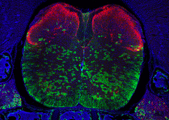
Image Credits
Neurodevelopmental Disorders: Courtesy of Lauren Orefice (MGH/HMS) Tools and Technology: Courtesy of Barbara Robens, lab of Ann Poduri (BCH) Sensory and Motor Systems: Courtesy of Lauren Orefice (MGH/HMS) Mental Health and Illness: Courtesy of Olga Alekseenko, Lab of Susan Dymecki (HMS) Neurodegenerative Disease: Courtesy of Jeff Lichtman (Harvard) and Takao Hensch (Harvard/BCH) Cellular and Molecular Neuroscience: Courtesy of Isle Bastille, lab of Lisa Goodrich (HMS) Theory and Computation: Courtesy of Tianyang Ye, lab of Hongkun Park (Harvard) Development Neuroscience: Courtesy of Katherine Morillo, lab of Christopher A. Walsh (BCH)
Johns Hopkins School of Medicine
The Solomon H. Snyder Department of Neuroscience
Areas of Research
Johns Hopkins has long been at the forefront of neuroscience research, combining scientific excellence, collegiality, and interdisciplinary collaborations to seed discovery and promote innovation.
Our researchers investigate some of the most perplexing mysteries of the brain to extend scientific advances in animal and human growth, health, and behavior throughout the lifespan.
Breakthroughs in key areas under investigation have enabled Hopkins scientists to translate fundamental discoveries into applications to improve health, extend life, and advance scientific understanding.
Division of Biology and Medicine
Department of neuroscience, research areas.
Research in neuroscience is collaborative, drawing upon the expertise of scientists across departments, institutes, and centers at Brown.
Cellular and Molecular Neurobiology
Cognition and behavior, computation in brain and mind, neural circuits, neuroengineering and neurotechnology, sensation and perception.
Princeton Neuroscience Institute
Research areas.

Researchers at PNI are currently working at the intersection of neuroscience and AI to gain new insights into neural data, generate new hypotheses for understanding the brain and behavior, and develop the next-generation AI architectures inspired by the brain.

This branch of neuroscience focuses on understanding how the brain processes information through the intricate networks of neurons and circuits. It aims to uncover the underlying mechanisms that govern complex brain functions such as perception, memory, decision-making, and behavior.

Human cognitive neuroscience is a field of study that investigates the neural basis of human cognition, which refers to various mental processes such as perception, attention, learning, memory, decision-making, language and problem-solving. It aims to understand how the brain supports these cognitive functions.

This field focuses on understanding the cellular and molecular mechanisms underlying the structure and function of the nervous system. It investigates how individual neurons and their various components such as synapses, receptors and ion channels contribute to the overall functioning of the brain and the nervous system.

Computation and theory neuroscience combines principles from neuroscience, computer science, and mathematics to understand how the brain processes information and performs complex computations. It aims to develop theoretical models and computational frameworks that can explain and predict the functions of the nervous system.

PNI is committed to maintaining leading-edge research computing infrastructure and support services which help support the creation of novel data collection and analysis methods.
- Primary Faculty
- Secondary Faculty
- Adjunct Faculty
- Emeritus Faculty
- Pierce Lab Faculty
- Administrative Staff
- Imaging Core Facility
- Swartz Center
- Graduate Program
- TraineeTuesday
- Past Retreats
- SYNAPSES: Seminars at Yale Neuroscience
- Kavli Institute
- Goals & Initiatives
- Our Members
- Opportunities
- Community Outreach
INFORMATION FOR
- Residents & Fellows
- Researchers
Areas of Research
The Yale Neuroscience Department aims to understand the fundamental mysteries of the nervous system, identify the neural underpinnings of psychiatric and neurological disorders, and develop novel diagnostic, treatment, and prevention approaches to improve human health. Our research explores the function of molecules and organelles, computation and communication by chemical and electrical signals, representation of the environment by neural circuits, and the generation of complex behaviors. We develop and apply a wide variety of experimental systems and approaches. Current interdisciplinary areas within the Department include neuronal cell biology, sensory processing, the development and function of the cerebral cortex, and neurodegeneration. We strive to facilitate connections between all aspects of neuroscience, including molecules/cells, circuits/systems, and disease/translation.
Cellular | Molecular | Developmental
- Disease | Injury
- Systems | Circuits | Cognition
Neurons perform highly complex and dynamic functions, are highly diverse, and are connected into precise networks that enable information processing. Researchers in the department seek to understand the biological mechanisms that enable neurons to function, differentiate, and wire together into networks.
Amy Arnsten Sreeganga Chandra Daniel Colón-Ramos Michael Crair Pietro De Camilli Emilia Favuzzi Elena Gracheva Junjie Guo Marc Hammarlund Michael Higley Liang Liang Janghoo Lim Pasko Rakic Michael Schwartz Nenad Sestan Heather Snell Stephen Strittmatter Susumu Tomita Shaul Yogev
An official website of the United States government
The .gov means it’s official. Federal government websites often end in .gov or .mil. Before sharing sensitive information, make sure you’re on a federal government site.
The site is secure. The https:// ensures that you are connecting to the official website and that any information you provide is encrypted and transmitted securely.
- Publications
- Account settings
The PMC website is updating on October 15, 2024. Learn More or Try it out now .
- Advanced Search
- Journal List
- v.40(1); 2020 Jan 2
The Next 50 Years of Neuroscience
Cara m. altimus.
1 Milken Institute,
Bianca Jones Marlin
2 Columbia University Zuckerman Institute,
Naomi Ekavi Charalambakis
3 University of Louisville,
Alexandra Colón-Rodriquez
4 University of California at Davis,
Elizabeth J. Glover
5 University of Illinois at Chicago,
Patricia Izbicki
6 Iowa State University,
Anthony Johnson
7 Veterans Administration Health Care System,
Mychael V. Lourenco
8 Federal University of Rio do Janeiro,
Ryan A. Makinson
9 Journal of DoD Research & Engineering,
Joseph McQuail
10 University of South Carolina School of Medicine,
Ignacio Obeso
11 Fundación de Investigación HM Hospitales,
Nancy Padilla-Coreano
12 Salk Institute, and
Michael F. Wells
13 Broad Institute
Associated Data
Selected influential advances in neuroscience over the past 50 years and predicted key discoveries that aim to support mission of the Society for Neuroscience, first articulated in 1969: “to advance [the] understanding of nervous systems and their role in behavior, to promote education in the neurosciences, and to inform the general public on results and implications of current research. Download Figure 1-1, JPG file
On the 50th anniversary of the Society for Neuroscience, we reflect on the remarkable progress that the field has made in understanding the nervous system, and look forward to the contributions of the next 50 years. We predict a substantial acceleration of our understanding of the nervous system that will drive the development of new therapeutic strategies to treat diseases over the course of the next five decades.
On the 50th anniversary of the Society for Neuroscience, we reflect on the remarkable progress that the field has made in understanding the nervous system, and look forward to the contributions of the next 50 years. We predict a substantial acceleration of our understanding of the nervous system that will drive the development of new therapeutic strategies to treat diseases over the course of the next five decades. We also see neuroscience at the nexus of many societal topics beyond medicine, including education, consumerism, and the justice system. In combination, advances made by basic, translational, and clinical neuroscience research in the next 50 years have great potential for lasting improvements in human health, the economy, and society.
Introduction
In 1969, the United States National Academies Committee on Brain Sciences agreed that a central organization was needed to “1) advance understanding of nervous systems and their role in behavior; 2) promote education in the neurosciences; and 3) inform the general public on results and implications of current research.” Thus, the Society for Neuroscience (SfN) was founded with the goal of serving as that central organization by bringing together neuroscientists across disciplines. Over the course of the last 50 years, members of the Society have been instrumental in driving the incredible growth and rapid technological advances that have accelerated our understanding of both healthy and pathological nervous system function.
As members of SfN's Trainee Advisory Committee, many of us joined the field within the last decade and recognize that it is our cohort's vision and drive that will advance the field through the next 50 years. Our Committee represents the vastness of neuroscience research, with members spanning broad scientific interests, ranging from neurodevelopment to neural correlates of behavior, and coming from countries around the world. As a diverse group of leaders within the Society, we think deeply about the next generation of neuroscientists and what their scientific world will look like because it is also what our world will look like. This article and timeline figure convey not only our vision of where the field will be, but are also a reflection of the research achievements that have driven us scientifically, and what we are most excited to see develop in the future (Extended Data Figure 1-1 ). It is our hope that this vision will spur enthusiasm among professionals in the field and increase public understanding of the remarkable potential that neuroscience research holds to improve human health and society.
Cellular and molecular neuroscience
The past 50 years produced monumental advancements in our understanding of the cellular and molecular processes that dictate our every thought, desire, and action. This progress was driven, in part, by technical innovations, such as patch-clamp electrophysiology, PCR, and genomic sequencing, which gifted neuroscientists with experimental opportunities that were inconceivable in the 1960s. By the time SfN celebrates its 100th anniversary, we anticipate even greater shifts in methodology and conceptual consensus that will push the field further toward answering such questions as follows: How do the billions of individual components of the brain work together to generate behavior? How do changes to the brain lead to disease? What makes the human brain unique?
Two notable accomplishments toward answering these questions will be the completion of the connectome and a comprehensive cellular atlas of the mammalian brain. Execution of these daunting tasks is fueled in part by funding from National Institutes of Health's BRAIN Initiative, a 10 year program initiated in 2016 in the United States aiming to support the development and implementation of innovative neurotechnologies to better understand the brain ( Bargmann, 2014 ), as well as the Human Brain Project funded by the European Union to foster research at the interface of neuroscience and computation and the Brain/MINDS project in Japan focused on mapping higher brain function in marmosets. Among these new technologies are ongoing advancements in single-cell transcriptomics/proteomics, which will play a pivotal role in revealing the immense diversity of cell types within brains across a wide range of species ( Saunders et al., 2018 ; Wang et al., 2018 ). In combination with the development of automated high-throughput and innovative optical electrophysiological approaches ( Priest et al., 2004 ; Zhang et al., 2016 ), neuroscientists will begin to understand how discrete cell populations are physiologically and phylogenetically distinct. In doing so, we will determine not only the roles of specific cell types in the healthy and diseased brain, but also the cellular mechanisms that separate humans from other mammalian species. The data obtained will be used in conjunction with recently developed approaches, such as optogenetics ( Boyden, 2011 ) and chemogenetics ( Sternson and Roth, 2014 ), as well as new methods for visualizing genetically encoded calcium indicators ( Resendez and Stuber, 2015 ), to revolutionize the ways in which we probe, perturb, and define distinct cell populations.
A cell's molecular makeup is vast and diverse, but neuroscientists' ability to resolve disease-induced alterations in molecular composition is currently laborious and imprecise. In the next 50 years, advances in microscopy ( Gao et al., 2019 ) will become broadly accessible and afford researchers the ability to visualize subcellular machinery with unprecedented resolution, catapulting our understanding of the interplay between changes at the transcriptional, molecular, and structural levels. The development of new tools facilitating in vivo measurement and manipulation of epigenetic and molecular endpoints will revolutionize our ability to reconcile the influence of changes to the epigenome, genome, transcriptome, and proteome with behavior ( Hayashi-Takagi et al., 2015 ). In vivo findings will be supplemented by studies performed in stem cell-derived cerebral organoids, which serve as a model of the developing human brain, and when combined with novel molecular and imaging tools, will begin to decipher the roles of specific cellular types and processes in the earliest stages of human neurodevelopment. Research over the next 50 years will further our understanding of the maturation of the synapse and the ways in which this critically important structure is regulated by complex signaling pathways, plasticity mechanisms, and non-neuronal elements, such as astrocytes, microglia, and the extracellular matrix (Dityatev et al., 2010; Prinz and Priller, 2014 ; Fields, 2015 ; Ben Haim and Rowitch, 2017 ).
Although initially these advancements will take a reductive approach, they will serve as a foundation upon which circuit and systems neuroscientists can build to gain a more thorough understanding of the brain across development, environment, and genetic background. Application of many of these tools in humans will be dependent on novel cellular targeting strategies that will make it easier for modified RNA, viral vectors, or small compounds to be directed to the cell types of choice, resulting in more accurate circuit manipulations and delivery of gene therapies and pharmaceuticals. These advances, combined with refined biomarkers of brain health, have the potential to vastly enhance our understanding of brain disease and open new avenues of therapeutic intervention.
Development
Building upon the advances in cellular and molecular neuroscience, the field of developmental neuroscience will be enabled to describe how internal and external factors shift the trajectory of individual neurons, circuits, and the brain to alter disease risk and behavior. Neurodevelopment spans intracellular study through systemwide analysis to allow an understanding of how individual neurons acquire specific function within the nervous system as well as how the brain develops over decades. While many avenues of research will be impactful, we see single-cell characterization, study of neurogenesis, and the use of organoids as key areas of focus in the next half century.
In particular, transcriptional characterization of neurons will be instrumental in providing a foundation by which researchers can study how cell fate, migratory paths, and connectivity are determined in unique cell types. Additionally, the use of whole genome sequencing to map cell lineage via the identification of somatic mutations ( Evrony et al., 2015 ; Lodato et al., 2015 ) will provide crucial insight into the similarities and differences in cell dispersal in humans relative to other species. Together with broad implementation of new techniques that build upon the development of the Brainbow mouse by selectively labeling neurons undergoing differentiation or division ( Gomez-Nicola et al., 2014 ; Loulier et al., 2014 ), these approaches will prove essential to allow neuroscientists to monitor the fate of individual neural progenitors and their trajectories as they form the complex circuits that define the nervous system.
The past 50 years of neuroscience research has played host to a decades-long debate over the existence of adult neurogenesis. This controversial concept was first introduced before SfN's formation in 1969 ( Altman, 1962 ) but failed to gain significant traction until the 1980s and 1990s when an increasing number of reports demonstrated the presence of newly born cells originating in the subventricular and subgranular zones of a number of species, including humans ( Eriksson et al., 1998 ; Knoth et al., 2010 ; Spalding et al., 2013 ). Nevertheless, despite convincing evidence, the debate has continued, with recent work suggesting that adult hippocampal neurogenesis is minimal, if not absent, at least in primates ( Sorrells et al., 2018 ). Yet, an even more recent study created additional controversy by demonstrating adult hippocampal neurogenesis is robust in healthy aged individuals ( Boldrini et al., 2018 ; Moreno-Jiménez et al., 2019 ). The reasons for the continued controversy likely relate to technical limitations arising from the use of postmortem tissues with variable fixation protocols, possibly measuring the wrong marker(s) for neural stem cells, and from attempting to generalize results from rodent models. Over the course of the next five decades, we expect that new technologies to definitively label new neurons in vivo via noninvasive imaging techniques or in ex vivo samples across mammalian species will move the field forward. These continuing attempts to resolve this issue will lead to even deeper insights into complex mechanisms of cortical development in primates. Additionally, by applying results from 'omic studies of developing neurons, we anticipate that tools may be developed to precisely control neurogenesis to modify disease processes and understand its roles in physiology and disease.
Since their introduction in 2013, brain organoids have presented neuroscientists with a model system that can be used to study a myriad of processes, including brain development and aging ( Lancaster et al., 2013 ; Gonzalez et al., 2018 ; Karzbrun and Reiner, 2019 ; Pollen et al., 2019 ). While protocols have been established to grow brain organoids from embryonic or induced pluripotent stem cells ( Lancaster et al., 2013 ; Sloan et al., 2017 ; Karzbrun and Reiner, 2019 ), several methodological drawbacks have prevented them from realizing their full research potential ( Karzbrun and Reiner, 2019 ; Yakoub, 2019 ; Yoon et al., 2019 ). Technological advances in the coming years will resolve the vascular and structural support difficulties researchers currently experience to allow the growth of larger, highly reproducible organoids that more closely resemble the complexity of the developing human brain. These advances will usher in a new era of in vitro research, enabling investigators to explore numerous facets of developmental neuroscience. In combination with live-cell imaging, brain organoids will vastly accelerate progress in understanding the complex signaling patterns that drive cell fate, neuronal migration, and neurite extension. Computational approaches, such as those developed for systems modeling, have thus far only seen limited application in developmental neuroscience; however, these methods will allow researchers to study the complex interplay between the seemingly infinite number of spatially and temporally distinct signals driving cell fate, neuronal migration, and circuit formation, which to date have largely been studied in isolation. Brain organoid experiments using viral strategies to measure and manipulate neuronal activity will also be pivotal in elucidating the role that experience-dependent plasticity plays in the formation and maintenance of neural circuits. Together with the development of more broadly accessible methods to manipulate cell structure in vivo ( Hayashi-Takagi et al., 2015 ), these advances will allow neuroscientists to better understand the mechanisms underlying synapse formation and link structural plasticity with synaptic plasticity and behavior.
In addition to improving our understanding of the mechanisms underlying neural development, organoids will afford researchers a system for studying characteristics of the nervous system that are uniquely human and contribute to our knowledge of neurodevelopmental diseases, including autism and schizophrenia, which have proven difficult to study in animal models ( Di Lullo and Kriegstein, 2017 ). With the ability to form functional circuits that could persist for months or potentially years, neuroscientists will be able to test the effects of genetics, age, and environment impact on brain function by comparing healthy cell lines to lines with genetic errors across time and in response to environmental stressors. Ultimately, brain organoids will become a standard model to screen pharmaceuticals and test the efficacy of gene editing techniques as therapies for neurological diseases. Furthermore, this technology may one day provide the means to correct damage resulting from injury or disease using self-derived replacement brain tissue.
From systems to behaviors
Historically, neuroscientists have taken a reductionist approach to understanding brain function. Our modern understanding of the brain has evolved over the past century, from the limited 47 brain regions known in 1909, to our current human brain map with 98 regions in the cortex, alone ( Glasser et al., 2016 ). Initially, neuroscientists relied on lesions or pharmacological manipulations in animals to determine the role of a given brain region. However, within the past two decades, new genetic tools have increased our ability to precisely manipulate circuits in animal models with increased precision. Such studies have heightened our understanding of the circuits underlying sensory processing, motor control, and memory. Questions still remain, as much of the work to date has focused on examining these circuits in isolation, therefore making limited progress in understanding how multiple regions or circuits interact to produce a behavioral output. For example, how do circuits for motor control, sensory processing, and decision making interact? How do manipulations of sensory processing affect the computations of planned motor movement?
Given that several brain systems are well understood individually, and the field has developed techniques with higher temporal and spatial resolution to monitor and manipulate neural activity, we are now better positioned to start deciphering how groups of neurons and distant regions work together to drive behavior. For this, the use of high-density multisite electrode recordings in the full brain will transform the field. Additionally, virtual reality environments, model-based analyses, and artificial intelligence approaches can be combined with these new recording and manipulation techniques to allow researchers to study how multiple sensory inputs are integrated and transformed into a behavioral output (e.g., action, thought, decision). Zebrafish and Caenorhabditis elegans will also prove instrumental in studying how multiple functional circuits operate in tandem because these animals offer researchers an opportunity to image the activity of the entire nervous system at once in conjunction with behavioral monitoring ( Cong et al., 2017 ). As neuroscientists sample more neurons with high-density electrodes or imaging methods, it will be important to attempt to understand what all the neurons are encoding, not just the neurons that are task-responsive or that support the specific hypotheses in the study. For this effort, statistical and computational methods, such as machine learning, will become essential and open up whole new areas of neuroengineering.
The recent development of virally mediated gene-editing strategies to optically measure and manipulate selected groups of neurons in vivo has been a boon for systems neuroscientists. These new technologies have moved circuit-based experiments into the limelight and are rapidly elucidating the connections, and the specific role of unique neural populations. Over the next 50 years, these techniques will provide the foundation for monumental breakthroughs in our understanding of how neural ensembles guide behavior ( Jennings et al., 2019 ), and perhaps even consciousness. Consciousness, in particular, is an important target of in-depth investigation as the very experience of awareness in ourselves and the world around us likely drives cognitive functioning (e.g., action planning or decision making) and may be modulated by diseases and conditions that affect the brain. Together with cellular-resolution functional human neuroimaging approaches, which are just beginning to be realized ( Koopmans and Yacoub, 2019 ), cognitive neuroscientists will begin to unlock the still poorly understood complexity of the distinct brain regions, such as the cerebellum, PFC, or hippocampus as well as how activity in multiple regions work in concert with each other. For example, higher-resolution imaging of the human brain will allow deciphering new understanding of circuit functionalities paving the way for neural modulatory interventions, such as transcranial magnetic stimulation and ultrasound neuromodulation. These circuit-based intervention strategies could be used to treat neuropsychiatric illness by manipulating a functionally distinct neural hub ( Diana et al., 2017 ).
In light of incredible strides, systems neuroscience has been limited by the way behavior is measured and correlated to neural activity. The ability of neuroscientists to resolve distinct functional circuits is limited by the precision with which behavior is defined and measured, often manually or semiautomatically by a human observer, resulting in oversimplified endpoints and frequently overlooked details ( Anderson and Perona, 2014 ). Additionally, behavioral measurements are especially rudimentary for social behaviors in animals. Over the next 50 years, approaches used in behavioral neuroscience will more closely resemble the sophisticated methods being used to functionally dissect neural circuits. Computer vision technology will enable fully automated, high-throughput, unbiased behavioral analysis, exponentially pushing the field forward ( Wiltschko et al., 2015 ; Mathis et al., 2018 ). Moreover, our ability to track behavior continuously and reliably in social settings will open the door to the development of new animal models of neuropsychiatric diseases, such as anxiety and depression, for which current models are overly simplistic. Similarly, approaches in humans (e.g., using in-home laboratories, online experiments, neurofeedback) ( Awolusi et al., 2018 ; Marins et al., 2019 ) have the potential to uncover previously unrealized symptomology and/or behavioral indices of risk for disease ( Anderson and Perona, 2014 ; Wiltschko et al., 2015 ; Cong et al., 2017 ; Mathis et al., 2018 ; Jennings et al., 2019 ).
Finally, through combining technologies to record and interact with neural circuits in real time, as well as incorporating unbiased methodologies to characterizing behavior and neural activity, we will see transformation on neural interface technologies that directly engage the nervous system. This technology is currently undergoing rapid advancement with brain–computer interfaces successfully allowing control of prosthetic limbs and perception of rudimentary visual imagery in the blind. As these technologies advance, there is hope that these neural interfaces will advance to allow broader application for prosthetic limbs, inclusion of sensory feedback, and perhaps memory improvement in individuals who experience cognitive decline.
Over the last 50 years, scientific discoveries have improved our understanding of how specific diseases disrupt nervous system function. Fortunately, we are moving past a time when individuals affected by conditions, such as autism, depression, schizophrenia, and dementia, are institutionalized, stigmatized, and marginalized. Today, policymakers and society rely heavily on neuroscientists to inform them about the role of the brain in these conditions and the advances in detection, prognosis, and treatment for patients affected by neurological and neuropsychiatric disorders. Over the next 50 years, we anticipate that disease research will address the following questions: What molecular/cellular changes happen in the brain before the onset of nervous system disorders? How can we harness the biological understanding of a disease to develop targeted therapeutics to tackle the full complexity and multifactorial nature of neurological disease? How can we intervene early to prevent disease manifestation and/or progression?
With this in mind, 50 years from now, we predict we will be celebrating an era of “neurotherapeutics.” The beginnings of this era are already upon us, as an impressive number of neuro-based therapies have recently gained FDA approval: examples are esketamine for major depressive disorder, brexanolone for postpartum depression, and siponimod for multiple sclerosis ( Urquhart, 2019 ). However, today drugs intended to treat nervous system disorders take longer and are less likely to gain FDA approval compared with other drugs ( Gaffney, 2014 ). Similar to the strong progress made in cancer treatment over the last 30 years, an increase in the number of successful therapies for the treatment of nervous system disorders will be driven largely by public and political support for funding directed toward such endeavors. The BRAIN Initiative has already played a fundamental role in the development of technologies that likely will greatly impact disease diagnosis and treatment. This funding, in addition to disease-specific funding, such as US Department of Health & Human Services' National Plan to Address AD and associated federal funding dedicated to AD research, as well as the dementia research initiatives led by the United Kingdom ( Fox and Petersen, 2013 ), has the potential to lead to faster translation and compounded discovery for prioritized classes of neural-based illness.
Beyond therapeutic development, we will also apply biological and mechanistic understanding to the diagnosis of neurological and psychiatric conditions. Specifically, we will transition from a symptom-based approach to one that also considers etiological agents and molecular intricacies. This realignment is exemplified by the use of genotype to define spinal muscular atrophy, as well as a new research criteria in which molecular alterations in the brain are used to classify dementias, including AD, even in the absence of clinical or postmortem neuropathology ( Jack and Vemuri, 2018 ; Khachaturian et al., 2018 ). Improved sensitivity and multiplexing of blood tests and other minimally invasive tests to detect changes in brain function will aid in extending this approach to diseases other than spinal muscular atrophy and AD. Technological advances, such as activity trackers and artificial intelligence, will have profound impacts on how we understand normal and abnormal function and treat neurological disorders. Artificial intelligence has already revealed specific plasma biomarker combinations to improve AD diagnostics ( Ashton et al., 2019 ) and will be used similarly in the future to more efficiently analyze the efficacy of pharmacotherapies in larger biobanks, thereby expediting the discovery of new therapies. Additionally, the development of new tracers compatible with positron emission tomography imaging hold promise as a valuable diagnostic and prognostic resource ( Leuzy et al., 2018 ).
Alongside the investment of time, resources, and effort in finding cures for brain illnesses, it will be imperative to foster research on preventive mechanisms. The high prevalence of neurological diseases worldwide is socially demanding and economically expensive. Therefore, defining the essential mechanisms by which tractable lifestyle interventions (physical exercise, diet, cognitive training, and engagement in social, cultural, and educational activities) could potentially modify disease risk should be an enduring research priority throughout the upcoming 50 years. Likewise, interrogation of genetic and environmental susceptibility factors (e.g., polymorphisms, exposure to toxins) may reveal important clues to inform health policies and medical practice in the future.
In total, we see the advancements in cellular, developmental, and systems neuroscience culminating in dramatic improvements for nervous system illnesses through improved understanding of underlying mechanisms of disease, identification of new diagnostic endpoints to detect disease before symptom onset, and ultimately, new methods for treatment and prevention.
An inclusive future
It is clear that the next 50 years will be marked not only by a more comprehensive understanding of the system that allows us to interact with the world around us, but also by fundamental changes in how neuroscience research is accomplished and the very topics that are studied. Among these changes, neuroscientists must acknowledge the importance of diversity. To date, research in male (across species) ( Shansky, 2019 ) and right-handed subjects has predominated. Additionally, clinical trials and genetic studies continue to overwhelmingly assess individuals of European descent. These systemic barriers to a comprehensive understanding of neuroscience, and the individual differences contained therein, are driven, in part, by a lack of diversity within neuroscientists themselves. As a result, the field suffers from a lack of understanding with respect to sex differences and the female brain, and FDA- or EMA-approved drugs frequently exhibit decreased therapeutic efficacy in nonwhite populations. Looking forward, we must prioritize greater diversity in both our researchers and our research subjects.
Neuroscience in society
The impacts of neuroscience research extend far beyond the clinic to the classroom, the courtroom, and even the grocery store. Indeed, neuro-technologies are already moving into our homes, promising to boost cognitive abilities, despite insufficient rigorous evidence of efficacy ( Nelson et al., 2016 ; Schuijer et al., 2017 ).
Neuroeducation, a field that combines research findings in developmental and cognitive neuroscience with educational strategies ( Sigman et al., 2014 ), has contributed greatly to our understanding of how students with dyslexia, attention deficit hyperactivity disorder, and other disorders learn. This knowledge has been used to implement changes in math, arts, and science curricula for students with these disorders. Recent evidence also shows that intertwining arts and science education allows students to find more creative and innovative approaches to solving problems. Despite this progress, cognitive psychology and neuroscience are not broadly implemented in standard educational practices of teachers in both primary and higher education ( Sigman et al., 2014 ). Further application of neuroscience and development of research in this space are beginning to change when mathematics concepts are taught and fundamentally change the way we schedule school days to align with circadian rhythms. Over the course of the next 50 years, we expect to see broader application of neuroeducational strategies across age and educational setting.
Neuroscience is becoming increasingly more common in the courtroom as it is used to explain criminal behavior ( Ward et al., 2018 ). Its use will increase over the next 50 years as researchers become more knowledgeable about the neurobiological mechanisms underlying decision making. Moreover, as diagnostic tools, human neuroimaging methods in particular, become more advanced and afford researchers greater insight into brain function, these strategies will be used to determine an individual's culpability and even likelihood for recidivism.
Although it may not be apparent in our everyday lives, companies all over the world are using the results of neuroscience research to inform their business practices from office structure to product placement and marketing strategies. This will likely increase over the course of the next five decades as our understanding of the neurobiology of cognition and attention matures ( Gottlieb and Oudeyer, 2018 ). In particular, wearable neurotechnology has the potential to play a prominent role in providing instant consumer feedback allowing personalized marketing strategies that update in real-time ( Awolusi et al., 2018 ). However, companies should exercise caution and follow ethical principles when developing new strategies to generate profit based on neurobiological understanding and techniques.
In conclusion, neuroscience is a vast field. With ∼86 billion neurons in the adult human brain, and approximately the same number of non-neuronal cells, it is not surprising that the study of this organ is complex. Furthermore, the nervous system extends far beyond the cranium with neurons projecting to the furthest reaches of the body collecting input and responding to the environment. The progress the field continues to reinforces its enormous potential.
Beyond examining the complexity of the nervous system itself, we must ask ourselves how we study this system of systems. When considering the approach that other scientific fields with seemingly infinite complexity have taken, the study of space comes to mind. While individual nations have embarked on space exploration over the last century, collaboration across disciplines and countries likely contributed to the great strides made thus far. Borrowing from this example, interdisciplinary approaches, with teams of mathematicians, engineers, computer scientists, biologists, and chemists, are key to the continued advancement of neuroscience. Presently, neuroscience is funded in many countries through numerous agencies; however, recent national and international initiatives facilitating large-scale interdisciplinary neuroscience are emerging. The BRAIN Initiative and the Human Brain Project, for example, have not focused on one specific area of neuroscience but instead embraced participation from researchers spanning science, engineering, math, and technology.
The vitality of SfN, whose annual meeting has grown from 1395 to >30,000 attendees per year, highlights its immense value as a central space for scientific dialog and collaboration ( Fields, 2018 ). Expansion of these centrally coordinated efforts to accelerate brain research as well as a strong community of scientists will be instrumental in elevating the quality and capability of neuroscience research as it continues to explore the unknown.
The Trainee Advisory Committee is a group of current and recent trainees (graduate students and postdoctoral fellows) charged with making recommendations for the Society of Neuroscience Council and committees on how Society initiatives, projects, and programming can be designed, enhanced, or modified to address the needs and concerns of the Society's trainee members. All authors participated in this perspective as members of the 2019 Society for Neuroscience, Trainee Advisory Committee.
The authors declare no competing financial interests.
- Altman J. (1962) Are new neurons formed in the brains of adult mammals? Science 135 :1127–1128. 10.1126/science.135.3509.1127 [ PubMed ] [ CrossRef ] [ Google Scholar ]
- Anderson DJ, Perona P (2014) Toward a science of computational ethology . Neuron 84 :18–31. 10.1016/j.neuron.2014.09.005 [ PubMed ] [ CrossRef ] [ Google Scholar ]
- Ashton NJ, Alejo J, Nevado-Holgado AJ, Barber IS, Lynham S, Gupta V, Chatterjee P, Goozee K, Hone E, Pedrini S, Blennow K, Schöll M, Zetterberg H, Ellis KA, Bush AI, Rowe CC, Villemagne VL, Ames D, Masters CL, Aarsland D, Powell J, et al. (2019) A plasma protein classifier for predicting amyloid burden for preclinical Alzheimer's disease . Sci Adv 5 :eaau7220. 10.1126/sciadv.aau7220 [ PMC free article ] [ PubMed ] [ CrossRef ] [ Google Scholar ]
- Awolusi I, Marks E, Hallowell M (2018) Wearable technology for personalized construction safety monitoring and trending: review of applicable devices . Automation Construction 85 :96–106. 10.1016/j.autcon.2017.10.010 [ CrossRef ] [ Google Scholar ]
- Bargmann C. (2014) BRAIN 2025: a scientific vision . [ Google Scholar ]
- Ben Haim L, Rowitch DH (2017) Functional diversity of astrocytes in neural circuit regulation . Nat Rev Neurosci 18 :31–41. 10.1038/nrn.2016.159 [ PubMed ] [ CrossRef ] [ Google Scholar ]
- Boldrini M, Fulmore CA, Tartt AN, Simeon LR, Pavlova I, Poposka V, Rosoklija GB, Stankov A, Arango V, Dwork AJ, Hen R, Mann JJ (2018) Human hippocampal neurogenesis persists throughout aging . Cell Stem Cell 22 : 589–599.e5. [ PMC free article ] [ PubMed ] [ Google Scholar ]
- Boyden ES. (2011) A history of optogenetics: the development of tools for controlling brain circuits with light . F1000 Biol Rep 3 :11. 10.3410/B3-11 [ PMC free article ] [ PubMed ] [ CrossRef ] [ Google Scholar ]
- Cong L, Wang Z, Chai Y, Hang W, Shang C, Yang W, Bai L, Du J, Wang K, Wen Q (2017) Rapid whole brain imaging of neural activity in freely behaving larval zebrafish ( Danio rerio ) . Elife 6 :e28158. 10.7554/eLife.28158 [ PMC free article ] [ PubMed ] [ CrossRef ] [ Google Scholar ]
- Diana M, Raij T, Melis M, Nummenmaa A, Leggio L, Bonci A (2017) Rehabilitating the addicted brain with transcranial magnetic stimulation . Nat Rev Neurosci 18 :685–693. 10.1038/nrn.2017.113 [ PubMed ] [ CrossRef ] [ Google Scholar ]
- Di Lullo E, Kriegstein AR (2017) The use of brain organoids to investigate neural development and disease . Nat Rev Neurosci 18 :573–584. 10.1038/nrn.2017.107 [ PMC free article ] [ PubMed ] [ CrossRef ] [ Google Scholar ]
- Dubinsky JM. (2010) Neuroscience education for prekindergarten-12 teachers . J Neurosci 30 :8057–8060. 10.1523/JNEUROSCI.2322-10.2010 [ PMC free article ] [ PubMed ] [ CrossRef ] [ Google Scholar ]
- Eriksson PS, Perfilieva E, Björk-Eriksson T, Alborn AM, Nordborg C, Peterson DA, Gage FH (1998) Neurogenesis in the adult human hippocampus . Nat Med 4 :1313–1317. 10.1038/3305 [ PubMed ] [ CrossRef ] [ Google Scholar ]
- Evrony GD, Lee E, Mehta BK, Benjamini Y, Johnson RM, Cai X, Yang L, Haseley P, Lehmann HS, Park PJ, Walsh CA (2015) Cell lineage analysis in human brain using endogenous retroelements . Neuron 85 :49–59. 10.1016/j.neuron.2014.12.028 [ PMC free article ] [ PubMed ] [ CrossRef ] [ Google Scholar ]
- Fields RD. (2015) A new mechanism of nervous system plasticity: activity-dependent myelination . Nat Rev Neurosci 16 :756–767. 10.1038/nrn4023 [ PMC free article ] [ PubMed ] [ CrossRef ] [ Google Scholar ]
- Fields RD. (2018) The first Annual Meeting of the Society for Neuroscience, 1971: reflections approaching the 50th anniversary of the Society's formation . J Neurosci 38 :9311–9317. 10.1523/JNEUROSCI.3598-17.2018 [ PMC free article ] [ PubMed ] [ CrossRef ] [ Google Scholar ]
- Fox NC, Petersen RC (2013) The G8 dementia research summit-a starter for eight? Lancet 382 :1968–1969. 10.1016/S0140-6736(13)62426-5 [ PubMed ] [ CrossRef ] [ Google Scholar ]
- Gaffney A. (2014) Report finds FDA slow to approve CNS drugs, but getting faster . November 5, 2014. https://www.raps.org/regulatory-focus%E2%84%A2/news-articles/2014/11/report-finds-fda-slow-to-approve-cns-drugs,-but-getting-faster .
- Gao R, Asano SM, Upadhyayula S, Pisarev I, Milkie DE, Liu TL, Singh V, Graves A, Huynh GH, Zhao Y, Bogovic J, Colonell J, Ott CM, Zugates C, Tappan S, Rodriguez A, Mosaliganti KR, Sheu SH, Pasolli HA, Pang S, et al. (2019) Cortical column and whole-brain imaging with molecular contrast and nanoscale resolution . Science 363 :eaau8302. 10.1126/science.aau8302 [ PMC free article ] [ PubMed ] [ CrossRef ] [ Google Scholar ]
- Glasser MF, Coalson TS, Robinson EC, Hacker CD, Harwell J, Yacoub E, Ugurbil K, Andersson J, Beckmann CF, Jenkinson M, Smith SM, Van Essen DC (2016) A multi-modal parcellation of human cerebral cortex . Nature 536 :171–178. 10.1038/nature18933 [ PMC free article ] [ PubMed ] [ CrossRef ] [ Google Scholar ]
- Gomez-Nicola D, Riecken K, Fehse B, Perry VH (2014) In-vivo RGB marking and multicolour single-cell tracking in the adult brain . Sci Rep 4 :7520. 10.1038/srep07520 [ PMC free article ] [ PubMed ] [ CrossRef ] [ Google Scholar ]
- Gonzalez C, Armijo E, Bravo-Alegria J, Becerra-Calixto A, Mays CE, Soto C (2018) Modeling amyloid beta and tau pathology in human cerebral organoids . Mol Psychiatry 23 :2363–2374. 10.1038/s41380-018-0229-8 [ PMC free article ] [ PubMed ] [ CrossRef ] [ Google Scholar ]
- Gottlieb J, Oudeyer PY (2018) Towards a neuroscience of active sampling and curiosity . Nat Rev Neurosci 19 :758–770. 10.1038/s41583-018-0078-0 [ PubMed ] [ CrossRef ] [ Google Scholar ]
- Hayashi-Takagi A, Yagishita S, Nakamura M, Shirai F, Wu YI, Loshbaugh AL, Kuhlman B, Hahn KM, Kasai H (2015) Labelling and optical erasure of synaptic memory traces in the motor cortex . Nature 525 :333–338. 10.1038/nature15257 [ PMC free article ] [ PubMed ] [ CrossRef ] [ Google Scholar ]
- Jack CR Jr, Vemuri P (2018) Amyloid-β: a reflection of risk or a preclinical marker? Nat Rev Neurol 14 :319–320. 10.1038/s41582-018-0008-9 [ PubMed ] [ CrossRef ] [ Google Scholar ]
- Jennings JH, Kim CK, Marshel JH, Raffiee M, Ye L, Quirin S, Pak S, Ramakrishnan C, Deisseroth K (2019) Interacting neural ensembles in orbitofrontal cortex for social and feeding behaviour . Nature 565 :645–649. 10.1038/s41586-018-0866-8 [ PMC free article ] [ PubMed ] [ CrossRef ] [ Google Scholar ]
- Karzbrun E, Reiner O (2019) Brain organoids: a bottom-up approach for studying human neurodevelopment . Bioengineering (Basel) 6 :E9. 10.3390/bioengineering6010009 [ PMC free article ] [ PubMed ] [ CrossRef ] [ Google Scholar ]
- Khachaturian AS, Hayden KM, Mielke MM, Tang Y, Lutz MW, Gustafson DR, Kukull WA, Mohs R, Khachaturian ZS (2018) Future prospects and challenges for Alzheimer's disease drug development in the era of the NIA-AA research framework . Alzheimers Dement 14 :532–534. 10.1016/j.jalz.2018.03.003 [ PubMed ] [ CrossRef ] [ Google Scholar ]
- Knoth R, Singec I, Ditter M, Pantazis G, Capetian P, Meyer RP, Horvat V, Volk B, Kempermann G (2010) Murine features of neurogenesis in the human hippocampus across the lifespan from 0 to 100 years . PLoS One 5 :e8809. 10.1371/journal.pone.0008809 [ PMC free article ] [ PubMed ] [ CrossRef ] [ Google Scholar ]
- Koopmans PJ, Yacoub E (2019) Strategies and prospects for cortical depth dependent T2 and T2* weighted BOLD fMRI studies . Neuroimage 197 :668–676. 10.1016/j.neuroimage.2019.03.024 [ PubMed ] [ CrossRef ] [ Google Scholar ]
- Lancaster MA, Renner M, Martin CA, Wenzel D, Bicknell LS, Hurles ME, Homfray T, Penninger JM, Jackson AP, Knoblich JA (2013) Cerebral organoids model human brain development and microcephaly . Nature 501 :373–379. 10.1038/nature12517 [ PMC free article ] [ PubMed ] [ CrossRef ] [ Google Scholar ]
- Leuzy A, Heurling K, Ashton NJ, Schöll M, Zimmer ER (2018) In vivo detection of Alzheimer's disease . Yale J Biol Med 91 :291–300. [ PMC free article ] [ PubMed ] [ Google Scholar ]
- Lodato MA, Woodworth MB, Lee S, Evrony GD, Mehta BK, Karger A, Lee S, Chittenden TW, D'Gama AM, Cai X, Luquette LJ, Lee E, Park PJ, Walsh CA (2015) Somatic mutation in single human neurons tracks developmental and transcriptional history . Science 350 :94–98. 10.1126/science.aab1785 [ PMC free article ] [ PubMed ] [ CrossRef ] [ Google Scholar ]
- Loulier K, Barry R, Mahou P, Le Franc Y, Supatto W, Matho KS, Ieng S, Fouquet S, Dupin E, Benosman R, Chédotal A, Beaurepaire E, Morin X, Livet J (2014) Multiplex cell and lineage tracking with combinatorial labels . Neuron 81 :505–520. 10.1016/j.neuron.2013.12.016 [ PubMed ] [ CrossRef ] [ Google Scholar ]
- Marins T, Rodrigues EC, Bortolini T, Melo B, Moll J, Tovar-Moll F (2019) Structural and functional connectivity changes in response to short-term neurofeedback training with motor imagery . Neuroimage 194 :283–290. 10.1016/j.neuroimage.2019.03.027 [ PubMed ] [ CrossRef ] [ Google Scholar ]
- Mathis A, Mamidanna P, Cury KM, Abe T, Murthy VN, Mathis MW, Bethge M (2018) DeepLabCut: markerless pose estimation of user-defined body parts with deep learning . Nat Neurosci 21 :1281–1289. 10.1038/s41593-018-0209-y [ PubMed ] [ CrossRef ] [ Google Scholar ]
- Moreno-Jiménez EP, Flor-García M, Terreros-Roncal J, Rábano A, Cafini F, Pallas-Bazarra N, Ávila J, Llorens-Martín M (2019) Adult hippocampal neurogenesis is abundant in neurologically healthy subjects and drops sharply in patients with Alzheimer's disease . Nat Med 25 :554–560. 10.1038/s41591-019-0375-9 [ PubMed ] [ CrossRef ] [ Google Scholar ]
- Nelson J, McKinley RA, Phillips C, McIntire L, Goodyear C, Kreiner A, Monforton L (2016) The effects of transcranial direct current stimulation (TDCS) on multitasking throughput capacity . Front Hum Neurosci 10 :589. 10.3389/fnhum.2016.00589 [ PMC free article ] [ PubMed ] [ CrossRef ] [ Google Scholar ]
- Pollen AA, Bhaduri A, Andrews MG, Nowakowski TJ, Meyerson OS, Mostajo-Radji MA, Di Lullo E, Alvarado B, Bedolli M, Dougherty ML, Fiddes IT, Kronenberg ZN, Shuga J, Leyrat AA, West JA, Bershteyn M, Lowe CB, Pavlovic BJ, Salama SR, Haussler D, et al. (2019) Establishing cerebral organoids as models of human-specific brain evolution . Cell 176 : 743–756.e17. [ PMC free article ] [ PubMed ] [ Google Scholar ]
- Priest BT, Cerne R, Krambis MJ, Schmalhofer WA, Wakulchik M, Wilenkin B, Burris KD (2004) Automated electrophysiology assays . In: Assay guidance manual (Sittampalam GS, Coussens NP, Brimacombe K, Grossman A, Arkin M, Auld D, Austin C, et al., eds). Bethesda, MD: Eli Lilly and the National Center for Advancing Translational Sciences. [ PubMed ] [ Google Scholar ]
- Prinz M, Priller J (2014) Microglia and brain macrophages in the molecular age: from origin to neuropsychiatric disease . Nat Rev Neurosci 15 :300–312. 10.1038/nrn3722 [ PubMed ] [ CrossRef ] [ Google Scholar ]
- Resendez SL, Stuber GD (2015) In vivo calcium imaging to illuminate neurocircuit activity dynamics underlying naturalistic behavior . Neuropsychopharmacology 40 :238–239. 10.1038/npp.2014.206 [ PMC free article ] [ PubMed ] [ CrossRef ] [ Google Scholar ]
- Saunders A, Macosko EZ, Wysoker A, Goldman M, Krienen FM, de Rivera H, Bien E, Baum M, Bortolin L, Wang S, Goeva A, Nemesh J, Kamitaki N, Brumbaugh S, Kulp D, McCarroll SA (2018) Molecular diversity and specializations among the cells of the adult mouse brain . Cell 174 :1015–1030.e16. 10.1016/j.cell.2018.07.028 [ PMC free article ] [ PubMed ] [ CrossRef ] [ Google Scholar ]
- Schuijer JW, de Jong IM, Kupper F, van Atteveldt NM (2017) Transcranial electrical stimulation to enhance cognitive performance of healthy minors: a complex governance challenge . Front Hum Neurosci 11 :142. 10.3389/fnhum.2017.00142 [ PMC free article ] [ PubMed ] [ CrossRef ] [ Google Scholar ]
- Shansky RM. (2019) Are hormones a 'female problem' for animal research? Science 364 :825–826. 10.1126/science.aaw7570 [ PubMed ] [ CrossRef ] [ Google Scholar ]
- Sigman M, Peña M, Goldin AP, Ribeiro S (2014) Neuroscience and education: prime time to build the bridge . Nat Neurosci 17 :497–502. 10.1038/nn.3672 [ PubMed ] [ CrossRef ] [ Google Scholar ]
- Sloan SA, Darmanis S, Huber N, Khan TA, Birey F, Caneda C, Reimer R, Quake SR, Barres BA, Pasca SP (2017) Human astrocyte maturation captured in 3D cerebral cortical spheroids derived from pluripotent stem cells . Neuron 95 :779–790.e6. 10.1016/j.neuron.2017.07.035 [ PMC free article ] [ PubMed ] [ CrossRef ] [ Google Scholar ]
- Sorrells SF, Paredes MF, Cebrian-Silla A, Sandoval K, Qi D, Kelley KW, James D, Mayer S, Chang J, Auguste KI, Chang EF, Gutierrez AJ, Kriegstein AR, Mathern GW, Oldham MC, Huang EJ, Garcia-Verdugo JM, Yang Z, Alvarez-Buylla A (2018) Human hippocampal neurogenesis drops sharply in children to undetectable levels in adults . Nature 555 :377–381. 10.1038/nature25975 [ PMC free article ] [ PubMed ] [ CrossRef ] [ Google Scholar ]
- Spalding KL, Bergmann O, Alkass K, Bernard S, Salehpour M, Huttner HB, Boström E, Westerlund I, Vial C, Buchholz BA, Possnert G, Mash DC, Druid H, Frisén J (2013) Dynamics of hippocampal neurogenesis in adult humans . Cell 153 :1219–1227. 10.1016/j.cell.2013.05.002 [ PMC free article ] [ PubMed ] [ CrossRef ] [ Google Scholar ]
- Sternson SM, Roth BL (2014) Chemogenetic tools to interrogate brain functions . Annu Rev Neurosci 37 :387–407. 10.1146/annurev-neuro-071013-014048 [ PubMed ] [ CrossRef ] [ Google Scholar ]
- Urquhart L. (2019) FDA new drug approvals in Q1 2019 . Nat Rev Drug Discov . Advance online publication. Retrieved Apr 10, 2019. doi: 10.1038/d41573-019-00070-3. 10.1038/d41573-019-00070-3 [ PubMed ] [ CrossRef ] [ CrossRef ] [ Google Scholar ]
- Wang X, Allen WE, Wright MA, Sylwestrak EL, Samusik N, Vesuna S, Evans K, Liu C, Ramakrishnan C, Liu J, Nolan GP, Bava FA, Deisseroth K (2018) Three-dimensional intact-tissue sequencing of single-cell transcriptional states . Science 361 :eaat5691. 10.1126/science.aat5691 [ PMC free article ] [ PubMed ] [ CrossRef ] [ Google Scholar ]
- Ward T, Wilshire C, Jackson L (2018) The contribution of neuroscience to forensic explanation . Psychol Crime Law 24 :195–209. 10.1080/1068316X.2018.1427746 [ CrossRef ] [ Google Scholar ]
- Wiltschko AB, Johnson MJ, Iurilli G, Peterson RE, Katon JM, Pashkovski SL, Abraira VE, Adams RP, Datta SR (2015) Mapping sub-second structure in mouse behavior . Neuron 88 :1121–1135. 10.1016/j.neuron.2015.11.031 [ PMC free article ] [ PubMed ] [ CrossRef ] [ Google Scholar ]
- Yakoub AM. (2019) Cerebral organoids exhibit mature neurons and astrocytes and recapitulate electrophysiological activity of the human brain . Neural Regen Res 14 :757–761. 10.4103/1673-5374.249283 [ PMC free article ] [ PubMed ] [ CrossRef ] [ Google Scholar ]
- Yoon SJ, Elahi LS, Pasca AM, Marton RM, Gordon A, Revah O, Miura Y, Walczak EM, Holdgate GM, Fan HC, Huguenard JR, Geschwind DH, Pasca SP (2019) Reliability of human cortical organoid generation . Nat Methods 16 :75–78. 10.1038/s41592-018-0255-0 [ PMC free article ] [ PubMed ] [ CrossRef ] [ Google Scholar ]
- Zhang H, Reichert E, Cohen AE (2016) Optical electrophysiology for probing function and pharmacology of voltage-gated ion channels . Elife 5 :e15202. 10.7554/eLife.15202 [ PMC free article ] [ PubMed ] [ CrossRef ] [ Google Scholar ]

Donate Apply
Research Areas
Aging leads to significant decline in cognition and memory and is a risk factor for neurodegenerative diseases. By understanding the molecular and cellular processes that drive normal aging of the brain, we hopefully can identify strategies to minimize aging-induced decline of brain function.
Faculty researching Aging
Biologically, behavior is the internally coordinated response of whole living organisms (individuals or groups) to internal and/or external stimuli. It can be innate or learned from the environment.
Faculty Researching Behavior
Cognitive Systems
The organized activities of neurons within and between brain regions underlie our capacity to form new memories, decide, act, and learn. We seek to understand the roles that neural activity within and between brain regions such as the hippocampus, amygdala, basal ganglia, and cortex play in cognition. Members of the cognitive systems group explore these questions using multiple approaches such as single-unit electrophysiology, fast-scan cyclic voltammetry, calcium imaging, optogenetics, and fMRI.
Faculty researching Cognitive Systems
Computational Modeling
Applications of mathematics and computing to problems in the development, structure, physiology and cognitive abilities of the nervous system at levels ranging from single membrane channels to operations of the entire nervous system.
Faculty researching Computational Modeling
Drug Development
The process of developing a new drug that effectively targets a specific weakness in a cell. This process involves specific pre-clinical development and testing, followed by trials in humans to determine the efficacy of the drug.
Faculty researching Drug Development
Emotions are brain states characterized by altered perception, memory, decision-making, and motor behavior. They arise from the coordinated activity of neurons that form brain-wide circuits. A major hub in this circuit is the amygdala. The cellular signals emitted by the amygdala during emotional states, and the interactions between the amygdala and other nodes of this circuit, provide an insight into how certain stimuli (e.g., a threatening face or a gentle touch) gain emotional significance and alter behavior.
Faculty researching Emotion
Homeostatic Regulation and Sleep
Sleep represents a set of states that are regulated by the brain and have implications for many areas of the body. Sleep is regulated by homeostatic processes that promote both sleep and wake states, as well as circadian processes that regulate the timing of physiologic and behavioral systems. Many of these systems interact with each other, as well as with tissues throughout the body, including immune, cardiovascular, metabolic, musculoskeletal, and sympathetic/parasympathetic systems.
Faculty researching Homeostatic Regulation and Sleep
Language and Neurolinguistics
Neurolinguistics studies the neural mechanisms in the human brain underlying comprehension, production, and acquisition of language. It draws methods and theories from fields such as neuroscience, linguistics, cognitive science, communication disorders and neuropsychology.
Faculty researching Language and Neurolinguistics
Learning & Memory
Neurobiological mechanisms of synaptic and neural plasticity mediate learning and memory Plasticity manifests itself as dynamic shifts in the strength and/or number of synaptic connections across neural circuits in response to changes in afferent input or efferent demand. Maladaptive changes in plasticity disturb neuronal circuits and cause pathological abnormalities that lead to neurological and/or psychiatric disorders.
Faculty Researching Learning & Memory
Molecular Mechanisms of Neural Function
The basic principles governing communication between neurons are determined by their molecular constituents, which work together in space and time to facilitate the functional properties of many types of neurons and/or glia. Key molecules include a neuronal-specific complement of cell-surface receptors, ion channels, transporters, transmitters, and signaling proteins facilitating excitability, synaptic function and plasticity, sensory transduction, cell-cell communication, and the formation of neural networks.
Faculty Researching Molecular Mechanisms of Neural Function
Motor Systems
To better understand how the brain and spinal cord coordinates the actions of multiple muscles to produce a host of movements – from breathing to object manipulation with the hand. Neuroscientists at Arizona study several aspects of the hierarchical motor system, using a variety of models and techniques, ranging from single neurons to behaving human subjects.
Faculty researching Motor Systems
Neural Engineering/Neurotechnology
Development and application of devices (invasive and noninvasive) that interface with the nervous system to restore or enhance function (e.g., visual prosthesis) or alleviate symptoms caused by a disease of the nervous system (e.g., spinal implant for relief of pain). Includes imaging – the development and application of tools and techniques to visualize neural activity and related processes on different spatial and temporal scales from noninvasive to molecular and milliseconds to seconds and beyond. Also includes electrophysiology - the branch of neuroscience that explores the electrical activity of living neurons and investigates the molecular and cellular processes that govern their signaling. Neurons communicate using electrical and chemical signals. Electrophysiology techniques listen in on these signals by measuring electrical activity, allowing scientists to decode intercellular and intracellular messages.
Faculty researching Neural Engineering/Neurotechnology
Neuro-immunology
Neuro-immunology is a relatively new area of neuroscience that studies the interactions of the immune system and the nervous system. Such interactions govern neurodevelopment and how the nervous system handles pathology such as infections, strokes, and neurodegeneration. In addition, the neuro-immune axis also influences systemic immune responses, and so understanding these interactions at the cellular and molecular level offers the opportunity for new insights into human health and disease.
Faculty researching Neuro-immunology
Neurodegeneration
Neurodegeneration is a progressive deterioration of neuronal structures and functions ultimately leading to physical and cognitive disability, dementia, and often premature death. Through the use of animal models, we seek a better understanding of the molecular and cellular causes underlying various neurodegenerative disease in the hope to develop biomarkers effectives therapies.
Faculty researching Neurodegeneration
Neurodevelopment and Regeneration
Molecular and cellular mechanism that generate, shape, and reshape the nervous system from embryonic development to adulthood and beyond. Defects lead to a wide variety of neurodevelopmental disorders affecting sensory, motor, and cognitive function. Regenerative mechanisms can drive the regrowth or repair of nervous tissues including the generation of new neurons or glia from stem cells, or the regeneration of axons or synapses.
Faculty researching Neurodevelopment and Regeneration
Pain is ordinarily experienced as an unpleasant sensory and emotional experience by events that result in, or have the potential to cause injury. In contrast, chronic pain is a maladaptive and debilitating condition that can result from nerve trauma, persistent inflammation, or disease. Currently available therapies have limited success in treating chronic pain and are associated with disabling, often intolerable, side effects.
Faculty researching Pain
Sensory Processing
The way a sensory stimulus is processed by the brain is not static but altered by experience. We try to elucidate the principles underlying these adaptive changes by examining how experience alters sensory processing.
Faculty researching Sensory Processing
Synaptic Function & Plasticity
Synapses are specialized cell-cell contact sites that facilitate communication and computation of information of neuronal circuits on a sub-millisecond scale. The accuracy of this process is vital as even subtle changes in synaptic function can disturb neuronal circuits and cause pathological abnormalities that lead to neurological and/or psychiatric disorders.
Faculty Researching Synaptic Function & Plasticity
Site Search
Research areas.
Neuroscience, the scientific study of the nervous system, is an exciting and growing field involving researchers from the physical, chemical, biological, computational, anthropological, and social sciences.
Some of the most fascinating and impactful research in the future likely will require scientists to have expertise in a variety of disciplines. Recognizing this, Penn State’s Neuroscience graduate program encourages students to gain a solid understanding of certain core fields through coursework and colloquia and to take multidisciplinary approaches to tackling research problems.
Student research is well supported by grants from private and public funds, particularly from the National Institutes of Health. In addition, all students admitted to the program receive financial aid (stipends and paid tuition costs). Students are usually admitted with the intent of obtaining a Ph.D. degree in Neuroscience; however, the M.S. degree may be sought as part of the doctoral program.
The Neuroscience graduate program provides students with interdisciplinary training in the growing field of neuroscience. Students conduct research alongside faculty members who are leaders in their fields. Research areas may include:
Molecular Neurobiology and Developmental Neuroscience
Investigating how and why the nervous system develops and functions at genetic, molecular and cellular levels.
Cognitive Neuroscience and Behavioral Neurobiology
Exploring how the nervous system processes information, controls autonomic functions, regulates states of consciousness, or determines behavior.
Neural Engineering
Using computer engineering, robotics and other technical disciplines to investigate how the nervous system works, and how it can be manipulated.
Systems Neuroscience
Examining how neural circuits function, are coordinated, and are controlled.
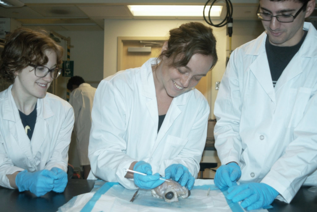
- Advanced search

Advanced Search
The Next 50 Years of Neuroscience
- Find this author on Google Scholar
- Find this author on PubMed
- Search for this author on this site
- ORCID record for Patricia Izbicki
- ORCID record for Anthony Johnson
- ORCID record for Mychael V. Lourenco
- ORCID record for Ignacio Obeso
- ORCID record for Michael F. Wells
- Figures & Data
- Info & Metrics

This article has a correction. Please see:
- Erratum: Altimus et al., “The Next 50 Years of Neuroscience” - May 11, 2020
On the 50th anniversary of the Society for Neuroscience, we reflect on the remarkable progress that the field has made in understanding the nervous system, and look forward to the contributions of the next 50 years. We predict a substantial acceleration of our understanding of the nervous system that will drive the development of new therapeutic strategies to treat diseases over the course of the next five decades. We also see neuroscience at the nexus of many societal topics beyond medicine, including education, consumerism, and the justice system. In combination, advances made by basic, translational, and clinical neuroscience research in the next 50 years have great potential for lasting improvements in human health, the economy, and society.
- Introduction
In 1969, the United States National Academies Committee on Brain Sciences agreed that a central organization was needed to “1) advance understanding of nervous systems and their role in behavior; 2) promote education in the neurosciences; and 3) inform the general public on results and implications of current research.” Thus, the Society for Neuroscience (SfN) was founded with the goal of serving as that central organization by bringing together neuroscientists across disciplines. Over the course of the last 50 years, members of the Society have been instrumental in driving the incredible growth and rapid technological advances that have accelerated our understanding of both healthy and pathological nervous system function.
As members of SfN's Trainee Advisory Committee, many of us joined the field within the last decade and recognize that it is our cohort's vision and drive that will advance the field through the next 50 years. Our Committee represents the vastness of neuroscience research, with members spanning broad scientific interests, ranging from neurodevelopment to neural correlates of behavior, and coming from countries around the world. As a diverse group of leaders within the Society, we think deeply about the next generation of neuroscientists and what their scientific world will look like because it is also what our world will look like. This article and timeline figure convey not only our vision of where the field will be, but are also a reflection of the research achievements that have driven us scientifically, and what we are most excited to see develop in the future (Extended Data Figure 1-1 ). It is our hope that this vision will spur enthusiasm among professionals in the field and increase public understanding of the remarkable potential that neuroscience research holds to improve human health and society.
Cellular and molecular neuroscience
The past 50 years produced monumental advancements in our understanding of the cellular and molecular processes that dictate our every thought, desire, and action. This progress was driven, in part, by technical innovations, such as patch-clamp electrophysiology, PCR, and genomic sequencing, which gifted neuroscientists with experimental opportunities that were inconceivable in the 1960s. By the time SfN celebrates its 100th anniversary, we anticipate even greater shifts in methodology and conceptual consensus that will push the field further toward answering such questions as follows: How do the billions of individual components of the brain work together to generate behavior? How do changes to the brain lead to disease? What makes the human brain unique?
Two notable accomplishments toward answering these questions will be the completion of the connectome and a comprehensive cellular atlas of the mammalian brain. Execution of these daunting tasks is fueled in part by funding from National Institutes of Health's BRAIN Initiative, a 10 year program initiated in 2016 in the United States aiming to support the development and implementation of innovative neurotechnologies to better understand the brain ( Bargmann, 2014 ), as well as the Human Brain Project funded by the European Union to foster research at the interface of neuroscience and computation and the Brain/MINDS project in Japan focused on mapping higher brain function in marmosets. Among these new technologies are ongoing advancements in single-cell transcriptomics/proteomics, which will play a pivotal role in revealing the immense diversity of cell types within brains across a wide range of species ( Saunders et al., 2018 ; Wang et al., 2018 ). In combination with the development of automated high-throughput and innovative optical electrophysiological approaches ( Priest et al., 2004 ; Zhang et al., 2016 ), neuroscientists will begin to understand how discrete cell populations are physiologically and phylogenetically distinct. In doing so, we will determine not only the roles of specific cell types in the healthy and diseased brain, but also the cellular mechanisms that separate humans from other mammalian species. The data obtained will be used in conjunction with recently developed approaches, such as optogenetics ( Boyden, 2011 ) and chemogenetics ( Sternson and Roth, 2014 ), as well as new methods for visualizing genetically encoded calcium indicators ( Resendez and Stuber, 2015 ), to revolutionize the ways in which we probe, perturb, and define distinct cell populations.
A cell's molecular makeup is vast and diverse, but neuroscientists' ability to resolve disease-induced alterations in molecular composition is currently laborious and imprecise. In the next 50 years, advances in microscopy ( Gao et al., 2019 ) will become broadly accessible and afford researchers the ability to visualize subcellular machinery with unprecedented resolution, catapulting our understanding of the interplay between changes at the transcriptional, molecular, and structural levels. The development of new tools facilitating in vivo measurement and manipulation of epigenetic and molecular endpoints will revolutionize our ability to reconcile the influence of changes to the epigenome, genome, transcriptome, and proteome with behavior ( Hayashi-Takagi et al., 2015 ). In vivo findings will be supplemented by studies performed in stem cell-derived cerebral organoids, which serve as a model of the developing human brain, and when combined with novel molecular and imaging tools, will begin to decipher the roles of specific cellular types and processes in the earliest stages of human neurodevelopment. Research over the next 50 years will further our understanding of the maturation of the synapse and the ways in which this critically important structure is regulated by complex signaling pathways, plasticity mechanisms, and non-neuronal elements, such as astrocytes, microglia, and the extracellular matrix (Dityatev et al., 2010; Prinz and Priller, 2014 ; Fields, 2015 ; Ben Haim and Rowitch, 2017 ).
Although initially these advancements will take a reductive approach, they will serve as a foundation upon which circuit and systems neuroscientists can build to gain a more thorough understanding of the brain across development, environment, and genetic background. Application of many of these tools in humans will be dependent on novel cellular targeting strategies that will make it easier for modified RNA, viral vectors, or small compounds to be directed to the cell types of choice, resulting in more accurate circuit manipulations and delivery of gene therapies and pharmaceuticals. These advances, combined with refined biomarkers of brain health, have the potential to vastly enhance our understanding of brain disease and open new avenues of therapeutic intervention.
Development
Building upon the advances in cellular and molecular neuroscience, the field of developmental neuroscience will be enabled to describe how internal and external factors shift the trajectory of individual neurons, circuits, and the brain to alter disease risk and behavior. Neurodevelopment spans intracellular study through systemwide analysis to allow an understanding of how individual neurons acquire specific function within the nervous system as well as how the brain develops over decades. While many avenues of research will be impactful, we see single-cell characterization, study of neurogenesis, and the use of organoids as key areas of focus in the next half century.
In particular, transcriptional characterization of neurons will be instrumental in providing a foundation by which researchers can study how cell fate, migratory paths, and connectivity are determined in unique cell types. Additionally, the use of whole genome sequencing to map cell lineage via the identification of somatic mutations ( Evrony et al., 2015 ; Lodato et al., 2015 ) will provide crucial insight into the similarities and differences in cell dispersal in humans relative to other species. Together with broad implementation of new techniques that build upon the development of the Brainbow mouse by selectively labeling neurons undergoing differentiation or division ( Gomez-Nicola et al., 2014 ; Loulier et al., 2014 ), these approaches will prove essential to allow neuroscientists to monitor the fate of individual neural progenitors and their trajectories as they form the complex circuits that define the nervous system.
The past 50 years of neuroscience research has played host to a decades-long debate over the existence of adult neurogenesis. This controversial concept was first introduced before SfN's formation in 1969 ( Altman, 1962 ) but failed to gain significant traction until the 1980s and 1990s when an increasing number of reports demonstrated the presence of newly born cells originating in the subventricular and subgranular zones of a number of species, including humans ( Eriksson et al., 1998 ; Knoth et al., 2010 ; Spalding et al., 2013 ). Nevertheless, despite convincing evidence, the debate has continued, with recent work suggesting that adult hippocampal neurogenesis is minimal, if not absent, at least in primates ( Sorrells et al., 2018 ). Yet, an even more recent study created additional controversy by demonstrating adult hippocampal neurogenesis is robust in healthy aged individuals ( Boldrini et al., 2018 ; Moreno-Jiménez et al., 2019 ). The reasons for the continued controversy likely relate to technical limitations arising from the use of postmortem tissues with variable fixation protocols, possibly measuring the wrong marker(s) for neural stem cells, and from attempting to generalize results from rodent models. Over the course of the next five decades, we expect that new technologies to definitively label new neurons in vivo via noninvasive imaging techniques or in ex vivo samples across mammalian species will move the field forward. These continuing attempts to resolve this issue will lead to even deeper insights into complex mechanisms of cortical development in primates. Additionally, by applying results from 'omic studies of developing neurons, we anticipate that tools may be developed to precisely control neurogenesis to modify disease processes and understand its roles in physiology and disease.
Since their introduction in 2013, brain organoids have presented neuroscientists with a model system that can be used to study a myriad of processes, including brain development and aging ( Lancaster et al., 2013 ; Gonzalez et al., 2018 ; Karzbrun and Reiner, 2019 ; Pollen et al., 2019 ). While protocols have been established to grow brain organoids from embryonic or induced pluripotent stem cells ( Lancaster et al., 2013 ; Sloan et al., 2017 ; Karzbrun and Reiner, 2019 ), several methodological drawbacks have prevented them from realizing their full research potential ( Karzbrun and Reiner, 2019 ; Yakoub, 2019 ; Yoon et al., 2019 ). Technological advances in the coming years will resolve the vascular and structural support difficulties researchers currently experience to allow the growth of larger, highly reproducible organoids that more closely resemble the complexity of the developing human brain. These advances will usher in a new era of in vitro research, enabling investigators to explore numerous facets of developmental neuroscience. In combination with live-cell imaging, brain organoids will vastly accelerate progress in understanding the complex signaling patterns that drive cell fate, neuronal migration, and neurite extension. Computational approaches, such as those developed for systems modeling, have thus far only seen limited application in developmental neuroscience; however, these methods will allow researchers to study the complex interplay between the seemingly infinite number of spatially and temporally distinct signals driving cell fate, neuronal migration, and circuit formation, which to date have largely been studied in isolation. Brain organoid experiments using viral strategies to measure and manipulate neuronal activity will also be pivotal in elucidating the role that experience-dependent plasticity plays in the formation and maintenance of neural circuits. Together with the development of more broadly accessible methods to manipulate cell structure in vivo ( Hayashi-Takagi et al., 2015 ), these advances will allow neuroscientists to better understand the mechanisms underlying synapse formation and link structural plasticity with synaptic plasticity and behavior.
In addition to improving our understanding of the mechanisms underlying neural development, organoids will afford researchers a system for studying characteristics of the nervous system that are uniquely human and contribute to our knowledge of neurodevelopmental diseases, including autism and schizophrenia, which have proven difficult to study in animal models ( Di Lullo and Kriegstein, 2017 ). With the ability to form functional circuits that could persist for months or potentially years, neuroscientists will be able to test the effects of genetics, age, and environment impact on brain function by comparing healthy cell lines to lines with genetic errors across time and in response to environmental stressors. Ultimately, brain organoids will become a standard model to screen pharmaceuticals and test the efficacy of gene editing techniques as therapies for neurological diseases. Furthermore, this technology may one day provide the means to correct damage resulting from injury or disease using self-derived replacement brain tissue.
From systems to behaviors
Historically, neuroscientists have taken a reductionist approach to understanding brain function. Our modern understanding of the brain has evolved over the past century, from the limited 47 brain regions known in 1909, to our current human brain map with 98 regions in the cortex, alone ( Glasser et al., 2016 ). Initially, neuroscientists relied on lesions or pharmacological manipulations in animals to determine the role of a given brain region. However, within the past two decades, new genetic tools have increased our ability to precisely manipulate circuits in animal models with increased precision. Such studies have heightened our understanding of the circuits underlying sensory processing, motor control, and memory. Questions still remain, as much of the work to date has focused on examining these circuits in isolation, therefore making limited progress in understanding how multiple regions or circuits interact to produce a behavioral output. For example, how do circuits for motor control, sensory processing, and decision making interact? How do manipulations of sensory processing affect the computations of planned motor movement?
Given that several brain systems are well understood individually, and the field has developed techniques with higher temporal and spatial resolution to monitor and manipulate neural activity, we are now better positioned to start deciphering how groups of neurons and distant regions work together to drive behavior. For this, the use of high-density multisite electrode recordings in the full brain will transform the field. Additionally, virtual reality environments, model-based analyses, and artificial intelligence approaches can be combined with these new recording and manipulation techniques to allow researchers to study how multiple sensory inputs are integrated and transformed into a behavioral output (e.g., action, thought, decision). Zebrafish and Caenorhabditis elegans will also prove instrumental in studying how multiple functional circuits operate in tandem because these animals offer researchers an opportunity to image the activity of the entire nervous system at once in conjunction with behavioral monitoring ( Cong et al., 2017 ). As neuroscientists sample more neurons with high-density electrodes or imaging methods, it will be important to attempt to understand what all the neurons are encoding, not just the neurons that are task-responsive or that support the specific hypotheses in the study. For this effort, statistical and computational methods, such as machine learning, will become essential and open up whole new areas of neuroengineering.
The recent development of virally mediated gene-editing strategies to optically measure and manipulate selected groups of neurons in vivo has been a boon for systems neuroscientists. These new technologies have moved circuit-based experiments into the limelight and are rapidly elucidating the connections, and the specific role of unique neural populations. Over the next 50 years, these techniques will provide the foundation for monumental breakthroughs in our understanding of how neural ensembles guide behavior ( Jennings et al., 2019 ), and perhaps even consciousness. Consciousness, in particular, is an important target of in-depth investigation as the very experience of awareness in ourselves and the world around us likely drives cognitive functioning (e.g., action planning or decision making) and may be modulated by diseases and conditions that affect the brain. Together with cellular-resolution functional human neuroimaging approaches, which are just beginning to be realized ( Koopmans and Yacoub, 2019 ), cognitive neuroscientists will begin to unlock the still poorly understood complexity of the distinct brain regions, such as the cerebellum, PFC, or hippocampus as well as how activity in multiple regions work in concert with each other. For example, higher-resolution imaging of the human brain will allow deciphering new understanding of circuit functionalities paving the way for neural modulatory interventions, such as transcranial magnetic stimulation and ultrasound neuromodulation. These circuit-based intervention strategies could be used to treat neuropsychiatric illness by manipulating a functionally distinct neural hub ( Diana et al., 2017 ).
In light of incredible strides, systems neuroscience has been limited by the way behavior is measured and correlated to neural activity. The ability of neuroscientists to resolve distinct functional circuits is limited by the precision with which behavior is defined and measured, often manually or semiautomatically by a human observer, resulting in oversimplified endpoints and frequently overlooked details ( Anderson and Perona, 2014 ). Additionally, behavioral measurements are especially rudimentary for social behaviors in animals. Over the next 50 years, approaches used in behavioral neuroscience will more closely resemble the sophisticated methods being used to functionally dissect neural circuits. Computer vision technology will enable fully automated, high-throughput, unbiased behavioral analysis, exponentially pushing the field forward ( Wiltschko et al., 2015 ; Mathis et al., 2018 ). Moreover, our ability to track behavior continuously and reliably in social settings will open the door to the development of new animal models of neuropsychiatric diseases, such as anxiety and depression, for which current models are overly simplistic. Similarly, approaches in humans (e.g., using in-home laboratories, online experiments, neurofeedback) ( Awolusi et al., 2018 ; Marins et al., 2019 ) have the potential to uncover previously unrealized symptomology and/or behavioral indices of risk for disease ( Anderson and Perona, 2014 ; Wiltschko et al., 2015 ; Cong et al., 2017 ; Mathis et al., 2018 ; Jennings et al., 2019 ).
Finally, through combining technologies to record and interact with neural circuits in real time, as well as incorporating unbiased methodologies to characterizing behavior and neural activity, we will see transformation on neural interface technologies that directly engage the nervous system. This technology is currently undergoing rapid advancement with brain–computer interfaces successfully allowing control of prosthetic limbs and perception of rudimentary visual imagery in the blind. As these technologies advance, there is hope that these neural interfaces will advance to allow broader application for prosthetic limbs, inclusion of sensory feedback, and perhaps memory improvement in individuals who experience cognitive decline.
Over the last 50 years, scientific discoveries have improved our understanding of how specific diseases disrupt nervous system function. Fortunately, we are moving past a time when individuals affected by conditions, such as autism, depression, schizophrenia, and dementia, are institutionalized, stigmatized, and marginalized. Today, policymakers and society rely heavily on neuroscientists to inform them about the role of the brain in these conditions and the advances in detection, prognosis, and treatment for patients affected by neurological and neuropsychiatric disorders. Over the next 50 years, we anticipate that disease research will address the following questions: What molecular/cellular changes happen in the brain before the onset of nervous system disorders? How can we harness the biological understanding of a disease to develop targeted therapeutics to tackle the full complexity and multifactorial nature of neurological disease? How can we intervene early to prevent disease manifestation and/or progression?
With this in mind, 50 years from now, we predict we will be celebrating an era of “neurotherapeutics.” The beginnings of this era are already upon us, as an impressive number of neuro-based therapies have recently gained FDA approval: examples are esketamine for major depressive disorder, brexanolone for postpartum depression, and siponimod for multiple sclerosis ( Urquhart, 2019 ). However, today drugs intended to treat nervous system disorders take longer and are less likely to gain FDA approval compared with other drugs ( Gaffney, 2014 ). Similar to the strong progress made in cancer treatment over the last 30 years, an increase in the number of successful therapies for the treatment of nervous system disorders will be driven largely by public and political support for funding directed toward such endeavors. The BRAIN Initiative has already played a fundamental role in the development of technologies that likely will greatly impact disease diagnosis and treatment. This funding, in addition to disease-specific funding, such as US Department of Health & Human Services' National Plan to Address AD and associated federal funding dedicated to AD research, as well as the dementia research initiatives led by the United Kingdom ( Fox and Petersen, 2013 ), has the potential to lead to faster translation and compounded discovery for prioritized classes of neural-based illness.
Beyond therapeutic development, we will also apply biological and mechanistic understanding to the diagnosis of neurological and psychiatric conditions. Specifically, we will transition from a symptom-based approach to one that also considers etiological agents and molecular intricacies. This realignment is exemplified by the use of genotype to define spinal muscular atrophy, as well as a new research criteria in which molecular alterations in the brain are used to classify dementias, including AD, even in the absence of clinical or postmortem neuropathology ( Jack and Vemuri, 2018 ; Khachaturian et al., 2018 ). Improved sensitivity and multiplexing of blood tests and other minimally invasive tests to detect changes in brain function will aid in extending this approach to diseases other than spinal muscular atrophy and AD. Technological advances, such as activity trackers and artificial intelligence, will have profound impacts on how we understand normal and abnormal function and treat neurological disorders. Artificial intelligence has already revealed specific plasma biomarker combinations to improve AD diagnostics ( Ashton et al., 2019 ) and will be used similarly in the future to more efficiently analyze the efficacy of pharmacotherapies in larger biobanks, thereby expediting the discovery of new therapies. Additionally, the development of new tracers compatible with positron emission tomography imaging hold promise as a valuable diagnostic and prognostic resource ( Leuzy et al., 2018 ).
Alongside the investment of time, resources, and effort in finding cures for brain illnesses, it will be imperative to foster research on preventive mechanisms. The high prevalence of neurological diseases worldwide is socially demanding and economically expensive. Therefore, defining the essential mechanisms by which tractable lifestyle interventions (physical exercise, diet, cognitive training, and engagement in social, cultural, and educational activities) could potentially modify disease risk should be an enduring research priority throughout the upcoming 50 years. Likewise, interrogation of genetic and environmental susceptibility factors (e.g., polymorphisms, exposure to toxins) may reveal important clues to inform health policies and medical practice in the future.
In total, we see the advancements in cellular, developmental, and systems neuroscience culminating in dramatic improvements for nervous system illnesses through improved understanding of underlying mechanisms of disease, identification of new diagnostic endpoints to detect disease before symptom onset, and ultimately, new methods for treatment and prevention.
An inclusive future
It is clear that the next 50 years will be marked not only by a more comprehensive understanding of the system that allows us to interact with the world around us, but also by fundamental changes in how neuroscience research is accomplished and the very topics that are studied. Among these changes, neuroscientists must acknowledge the importance of diversity. To date, research in male (across species) ( Shansky, 2019 ) and right-handed subjects has predominated. Additionally, clinical trials and genetic studies continue to overwhelmingly assess individuals of European descent. These systemic barriers to a comprehensive understanding of neuroscience, and the individual differences contained therein, are driven, in part, by a lack of diversity within neuroscientists themselves. As a result, the field suffers from a lack of understanding with respect to sex differences and the female brain, and FDA- or EMA-approved drugs frequently exhibit decreased therapeutic efficacy in nonwhite populations. Looking forward, we must prioritize greater diversity in both our researchers and our research subjects.
Neuroscience in society
The impacts of neuroscience research extend far beyond the clinic to the classroom, the courtroom, and even the grocery store. Indeed, neuro-technologies are already moving into our homes, promising to boost cognitive abilities, despite insufficient rigorous evidence of efficacy ( Nelson et al., 2016 ; Schuijer et al., 2017 ).
Neuroeducation, a field that combines research findings in developmental and cognitive neuroscience with educational strategies ( Sigman et al., 2014 ), has contributed greatly to our understanding of how students with dyslexia, attention deficit hyperactivity disorder, and other disorders learn. This knowledge has been used to implement changes in math, arts, and science curricula for students with these disorders. Recent evidence also shows that intertwining arts and science education allows students to find more creative and innovative approaches to solving problems. Despite this progress, cognitive psychology and neuroscience are not broadly implemented in standard educational practices of teachers in both primary and higher education ( Sigman et al., 2014 ). Further application of neuroscience and development of research in this space are beginning to change when mathematics concepts are taught and fundamentally change the way we schedule school days to align with circadian rhythms. Over the course of the next 50 years, we expect to see broader application of neuroeducational strategies across age and educational setting.
Neuroscience is becoming increasingly more common in the courtroom as it is used to explain criminal behavior ( Ward et al., 2018 ). Its use will increase over the next 50 years as researchers become more knowledgeable about the neurobiological mechanisms underlying decision making. Moreover, as diagnostic tools, human neuroimaging methods in particular, become more advanced and afford researchers greater insight into brain function, these strategies will be used to determine an individual's culpability and even likelihood for recidivism.
Although it may not be apparent in our everyday lives, companies all over the world are using the results of neuroscience research to inform their business practices from office structure to product placement and marketing strategies. This will likely increase over the course of the next five decades as our understanding of the neurobiology of cognition and attention matures ( Gottlieb and Oudeyer, 2018 ). In particular, wearable neurotechnology has the potential to play a prominent role in providing instant consumer feedback allowing personalized marketing strategies that update in real-time ( Awolusi et al., 2018 ). However, companies should exercise caution and follow ethical principles when developing new strategies to generate profit based on neurobiological understanding and techniques.
In conclusion, neuroscience is a vast field. With ∼86 billion neurons in the adult human brain, and approximately the same number of non-neuronal cells, it is not surprising that the study of this organ is complex. Furthermore, the nervous system extends far beyond the cranium with neurons projecting to the furthest reaches of the body collecting input and responding to the environment. The progress the field continues to reinforces its enormous potential.
Beyond examining the complexity of the nervous system itself, we must ask ourselves how we study this system of systems. When considering the approach that other scientific fields with seemingly infinite complexity have taken, the study of space comes to mind. While individual nations have embarked on space exploration over the last century, collaboration across disciplines and countries likely contributed to the great strides made thus far. Borrowing from this example, interdisciplinary approaches, with teams of mathematicians, engineers, computer scientists, biologists, and chemists, are key to the continued advancement of neuroscience. Presently, neuroscience is funded in many countries through numerous agencies; however, recent national and international initiatives facilitating large-scale interdisciplinary neuroscience are emerging. The BRAIN Initiative and the Human Brain Project, for example, have not focused on one specific area of neuroscience but instead embraced participation from researchers spanning science, engineering, math, and technology.
The vitality of SfN, whose annual meeting has grown from 1395 to >30,000 attendees per year, highlights its immense value as a central space for scientific dialog and collaboration ( Fields, 2018 ). Expansion of these centrally coordinated efforts to accelerate brain research as well as a strong community of scientists will be instrumental in elevating the quality and capability of neuroscience research as it continues to explore the unknown.
The Trainee Advisory Committee is a group of current and recent trainees (graduate students and postdoctoral fellows) charged with making recommendations for the Society of Neuroscience Council and committees on how Society initiatives, projects, and programming can be designed, enhanced, or modified to address the needs and concerns of the Society's trainee members. All authors participated in this perspective as members of the 2019 Society for Neuroscience, Trainee Advisory Committee.
The authors declare no competing financial interests.
- Correspondence should be addressed to Bianca Jones Marlin at bjm2174{at}columbia.edu
- Anderson DJ ,
- Ashton NJ ,
- Nevado-Holgado AJ ,
- Barber IS ,
- Chatterjee P ,
- Pedrini S ,
- Blennow K ,
- Zetterberg H ,
- Villemagne VL ,
- Masters CL ,
- Aarsland D ,
- Powell J , et al
- Awolusi I ,
- Hallowell M
- Ben Haim L ,
- Boldrini M ,
- Fulmore CA ,
- Simeon LR ,
- Pavlova I ,
- Poposka V ,
- Rosoklija GB ,
- Stankov A ,
- Nummenmaa A ,
- Di Lullo E ,
- Kriegstein AR
- Dubinsky JM
- Eriksson PS ,
- Perfilieva E ,
- Björk-Eriksson T ,
- Alborn AM ,
- Nordborg C ,
- Peterson DA ,
- Evrony GD ,
- Benjamini Y ,
- Johnson RM ,
- Haseley P ,
- Lehmann HS ,
- Petersen RC
- Upadhyayula S ,
- Pisarev I ,
- Milkie DE ,
- Bogovic J ,
- Colonell J ,
- Zugates C ,
- Rodriguez A ,
- Mosaliganti KR ,
- Pasolli HA ,
- Pang S , et al
- Glasser MF ,
- Coalson TS ,
- Robinson EC ,
- Hacker CD ,
- Harwell J ,
- Ugurbil K ,
- Andersson J ,
- Beckmann CF ,
- Jenkinson M ,
- Van Essen DC
- Gomez-Nicola D ,
- Riecken K ,
- Gonzalez C ,
- Bravo-Alegria J ,
- Becerra-Calixto A ,
- Gottlieb J ,
- Hayashi-Takagi A ,
- Yagishita S ,
- Nakamura M ,
- Loshbaugh AL ,
- Kuhlman B ,
- Jack CR Jr . ,
- Jennings JH ,
- Marshel JH ,
- Raffiee M ,
- Ramakrishnan C ,
- Deisseroth K
- Karzbrun E ,
- Khachaturian AS ,
- Hayden KM ,
- Mielke MM ,
- Gustafson DR ,
- Kukull WA ,
- Khachaturian ZS
- Pantazis G ,
- Capetian P ,
- Kempermann G
- Koopmans PJ ,
- Lancaster MA ,
- Martin CA ,
- Bicknell LS ,
- Hurles ME ,
- Homfray T ,
- Penninger JM ,
- Jackson AP ,
- Knoblich JA
- Heurling K ,
- Lodato MA ,
- Woodworth MB ,
- Chittenden TW ,
- D'Gama AM ,
- Luquette LJ ,
- Loulier K ,
- Le Franc Y ,
- Supatto W ,
- Fouquet S ,
- Benosman R ,
- Chédotal A ,
- Beaurepaire E ,
- Rodrigues EC ,
- Bortolini T ,
- Tovar-Moll F
- Mamidanna P ,
- Murthy VN ,
- Mathis MW ,
- Moreno-Jiménez EP ,
- Flor-García M ,
- Terreros-Roncal J ,
- Pallas-Bazarra N ,
- Llorens-Martín M
- McKinley RA ,
- Phillips C ,
- McIntire L ,
- Goodyear C ,
- Kreiner A ,
- Monforton L
- Pollen AA ,
- Bhaduri A ,
- Andrews MG ,
- Nowakowski TJ ,
- Meyerson OS ,
- Mostajo-Radji MA ,
- Alvarado B ,
- Bedolli M ,
- Dougherty ML ,
- Fiddes IT ,
- Kronenberg ZN ,
- Leyrat AA ,
- Bershteyn M ,
- Pavlovic BJ ,
- Salama SR ,
- Haussler D , et al
- Priest BT ,
- Krambis MJ ,
- Schmalhofer WA ,
- Wakulchik M ,
- Wilenkin B ,
- Resendez SL ,
- Saunders A ,
- Macosko EZ ,
- Wysoker A ,
- Goldman M ,
- Krienen FM ,
- de Rivera H ,
- Bortolin L ,
- Kamitaki N ,
- Brumbaugh S ,
- McCarroll SA
- Schuijer JW ,
- de Jong IM ,
- van Atteveldt NM
- Goldin AP ,
- Darmanis S ,
- Barres BA ,
- Sorrells SF ,
- Paredes MF ,
- Cebrian-Silla A ,
- Sandoval K ,
- Kelley KW ,
- Auguste KI ,
- Gutierrez AJ ,
- Kriegstein AR ,
- Mathern GW ,
- Oldham MC ,
- Garcia-Verdugo JM ,
- Alvarez-Buylla A
- Spalding KL ,
- Bergmann O ,
- Bernard S ,
- Salehpour M ,
- Huttner HB ,
- Boström E ,
- Westerlund I ,
- Buchholz BA ,
- Possnert G ,
- Sternson SM ,
- Wright MA ,
- Sylwestrak EL ,
- Samusik N ,
- Wilshire C ,
- Wiltschko AB ,
- Johnson MJ ,
- Iurilli G ,
- Peterson RE ,
- Pashkovski SL ,
- Abraira VE ,
- Marton RM ,
- Walczak EM ,
- Holdgate GM ,
- Huguenard JR ,
- Geschwind DH ,
- Reichert E ,
In this issue

- Table of Contents
- Table of Contents (PDF)
- About the Cover
- Index by author
- Advertising (PDF)
- Ed Board (PDF)
Thank you for sharing this Journal of Neuroscience article.
NOTE: We request your email address only to inform the recipient that it was you who recommended this article, and that it is not junk mail. We do not retain these email addresses.
Citation Manager Formats
- EndNote (tagged)
- EndNote 8 (xml)
- RefWorks Tagged
- Ref Manager

- Tweet Widget
- Facebook Like
- Google Plus One
Jump to section
Responses to this article, jump to comment:, related articles, cited by..., more in this toc section.
- A Reconsideration of the Core and Matrix Classification of Thalamocortical Projections
- Pavlovian Fear Conditioning Is More than You Think It Is
- Synaptic Compensatory Plasticity in Alzheimer’s Disease
- 50th Anniversary
Thank you for visiting nature.com. You are using a browser version with limited support for CSS. To obtain the best experience, we recommend you use a more up to date browser (or turn off compatibility mode in Internet Explorer). In the meantime, to ensure continued support, we are displaying the site without styles and JavaScript.
- View all journals
- Explore content
- About the journal
- Publish with us
- Sign up for alerts
- Published: 02 September 2020
Reflections on the past two decades of neuroscience
- Danielle S. Bassett ORCID: orcid.org/0000-0002-6183-4493 1 , 2 , 3 , 4 , 5 , 6 ,
- Kathleen E. Cullen ORCID: orcid.org/0000-0002-9348-0933 7 , 8 , 9 ,
- Simon B. Eickhoff ORCID: orcid.org/0000-0001-6363-2759 10 , 11 ,
- Martha J. Farah ORCID: orcid.org/0000-0002-6683-0352 12 ,
- Yukiko Goda ORCID: orcid.org/0000-0003-0352-9498 13 ,
- Patrick Haggard ORCID: orcid.org/0000-0001-7798-793X 14 ,
- Hailan Hu ORCID: orcid.org/0000-0002-0425-5057 15 , 16 ,
- Yasmin L. Hurd ORCID: orcid.org/0000-0003-0808-2832 17 , 18 ,
- Sheena A. Josselyn ORCID: orcid.org/0000-0001-5451-489X 19 , 20 , 21 ,
- Baljit S. Khakh ORCID: orcid.org/0000-0002-0939-1218 22 ,
- Jürgen A. Knoblich ORCID: orcid.org/0000-0002-6751-3404 23 ,
- Panayiota Poirazi ORCID: orcid.org/0000-0001-6152-595X 24 ,
- Russell A. Poldrack ORCID: orcid.org/0000-0001-6755-0259 25 ,
- Marco Prinz ORCID: orcid.org/0000-0002-0349-1955 26 , 27 , 28 ,
- Pieter R. Roelfsema ORCID: orcid.org/0000-0002-1625-0034 29 , 30 , 31 ,
- Tara L. Spires-Jones ORCID: orcid.org/0000-0003-2530-0598 32 ,
- Mriganka Sur ORCID: orcid.org/0000-0003-2442-5671 33 &
- Hiroki R. Ueda ORCID: orcid.org/0000-0001-8342-9176 34 , 35
Nature Reviews Neuroscience volume 21 , pages 524–534 ( 2020 ) Cite this article
25k Accesses
28 Citations
230 Altmetric
Metrics details
- Nervous system
- Neuroscience
The first issue of Nature Reviews Neuroscience was published 20 years ago, in 2000. To mark this anniversary, in this Viewpoint article we asked a selection of researchers from across the field who have authored pieces published in the journal in recent years for their thoughts on notable and interesting developments in neuroscience, and particularly in their areas of the field, over the past two decades. They also provide some thoughts on current lines of research and questions that excite them.
The contributors
Danielle S. Bassett is the J. Peter Skirkanich Professor at the University of Pennsylvania, Departments of Bioengineering, Electrical and Systems Engineering, Physics and Astronomy, Neurology, and Psychiatry. Her expertise centres on the architecture, function, design and control of complex systems, with a particular focus on large-scale neural systems in humans.
Kathleen E. Cullen is a professor of biomedical engineering at the Johns Hopkins University School of Medicine. She holds joint appointments in neuroscience and in otolaryngology. Her expertise in systems and computational neuroscience uses state-of-the-art methodologies that manipulate and monitor neural circuits to understand the neural computations that predict the consequences of natural self-motion and the implications for perception and action.
Simon B. Eickhoff is a neuroimaging researcher at the Heinrich-Heine University Düsseldorf and the Forschungszentrum Jülich. At the interface between neuroanatomy, data science and medicine, his work combines brain mapping, modelling of inter-individual differences and brain–phenotype relationships as well as machine learning for single subject prediction of behavioural traits and precision medicine.
Martha J. Farah is a cognitive neuroscientist at the University of Pennsylvania, where she directs the Center for Neuroscience & Society. Her current research is focused on the relations between socio-economic status and the brain.
Yukiko Goda is Deputy Director and a team leader at the RIKEN Center for Brain Science. Her research addresses the cellular principles by which synaptic strengths are set and dynamically modified at individual synapses in relationship to other synapses in defined neural circuits. Recent efforts in the group also target roles for astrocytes in synapse regulation.
Patrick Haggard leads the ‘Action and Body’ research group at the Institute of Cognitive Neuroscience, University College London.
Hailan Hu is a professor at Zhejiang University School of Medicine, China. Her research interest focuses on how emotional and social behaviours are encoded and regulated in the brain, with a main focus on the neural circuitry underlying depression and social dominance.
Yasmin L. Hurd is Professor of Psychiatry, Neuroscience and Pharmacological Sciences at the Icahn School of Medicine in New York and Director of the Addiction Institute in the Mount Sinai Behavioural Health System. Her research focuses on opioid abuse and the developmental consequences of early drug exposure (particularly cannabis) using multidisciplinary and translational approaches in humans and animal models.
Sheena A. Josselyn is Senior Scientist at the Hospital for Sick Children (SickKids), a Professor at the University of Toronto in Canada and a Senior Fellow in the Canadian Institute for Advanced Research (CIFAR). She is interested in understanding how the brain uses information. Her laboratory uses a variety of experimental techniques to probe memory function in mice.
Baljit S. Khakh is a Professor of Physiology and Neurobiology at University of California Los Angeles. His expertise centres on glial biology and the role of astrocytes in neural circuits. The Khakh laboratory develops and uses novel genetic and optical methods to explore glial biology in vivo.
Jürgen A. Knoblich is Director of the Institute of Molecular Biotechnology of the Austrian Academy of Sciences in Vienna. He is a developmental neuroscientist studying human brain development and psychiatric disorders. Researchers in his group have developed a method for growing human brain tissue in the laboratory. They can recapitulate human embryonic brain development during the first trimester and analyse the developmental defects leading to neurological disorders.
Panayiota Poirazi is a Research Director at the Foundation for Research and Technology-Hellas (FORTH) in Heraklion, Crete, Greece. She uses mathematical and computational techniques to develop experimentally constrained models of single neurons and neuronal networks, aiming at inferring the contributions of dendritic computations to complex brain functions.
Russell A. Poldrack is the Albert Ray Lang Professor in the Department of Psychology and Professor (by courtesy) of Computer Science at Stanford University, and Director of the Stanford Center for Reproducible Neuroscience. His research uses neuroimaging to understand the brain systems underlying decision-making and executive function. His laboratory also develops neuroinformatics tools to help improve the reproducibility and transparency of neuroscience.
Marco Prinz is a neuropathologist at the University of Freiburg, Germany. Currently, his research group aims to understand myeloid cell biology in the CNS during health and disease and studies the impact of the CNS endogenous immune system on the pathogenesis of neurological disorders such as neurodegenerative and neuroinflammatory diseases.
Pieter R. Roelfsema has been Director of the Netherlands Institute for Neuroscience, Amsterdam, since 2007. He studies visual perception and perceptual organization, as well as plasticity rules. He aims to develop a neuroprosthesis for the visual cortex to restore a rudimentary form of vision in blind individuals.
Tara Spires-Jones is Professor of Neurodegeneration and a UK Dementia Research Institute Group Leader at the University of Edinburgh. Her group studies the role of synapses in brain degeneration and resilience. Using pioneering imaging techniques, her group has discovered links between genetic risk factors for Alzheimer disease and synaptic degeneration.
Mriganka Sur is a neuroscientist at the Massachusetts Institute of Technology, where he directs the Simons Center for the Social Brain. His laboratory studies mechanisms of brain wiring and processing, especially in the cerebral cortex, and how they go awry in disorders of brain development.
Hiroki R. Ueda is a professor at the University of Tokyo and a team leader in RIKEN BDR. He has an expertise in systems biology and focuses on mammalian sleep–wake cycles. He invented the whole-brain and whole-body clearing and imaging methods called CUBIC and the next-generation mammalian genetics for one-step production and analysis of knockout and knock-in mice without crossing.
This is a preview of subscription content, access via your institution
Access options
Access Nature and 54 other Nature Portfolio journals
Get Nature+, our best-value online-access subscription
24,99 € / 30 days
cancel any time
Subscribe to this journal
Receive 12 print issues and online access
176,64 € per year
only 14,72 € per issue
Buy this article
- Purchase on SpringerLink
- Instant access to full article PDF
Prices may be subject to local taxes which are calculated during checkout
If deep learning is the answer, what is the question?
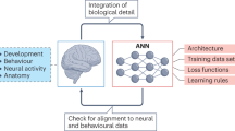
The neuroconnectionist research programme
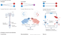
Evolution of central neural circuits: state of the art and perspectives
Acknowledgements.
D.S.B. is grateful to the mentees, colleagues and mentors who constantly broaden her appreciation of the diversity of scientific inquiry and deepen her excitement to pursue the most puzzling questions. K.E.C. was supported by the National Institutes of Health (NIH; grant numbers DC002390, DC018061 and UF1 NS111695). S.B.E. is supported by the Deutsche Forschungsgemeinschaft (DFG; EI 816/16-1 and EI 816/21-1) and the European Union’s Horizon 2020 Research and Innovation Programme under Grant Agreement No. 945539 (HBP SGA3) and 826421 (Virtual Brain Cloud). Y.G. is supported by funds from the RIKEN Center for Brain Science, The Uehara Memorial Foundation and the Japan Society for the Promotion of Science (JSPS) Core-to-Core Program A Advanced Research Networks. P.H. is partly supported by a joint grant from the John Templeton Foundation and the Fetzer Institute. The opinions expressed in this publication are those of the author(s) and do not necessarily reflect the views of the John Templeton Foundation or the Fetzer Institute. Furthermore, P.H. was supported by a Chaire de Recherche Jean D’Alembert held at Paris-Saclay University, and from a fellowship at the Paris Institute for Advanced Study (France), with the financial support of the French State, programme ‘Investissements d’avenir’ managed by the Agence Nationale de la Recherche (ANR-11-LABX-0027-01 Labex RFIEA+). H.H. is supported by grants from the National Natural Science Foundation of China (31830032 and 81527901) and the Fountain-Valley Life Sciences Fund of University of Chinese Academy of Sciences Education Foundation. Y.L.H. is supported by grants DA050323, DA048613, DA043247 and DA030359 from the National Institute of Drug Abuse (NIDA). S.A.J. acknowledges discussions with P. W. Frankland (SickKids) and members of the Josselyn and Frankland laboratories for continued inspiration. B.S.K. is supported by the NIH (NS111583 and MH104069), CHDI Inc., an Allen Distinguished Investigator Award through The Paul G. Allen Frontiers Group, a UCLA David Geffen School of Medicine Seed Grant and the Ressler Family Foundation. J.A.K. would like to thank all his lab members for exciting discussions. Work in J.A.K.’s laboratory is supported by the Austrian Federal Ministry of Education, Science and Research, the Austrian Academy of Sciences, the Austrian Science Fund (Z_153_B09), the City of Vienna and the European Research Council (ERC, grants 695642 and 693184). P.P. thanks the members of her laboratory ( www.dendrites.gr ) for their dedication and hard work. Her laboratory is funded by the European Union’s Horizon 2020 research and innovation programme (FET Open RIA GA 863245: NEUREKA, MC RISE GA 873178 INAVIGATE), the EINSTEIN Foundation-Berlin and the Fondation Santé. M.P. is supported by the Sobek Foundation, the Ernst-Jung Foundation, the DFG (SFB 992, SFB1160, SFB/TRR167, Reinhart-Koselleck Grant and Gottfried Wilhelm Leibniz Prize) and the Ministry of Science, Research and Arts, Baden-Wuerttemberg (Sonderlinie ‘Neuroinflammation’). M.P. is further supported by the DFG under Germany’s Excellence Strategy (CIBSS — EXC-2189 — Project ID390939984). P.R.R. was supported by the Netherlands Organization for Scientific Research (NWO; grant P15-42 ‘NESTOR’), the European Union’s Horizon 2020 Framework Programme Agreement No. 650003 (HBP FPA) and the Friends Foundation of the Netherlands Institute for Neuroscience. T.L.S.-J. is funded by the European Research Council (ERC) under the European Union’s Horizon 2020 research and innovation programme (Grant agreement No. 681181), the UK Dementia Research Institute — which receives its funding from DRI Ltd, funded by the UK Medical Research Council, Alzheimer’s Society and Alzheimer’s Research UK — and the University of Edinburgh. M.S. thanks the members of his laboratory for their comments. Research in the laboratory of M.S. is supported by NIH grants EY028219, MH085802 and DA049005, and the Simons Foundation Autism Research Initiative. H.R.U. is supported by Brain/MINDS JP21dm0207049, Science and Technology Platform Program for Advanced Biological Medicine JP21am0401011, Grant-in-Aid for Scientific Research (S) JP25221004 (JSPS KAKENHI) and HFSP Research Grant Program (HFSP RGP0019/2018).
Author information
Authors and affiliations.
Department of Bioengineering, University of Pennsylvania, Philadelphia, PA, USA
Danielle S. Bassett
Department of Electrical and Systems Engineering, University of Pennsylvania, Philadelphia, PA, USA
Department of Physics and Astronomy, University of Pennsylvania, Philadelphia, PA, USA
Department of Neurology, University of Pennsylvania, Philadelphia, PA, USA
Department Psychiatry, University of Pennsylvania, Philadelphia, PA, USA
Santa Fe Institute, Santa Fe, NM, USA
Department of Biomedical Engineering, Johns Hopkins University, Baltimore, MD, USA
Kathleen E. Cullen
Department of Otolaryngology — Head and Neck Surgery, Johns Hopkins University, Baltimore, MD, USA
Department of Neuroscience, Johns Hopkins University, Baltimore, MD, USA
Institute of Systems Neuroscience, Medical Faculty, Heinrich Heine University Düsseldorf, Düsseldorf, Germany
Simon B. Eickhoff
Institute of Neuroscience and Medicine, Brain & Behaviour (INM-7), Research Centre Jülich, Jülich, Germany
Center for Neuroscience & Society, University of Pennsylvania, Philadelphia, PA, USA
Martha J. Farah
Laboratory for Synaptic Plasticity and Connectivity, RIKEN Center for Brain Science, Wako-shi, Saitama, Japan
Yukiko Goda
Institute of Cognitive Neuroscience, University College London, London, UK
Patrick Haggard
Department of Psychiatry of First Affiliated Hospital, Zhejiang University School of Brain Science and Brain Medicine, Zhejiang University School of Medicine, Hangzhou, China
The MOE Frontier Research Center of Brain & Brain–Machine Integration, Zhejiang Laboratory for Systems & Precision Medicine, Hangzhou, China
Department of Psychiatry, Icahn School of Medicine at Mount Sinai, Addiction Institute of Mount Sinai, New York, NY, USA
Yasmin L. Hurd
Department of Neuroscience, Icahn School of Medicine at Mount Sinai, Addiction Institute of Mount Sinai, New York, NY, USA
Program in Neurosciences & Mental Health, Hospital for Sick Children, Toronto, Ont., Canada
Sheena A. Josselyn
Department of Psychology, Physiology & Institute of Medical Sciences, University of Toronto, Toronto, Ont., Canada
Brain, Mind & Consciousness Program, Canadian Institute for Advanced Research (CIFAR), Toronto, Ont., Canada
Department of Physiology, David Geffen School of Medicine, University of California Los Angeles, Los Angeles, CA, USA
Baljit S. Khakh
Institute of Molecular Biotechnology of the Austrian Academy of Sciences (IMBA), Vienna BioCenter (VBC), Vienna, Austria
Jürgen A. Knoblich
Institute of Molecular Biology and Biotechnology (IMBB), Foundation of Research and Technology-Hellas (FORTH), Heraklion, Crete, Greece
Panayiota Poirazi
Department of Psychology, Stanford University, Stanford, CA, USA
Russell A. Poldrack
Institute of Neuropathology, Medical Faculty, University of Freiburg, Freiburg, Germany
Marco Prinz
Signalling Research Centres BIOSS and CIBSS, University of Freiburg, Freiburg, Germany
Center for Basics in NeuroModulation (NeuroModulBasics), Faculty of Medicine, University of Freiburg, Freiburg, Germany
Department of Vision and Cognition, Netherlands Institute for Neuroscience, Amsterdam, Netherlands
Pieter R. Roelfsema
Department of Integrative Neurophysiology, VU University, Amsterdam, Netherlands
Department of Psychiatry, Academic Medical Centre, Amsterdam, Netherlands
Centre for Discovery Brain Sciences and UK Dementia Research Institute, University of Edinburgh, Edinburgh, UK
Tara L. Spires-Jones
Picower Institute for Learning and Memory, Department of Brain and Cognitive Sciences, Massachusetts Institute of Technology, Cambridge, MA, USA
Mriganka Sur
Department of Systems Pharmacology, The University of Tokyo, Tokyo, Japan
Hiroki R. Ueda
Laboratory for Synthetic Biology, RIKEN BDR, Suita, Osaka, Japan
You can also search for this author in PubMed Google Scholar
Contributions
The authors contributed equally to all aspects of the article.
Corresponding authors
Correspondence to Danielle S. Bassett , Kathleen E. Cullen , Simon B. Eickhoff , Martha J. Farah , Yukiko Goda , Patrick Haggard , Hailan Hu , Yasmin L. Hurd , Sheena A. Josselyn , Baljit S. Khakh , Jürgen A. Knoblich , Panayiota Poirazi , Russell A. Poldrack , Marco Prinz , Pieter R. Roelfsema , Tara L. Spires-Jones , Mriganka Sur or Hiroki R. Ueda .
Ethics declarations
Competing interests.
P.R.R. is a co-founder of and shareholder in a neurotechnology start-up, Phosphoenix (Netherlands). T.L.S.-J. receives collaborative funding from three industry partners (Autifony, Cognition Therapeutics and one anonymous industry donor), is on the Scientific Advisory Board of Cognition Therapeutics and is an editor for several journals, including founding Editor in Chief of Brain Communications . None of these influenced this piece. D.S.B., K.E.C., S.B.E., M.J.F., Y.G., P.H., H.H., Y.L.H, S.A.J., B.S.K., J.A.K., P.P., R.A.P., M.P., M.S. and H.R.U. declare no competing interests.
Additional information
Publisher’s note.
Springer Nature remains neutral with regard to jurisdictional claims in published maps and institutional affiliations.
Related links
anneslist : https://anneslist.net/
BiasWatchNeuro : https://biaswatchneuro.com/
Women in Neuroscience Repository : https://www.winrepo.org/
Rights and permissions
Reprints and permissions
About this article
Cite this article.
Bassett, D.S., Cullen, K.E., Eickhoff, S.B. et al. Reflections on the past two decades of neuroscience. Nat Rev Neurosci 21 , 524–534 (2020). https://doi.org/10.1038/s41583-020-0363-6
Download citation
Accepted : 03 August 2020
Published : 02 September 2020
Issue Date : October 2020
DOI : https://doi.org/10.1038/s41583-020-0363-6
Share this article
Anyone you share the following link with will be able to read this content:
Sorry, a shareable link is not currently available for this article.
Provided by the Springer Nature SharedIt content-sharing initiative
This article is cited by
Bridging functional and anatomical neural connectivity through cluster synchronization.
- Valentina Baruzzi
- Matteo Lodi
- Marco Storace
Scientific Reports (2023)
State-dependent effects of neural stimulation on brain function and cognition
- Claire Bradley
- Abbey S. Nydam
- Jason B. Mattingley
Nature Reviews Neuroscience (2022)
Hemispheric asymmetries in visual mental imagery
- Jianghao Liu
- Alfredo Spagna
- Paolo Bartolomeo
Brain Structure and Function (2022)
Turning twenty
Nature Reviews Neuroscience (2020)
Quick links
- Explore articles by subject
- Guide to authors
- Editorial policies
Sign up for the Nature Briefing newsletter — what matters in science, free to your inbox daily.

Neuroscience at NIH Research Areas
The NIH has over 180 laboratories spanning 14 Institutes, all conducting research in the basic, translational, and clinical neurosciences. Many of these laboratories and facilities are clustered together on the collegiate NIH campus in Bethesda; over 80 can be found in the The John Edward Porter Neuroscience Research Center alone. On this page are investigators throughout the NIH listed by neuroscience-research areas. Expand the research areas and click on the investigator names to learn more or click to see an alphabetical listing of neuroscience investigators.
This vibrant, highly collaborative, interactive, and multidisciplinary environment offers researchers:
- Access to state-of-the-art core research facilities.
- Opportunities for trainees and researchers at every level, including pre- and postdoctoral research and clinical training programs.
- Scientific retreats; grant-writing and career development workshops.
- Dozens of topical seminars and events , as well as Special Interest Groups .
To learn more about training opportunities in neuroscience at NIH , please reach out directly to investigators . You can also visit the NIH Office of Training and Education for more information about NIH training programs in general and search current Postdoctoral Opportunities in Neuroscience .
Explore webpages for NIH Institutes currently conducting neuroscience research to learn about their individual programs: NCCIH • NEI • NHGRI • NIA • NIAAA • NIAID • NICHD • NIDA • NIDCD • NIDCR • NIDDK • NIEHS • NIMH • NINDS.
Investigators by Research Areas
Ion channels, transporters and neurotransmitter receptors.
Cell Biology of Neurons, Muscle and Glia
Neurogenetics
Integrative Neuroscience
|
| ||
Neurological Disorders
Behavioral Neuroscience
|
| ||
Synapses and Circuits
|
| ||
Neural Development and Plasticity
|
| ||
Functional and Molecular Imaging
Neuroimmunology and Virology
Neuroendocrinology
Clinical Neuroscience
PhD Program Admissions

Visualization of copper in normal (top row) and mutant (bottom row) zebrafish brains. Image by Tong Xiao, Chang lab.
The application for Fall 2025 admission will open September 12, 2024 and is due on December 2nd, 2024 (by 8:59 pm Pacific Standard Time).
The Neuroscience PhD Program grants PhDs only. We do not offer a master’s degree. Applications are accepted from the middle of September through early December for admission for Fall of the following year. We do not accept applications for spring semester. All application materials must be received by the deadline. Late applications are not accepted or reviewed.
Applications will be reviewed holistically, using a rubric that considers academic preparation, research experience, contributions to diversity and community, initiative and motivation, and synergy with the program, each evaluated in the context of the individual applicant.
For more information please visit:
- Which Program is Right For You
- Steps to a PhD
- Application Requirements
- Review Process
Areas of Neuroscience
The Neuroscience PhD Program provides research training in four broad areas of neuroscience: cellular & molecular neuroscience, circuit, systems & behavioral neuroscience, human cognition, and computational neuroscience. Read more about each below.
Molecular & Cellular Neuroscience
Molecular and cellular neuroscientists at Berkeley study neuronal cell biology, cellular physiology, and molecular and genetic basis of neuron, synapse, and glial function. Specific topics include sensory transduction, cellular-level neuronal development, synaptic transmission and plasticity, ion channel physiology, neurodegenerative disease, and neurodevelopmental disorders. Many faculty develop novel molecular genetic tools to more precisely measure cellular physiology or to develop new therapeutical approaches to disease. Methods from molecular biology, computational biology (bioinformatics), and cellular physiology are often used in this research.
Circuit, Systems & Behavioral Neuroscience
Circuit, systems and behavioral neuroscientists at UC Berkeley study how neural circuits, ensembles, and large-scale neural systems process information in order to interpret the sensory world, make and recall memories, and produce specific behaviors. Our faculty study neural systems for sensory processing (vision, audition, touch), innate behaviors, memory, navigation, motivated behaviors, sleep, circadian rhythms, social behaviors, decision making, and more. This research often involves neurophysiology, imaging, and optogenetics experiments, usually in behaving animals. Computational models of neural circuits, and sophisticated data analysis involving modeling and machine learning, are often used in this research.
Cognitive Neuroscience
Cognitive neuroscience at UC Berkeley focuses on human cognition and its brain correlates. Our faculty study the human cognitive abilities and neural mechanisms underlying learning and memory, decision making, perception, reasoning, attention, sleep, motor control, etc. Berkeley human cognition labs employ a broad range of experimental techniques, including functional and structural neuroimaging, electrophysiology, brain stimulation, pharmacology, computational modeling, and quantitative behavioral analyses.
Computational Neuroscience
Although quantitative methods are used in all sub areas of neuroscience for analyzing complex data sets, the focus of Computational Neuroscience is to model the brain or brain function: computational models can attempt to model experimental data obtained in neurophysiological experiments (biophysically plausible models) or model functions achieved by the brain such as object recognition, language comprehension, symbolic manipulations, etc. A strong mathematical and programming background is required for research in Computational Neuroscience.
Please see the Neuroscience Department page: Diversity, Equity & Inclusion .
Upcoming Info Sessions
Thursday, October 10, 2024 at 2-3 pm Pacific Time Neuroscience PhD Program – Diversity Admissions Fair Info Session Hosted by Program Director with Current Students | Register Here
Thursday, October 31, 2024 at 1-2 pm Pacific Time AMA Grad Student Panel for Prospective Applicants Hosted by Current Students Registration Link coming soon
Previously Recorded Info Sessions:
Friday, November 3, 2023 11am-12pm Pacific Time Neuroscience PhD Program – Diversity Admissions Fair Info Session Hosted by Program Faculty with Current Students Session Recording
Friday, November 4, 2022 10-11am Pacific Time AMA Grad Student Panel for Prospective Applicants Hosted by Current Students Session Recording

- People Directory
- Safety at UD

Jaclyn Schwarz
- PSYC100 Research Requirement
- Neuroscience 4+1 (B.S./M.S.)
- Behavioral Neuroscience Requirements
- Clinical Science Requirements
- Cognitive Psychology Requirements
- Social Psychology Requirements
- Delaware Bridge Program
- Psychological & Brain Sciences Labs
- Make a Gift
- Institute for Community Mental Health Clinic

Office location
University of Delaware, 105 The Green, Room 110, Wolf Hall, Newark, DE 19716
302-831-7623 / Delaware Biotechnology Institute, 15 Innovation Way, Newark, DE
- Ph.D. – University of Maryland, School of Medicine
- B.A. – Boston College
Jaclyn Schwarz, Ph.D., is an associate professor in the Department of Psychological and Brain Sciences at the University of Delaware. Her research area is behavioral neuroscience with a focus on development and health.
Schwarz received her Ph.D. from the University of Maryland Medical School, where she examined the mechanisms by which testosterone masculinizes neural circuits in the neonatal brain. She continued her training as a postdoctoral fellow at Duke University, where she studied how early-life experiences, including parental care, can program the function of the immune system, and thereby affect later-life brain and behaviors.
In her own lab at the University of Delaware, Schwarz is currently funded by three NIH grants to study the neural-immune mechanisms by which early-life immune activation (both bacterial and viral) can disrupt the development of important neural circuits that control learning, and how these mechanisms and effects may be different between males and females.
Schwarz received the Frank Beach Young Investigator Award from the Society for Behavioral Neuroendocrinology, as well as a NARSAD Young Investigator Award from the Brain and Behavior Research Foundation.
Research Projects
Area: behavioral neuroscience, development, health, impact of zika virus infection on the developing brain.
Zika virus (ZIKV) infection in pregnant women has been linked to a neurological disorder in the fetus called microcephaly; and we hypothesize this is due to robust inflammatory response to ZIKV in the fetal brain that precipitates neurological damage. The ongoing experiments in our lab will examine the impact of maternal ZIKV infection on inflammation, microglial activation, and associated neural cell death in the fetal brain using a rat model of ZIKV infection. The immediate goal of these experiments is to determine whether ZIKV activates microglia within the developing fetal brain, and to identify key inflammatory and cellular targets for potential therapeutic interventions for ZIKV associated neurological disorder. Using the data obtained in these experiments, future experiments can also examine the impact of prenatal ZIKV infection on long-term neural, immune, and behavioral outcomes in infected offspring that do not have severe neural deformations.
Pregnancy, Microglia, and Postpartum Depression
This project examines how pregnancy impacts the immune system, in particular the immune cells of the brain, microglia. It is well known that the peripheral immune system undergoes significant changes in function throughout pregnancy. However, no one has ever examined whether pregnancy also impacts the immune cells of the brain in a similar manner and how these changes in the brain may impact the risk of depression during pregnancy or the immediate postpartum period.
Sex Differences in Response to Neonatal Immune Activation
We are currently funded to examine how neonatal males and females respond to events that challenge the immune system and what the consequences of this immune activation may be on later-life brain and behavior. Are males more vulnerable to immune activation early in development? Does this vulnerability increase the risk of learning disabilities and other developmental disorders, which are more prevalent in boys than in girls?
Resources and Links

Outstanding faculty
College of arts & sciences.
- Prospective Students
- Current Students
- News & Events
- Research & Innovation
- Alumni & Friends
- College Operations

IMAGES
VIDEO
COMMENTS
There are many different branches of neuroscience. Each focuses on a specific topic, body system, or function: Developmental neuroscience describes how the brain forms, grows, and changes.; Cognitive neuroscience is about how the brain creates and controls thought, language, problem-solving, and memory.; Molecular and cellular neuroscience explores the genes, proteins, and other molecules that ...
Computational neuroscience is an area of brain research that makes use of the power of computer modeling and mathematical tools to unpack how the brain works in all of its complexity. The ...
Research Areas. Neuroscience spans from the microscopic to the macroscopic, from genes and proteins all the way to behavior, thought, and computation. Our faculty laboratories study nervous system function at multiple levels, using approaches from biology, chemistry, physics, physiology, psychology, computation, engineering, and other fields. ...
The broad goal of cognitive science is to characterize the nature of human knowledge - its forms and content - and how that knowledge is used, processed, and acquired. Active areas of cognitive research in the Department include language, memory, visual perception and cognition, thinking and reasoning, social cognition, decision making, and ...
Neurology research is an incredibly dynamic area of medicine. We invite you to follow our progress as we continue to explore new scientific and clinical frontiers in neuroscience. Neurology & Neurological Sciences
Neuroscience is a multidisciplinary science that is concerned with the study of the structure and function of the nervous system. ... Here the authors show that these critical areas function as ...
Neuro Topics. Harvard researchers are deeply committed to understanding nervous system development and function, in both healthy and disease states. Basic scientists and clinician-researchers work together across departments, programs and centers to study the nervous system from diverse perspectives, as shown in the overlapping subfields below.
Neuroscience Research Areas. To advance our understanding of the underlying mechanisms of healthy brain function and neurological and psychological disease, Neuroscience Institute faculty at NYU Langone lead research projects in five broad, overlapping areas of neuroscience: cellular and molecular neuroscience, systems neuroscience, cognitive ...
Areas of Research. Johns Hopkins has long been at the forefront of neuroscience research, combining scientific excellence, collegiality, and interdisciplinary collaborations to seed discovery and promote innovation.
Classes cover neuroscience at all its levels, including molecular, cellular, biophysical, developmental, circuits, systems, computational, behavioral, and human cognitive neuroscience. Thesis research is available in all these areas, studying normal brain function from cellular to systems levels, behavior, cognition, and disease.
Research in neuroscience is collaborative, drawing upon the expertise of scientists across departments, institutes, and centers at Brown. Cellular and Molecular Neurobiology Projects in cellular and molecular neurobiology range from basic science investigations of development, function, and maintenance of synapses to translational research ...
Nature Neuroscience presents a special focus issue that highlights advances in methods, analyses and practices across scales of investigation and subfields of neuroscience. Single-cell ...
During training, students are expected to participate in a range of activities to increase their exposure to neuroscience research within and outside their specialty areas. These include the annual Neuroscience Conference, the Neuroscience Seminar Series, as well as other affiliated seminar series and lectures.
Research Areas. Researchers at PNI are currently working at the intersection of neuroscience and AI to gain new insights into neural data, generate new hypotheses for understanding the brain and behavior, and develop the next-generation AI architectures inspired by the brain. This branch of neuroscience focuses on understanding how the brain ...
Areas of Research. The Yale Neuroscience Department aims to understand the fundamental mysteries of the nervous system, identify the neural underpinnings of psychiatric and neurological disorders, and develop novel diagnostic, treatment, and prevention approaches to improve human health. Our research explores the function of molecules and ...
Selected influential advances in neuroscience over the past 50 years and predicted key discoveries that aim to support mission of the Society for Neuroscience, first articulated in 1969: "to advance [the] understanding of nervous systems and their role in behavior, to promote education in the neurosciences, and to inform the general public on results and implications of current research.
Neuro-immunology is a relatively new area of neuroscience that studies the interactions of the immune system and the nervous system. Such interactions govern neurodevelopment and how the nervous system handles pathology such as infections, strokes, and neurodegeneration. In addition, the neuro-immune axis also influences systemic immune ...
Research Areas. Neuroscience, the scientific study of the nervous system, is an exciting and growing field involving researchers from the physical, chemical, biological, computational, anthropological, and social sciences. Some of the most fascinating and impactful research in the future likely will require scientists to have expertise in a ...
The REP-Neuroscience Program (REP Neuro) is an inclusive undergraduate research program focused on connecting work-study eligible Berkeley undergrads with Berkeley neuroscience laboratories for research experience, career mentorship, and scientific training. REP is a year-long program. Students apply to REP, and each accepted student is matched ...
Selected influential advances in neuroscience over the past 50 years and predicted key discoveries that aim to support mission of the Society for Neuroscience, first articulated in 1969: "to advance [the] understanding of nervous systems and their role in behavior, to promote education in the neurosciences, and to inform the general public on results and implications of current research.
Which lines of research in your area of neuroscience are you particularly excited about? Danielle S. Bassett. Science is a culture as much as it is a practice in the acquisition and curation of ...
Neuroscience at NIH Research Areas. The NIH has over 180 laboratories spanning 14 Institutes, all conducting research in the basic, translational, and clinical neurosciences. Many of these laboratories and facilities are clustered together on the collegiate NIH campus in Bethesda; over 80 can be found in the The John Edward Porter Neuroscience ...
The Neuroscience PhD Program provides research training in four broad areas of neuroscience: cellular & molecular neuroscience, circuit, systems & behavioral neuroscience, human cognition, and computational neuroscience. ... Although quantitative methods are used in all sub areas of neuroscience for analyzing complex data sets, the focus of ...
Find research information for specific courses in Neuroscience. Learn about important scholarly skills such as research data management and citation managers. As always, if you have any questions about this guide or about library services in general, please reach out to me, your subject specialist by emailing me from the box on the left, or ...
Her research area is behavioral neuroscience with a focus on development and health. Schwarz received her Ph.D. from the University of Maryland Medical School, where she examined the mechanisms by which testosterone masculinizes neural circuits in the neonatal brain. She continued her training as a postdoctoral fellow at Duke University, where ...