- Patient Care & Health Information
- Diseases & Conditions
Chronic obstructive pulmonary disease (COPD) is an ongoing lung condition caused by damage to the lungs. The damage results in swelling and irritation, also called inflammation, inside the airways that limit airflow into and out of the lungs. This limited airflow is known as obstruction. Symptoms include trouble breathing, a daily cough that brings up mucus and a tight, whistling sound in the lungs called wheezing.
COPD is most often caused by long-term exposure to irritating smoke, fumes, dust or chemicals. The most common cause is cigarette smoke.
Emphysema and chronic bronchitis are the two most common types of COPD. These two conditions usually occur together and can vary in severity among people with COPD.
Chronic bronchitis is inflammation of the lining of the tubes that bring air into the lungs. These tubes are called bronchi. The inflammation prevents good airflow into and out of the lungs and makes extra mucus. In emphysema, the small air sacs of the lungs, called alveoli, are damaged. The damaged alveoli can't pass enough oxygen into the bloodstream.
Although COPD is a condition that can get worse over time, COPD is treatable. With proper management, most people with COPD can control symptoms and improve their quality of life. Proper management also can lower the risk of other conditions linked to COPD, such as heart disease and lung cancer.

Products & Services
- A Book: Mayo Clinic Family Health Book
- Newsletter: Mayo Clinic Health Letter — Digital Edition
COPD symptoms often don't appear until a lot of lung damage has occurred. Symptoms usually worsen over time, especially if smoking or other irritating exposure continues.
Symptoms of COPD may include:
- Trouble catching your breath, especially during physical activities.
- Wheezing or whistling sounds when breathing.
- Ongoing cough that may bring up a lot of mucus. The mucus may be clear, white, yellow or greenish.
- Chest tightness or heaviness.
- Lack of energy or feeling very tired.
- Frequent lung infections.
- Losing weight without meaning to. This may happen as the condition worsens.
- Swelling in ankles, feet or legs.
People with COPD also are likely to have times when their symptoms become worse than the usual day-to-day variation. This time of worsening symptoms is called an exacerbation (eg-zas-er-bay-shun). Exacerbations can last for several days to weeks. They can be caused by triggers such as smells, cold air, air pollution, colds or infections. Symptoms may include:
- Working harder than usual to breathe or having trouble breathing.
- Chest tightness.
- Coughing more often.
- More mucus or changes in mucus color or thickness.
When to see a doctor
Talk with your doctor or other healthcare professional if your symptoms don't get better with treatment or if symptoms get worse. Also talk with your healthcare professional if you notice symptoms of an infection, such as fever or a change in the mucus you cough up.
In the U.S., call 911 or your local emergency number for help or go to the emergency department at a hospital right away if you can't catch your breath, your lips or fingernail beds are blue, you have a fast heartbeat, or you feel foggy and have trouble concentrating.
There is a problem with information submitted for this request. Review/update the information highlighted below and resubmit the form.
From Mayo Clinic to your inbox
Sign up for free and stay up to date on research advancements, health tips, current health topics, and expertise on managing health. Click here for an email preview.
Error Email field is required
Error Include a valid email address
To provide you with the most relevant and helpful information, and understand which information is beneficial, we may combine your email and website usage information with other information we have about you. If you are a Mayo Clinic patient, this could include protected health information. If we combine this information with your protected health information, we will treat all of that information as protected health information and will only use or disclose that information as set forth in our notice of privacy practices. You may opt-out of email communications at any time by clicking on the unsubscribe link in the e-mail.
Thank you for subscribing!
You'll soon start receiving the latest Mayo Clinic health information you requested in your inbox.
Sorry something went wrong with your subscription
Please, try again in a couple of minutes
The main cause of COPD in developed countries is tobacco smoking. In the developing world, COPD often occurs in people exposed to fumes from burning fuel for cooking and heating in homes that don't have good airflow. Long-term exposure to chemical fumes, vapors and dusts in the workplace is another cause of COPD.
Not all people who have smoked for a long time have COPD symptoms, but they may still have lung damage, so their lungs don't work as well as they used to. Some people who smoke get less common lung conditions that may be diagnosed as COPD until a more thorough exam shows a different diagnosis.
How the lungs are affected
Air travels down the windpipe called the trachea and into the lungs through two large tubes called bronchi. Inside the lungs, these tubes divide many times like the branches of a tree. Many smaller tubes called bronchioles end in clusters of tiny air sacs called alveoli.
The alveoli have very thin walls full of tiny blood vessels. The oxygen in the air breathed in passes into these blood vessels and goes into the bloodstream. At the same time, carbon dioxide, a gas that is a waste product from the body, passes into the alveoli and is breathed out.
When breathing out, the natural stretchiness of the alveoli forces old air out, allowing new air to get in. This stretchiness is also called elasticity.
Causes of airway obstruction

In emphysema, the inner walls of the lungs' air sacs (alveoli) are damaged, causing them to eventually rupture. This creates one larger air space instead of many small ones and reduces the surface area available for the exchange of oxygen and carbon dioxide.

Bronchitis is an inflammation of the lining of the bronchial tubes, which carry air to and from the lungs. People who have bronchitis often cough up thickened mucus, which can be discolored.
Long-term exposure to irritants, such as from smoking, injures the lungs. This damage keeps air from moving in and out of the lungs freely, limiting their ability to provide oxygen to the bloodstream and take away carbon dioxide. The two main conditions that prevent effective airflow in the lungs are:
- Emphysema. This lung condition causes destruction of the fragile walls and elastic fibers of the alveoli. The damaged inner walls of the alveoli may be destroyed, creating one large air space that is hard to empty compared with the many healthy small ones. The alveoli now have less surface area that can be used to exchange oxygen and carbon dioxide. Also, old air gets trapped in the large alveoli so there isn't room for enough new air to get in.
- Chronic bronchitis. In this condition, the bronchial tubes become inflamed and narrowed. As a result, the tubes thicken, making less room for air to pass through. Extra mucus caused by the irritation blocks the narrowed tubes even more. An ongoing cough results from trying to clear mucus from the airways.
Cigarette smoke and other irritants
In the vast majority of people with COPD in the United States, the lung damage that leads to COPD is caused by long-term cigarette smoking. But there are likely other factors at play in developing COPD because not everyone who smokes gets COPD. One such factor may be gene changes that make some people more likely to develop the condition.
Other irritants can cause COPD, including cigar smoke, secondhand smoke, pipe smoke, air pollution, and workplace exposure to dust, smoke or fumes.
Alpha-1-antitrypsin deficiency
In about 1% of people with COPD, the condition results from a gene change passed down in families. This is a genetic form of emphysema. This gene lessens the levels of a protein called alpha-1-antitrypsin (AAT) in the body. AAT is made in the liver and released into the bloodstream to help protect the lungs from damage caused by smoke, fumes and dust.
Low levels of this protein, a condition called alpha-1-antitrypsin (AAT) deficiency, can cause liver damage, lung conditions such as COPD or both. With AAT deficiency, there is usually a family history of COPD, and symptoms begin at a younger age.
Risk factors
Risk factors for COPD include:
- Tobacco smoke. The biggest risk factor for COPD is long-term cigarette smoking. The more years you smoke and the more packs you smoke, the greater your risk. Pipe, cigar and marijuana smoking also may raise your risk. People who breathe in large amounts of secondhand smoke are at risk of COPD too.
- Asthma. Asthma is a condition in which the airways narrow and swell and may produce extra mucus. Asthma may be a risk factor for developing COPD. The mix of asthma and smoking raises the risk of COPD even more.
- Workplace exposure. Long-term exposure to chemical fumes, smoke, vapors and dusts in the workplace can irritate and cause swelling in the lungs. This can raise the risk of COPD.
- Fumes from burning fuel. In the developing world, people exposed to fumes from burning fuel for cooking and heating in homes with poor airflow are at higher risk of COPD.
- Genetics. AAT deficiency caused by a gene change passed down in families is the cause of COPD in some people. This genetic form of emphysema is not common. Other genetic factors may make certain people who smoke more likely to get COPD.
Complications
COPD can cause many complications, including:
- Respiratory infections. People with COPD are more likely to have colds, the flu and pneumonia. Any respiratory infection can make it much harder to breathe and could cause more damage to lung tissue.
- Heart problems. For reasons that aren't fully understood, COPD can raise the risk of heart disease, including heart attack.
- Lung cancer. People with COPD have a higher risk of getting lung cancer.
- High blood pressure in lung arteries. COPD may cause high blood pressure in the arteries that bring blood to the lungs. This condition is called pulmonary hypertension.
- Anxiety and depression. Difficulty breathing can keep you from doing activities that you enjoy. And having a serious medical condition such as COPD can sometimes cause anxiety and depression.
Unlike some other medical conditions, COPD often has a clear cause and a clear way to prevent it. Most of the time, COPD is directly linked to cigarette smoking. The best way to prevent COPD is to never smoke. If you smoke and have COPD, stopping now can slow how fast the condition worsens.
If you've smoked for a long time, quitting can be hard, especially if you've tried quitting once, twice or many times before. But keep trying to quit. It's critical to find a stop-smoking program that can help you quit for good. It's your best chance for lessening damage to your lungs. Talk with your healthcare professional about options that might work best for you.
Workplace exposure to chemical fumes, vapors and dusts is another risk factor for COPD. If you work with these types of lung irritants, talk with your supervisor about the best ways to protect yourself. This may include wearing equipment that prevents you from breathing in these substances.
Here are some steps you can take to help prevent complications linked with COPD:
- Quit smoking to help lower your risk of heart disease and lung cancer.
- Get an annual flu vaccination and vaccination against pneumococcal pneumonia to lower your risk of or prevent some infections. Also talk with your doctor or other healthcare professional about when you need the COVID-19 vaccine and the RSV vaccine.
- Talk with your healthcare professional or a mental health professional if you feel sad or hopeless or think that you may have depression.
COPD care at Mayo Clinic
- COPD. National Heart, Lung, and Blood Institute. https://www.nhlbi.nih.gov/health/copd. Accessed March 13, 2024.
- Nici L, et al. Pharmacologic management of chronic obstructive pulmonary disease: An official American Thoracic Society clinical practice guideline. American Journal of Respiratory and Critical Care Medicine. 2020; doi:10.1164/rccm.202003-0625ST.
- Ferri FF. Chronic obstructive pulmonary disease. In: Ferri's Clinical Advisor 2024. Elsevier; 2024. https://www.clinicalkey.com. Accessed March 13, 2024.
- Park HM, et al. In vitro delivery efficiencies of nebulizers for different breathing patterns. BioMedical Engineering OnLine. 2021; doi:10.1186/s12938-021-00895-3.
- Goldman L, et al., eds. Chronic obstructive pulmonary disease. In: Goldman-Cecil Medicine. 27th ed. Elsevier; 2024. https://www.clinicalkey.com. Accessed March 13, 2024.
- Wingardh ASL, et al. Effectiveness of energy conservation techniques in patients with COPD. Respiration. 2020; doi:10.1159/000506816.
- Broaddus VC, et al., eds. COPD: Pathogenesis and natural history. In: Murray and Nadel's Textbook of Respiratory Medicine. 7th ed. Elsevier; 2022. https://www.clinicalkey.com. Accessed March 13, 2024.
- Broaddus VC, et al., eds. COPD: Diagnosis and management. In: Murray and Nadel's Textbook of Respiratory Medicine. 7th ed. Elsevier; 2022. https://www.clinicalkey.com. Accessed March 13, 2024.
- Janjua S, et al. Prophylactic antibiotics for adults with chronic obstructive pulmonary disease: A network meta-analysis. Cochrane Database of Systematic Reviews. 2021; doi:10.1002/14651858.CD013198.pub2.
- Agustí A, et al. Global initiative for chronic obstructive lung disease 2023 report: GOLD executive summary. American Journal of Respiratory and Critical Care Medicine. 2023; doi:10.1164/rccm.202301-0106PP.
- Nagata K, et al. Home high-flow nasal cannula oxygen therapy for stable hypercapnic COPD. American Journal of Respiratory and Critical Care Medicine. 2022; doi:10.1164/rccm.202201-0199OC.
- Allscripts EPSi. Mayo Clinic.
- Mallea JM (expert opinion). Mayo Clinic. June 5, 2024.
- Yost KJ (expert opinion). Mayo Clinic. July 17, 2024.
Associated Procedures
- Chest X-rays
- Lung transplant
- Lung volume reduction surgery
- Stop-smoking services
- Symptoms & causes
- Diagnosis & treatment
- Doctors & departments
- Care at Mayo Clinic
Mayo Clinic does not endorse companies or products. Advertising revenue supports our not-for-profit mission.
- Opportunities
Mayo Clinic Press
Check out these best-sellers and special offers on books and newsletters from Mayo Clinic Press .
- Mayo Clinic on Incontinence - Mayo Clinic Press Mayo Clinic on Incontinence
- The Essential Diabetes Book - Mayo Clinic Press The Essential Diabetes Book
- Mayo Clinic on Hearing and Balance - Mayo Clinic Press Mayo Clinic on Hearing and Balance
- FREE Mayo Clinic Diet Assessment - Mayo Clinic Press FREE Mayo Clinic Diet Assessment
- Mayo Clinic Health Letter - FREE book - Mayo Clinic Press Mayo Clinic Health Letter - FREE book
5X Challenge
Thanks to generous benefactors, your gift today can have 5X the impact to advance AI innovation at Mayo Clinic.
It seems you are using an outdated browser. Please upgrade to a modern browser to improve your experience on this website.

COPD 101 is an overview of key clinical concepts for COPD, including risk factors, epidemiology, screening and diagnostics, and treatment strategies. This presentation is the starting point for anyone new to COPD or seeking to improve overall care for their COPD population. Patient education materials, created specifically to assist clear communication of disease management concepts, are also included to help optimize therapy plans.
Copd 101 is available in basic pdf, enhanced pdf (including speaker notes), and full powerpoint (presentation-ready) formats., copd 101 basic.

COPD 101 (basic): This PDF version of COPD 101 covers the core concepts (including patient-facing materials) and is suitable for viewing on all platforms.
COPD 101 Enhanced

COPD 101 (enhanced): This PDF version of COPD 101 includes additional information and context for educational purposes. It is best viewed using a standalone PDF program, such as Adobe Acrobat.
COPD 101 Full

COPD 101 (full): This PPT version of COPD 101 is a complete slideshow, ready for immediate presentation. It can be viewed in common presentation viewer software such as Microsoft PowerPoint or Google Slides.
Join Us on COPD360social
Join the Conversation
Already a Member?
This page was reviewed on February 7, 2023 by the COPD Foundation Content Review and Evaluation Committee .
Chronic Obstructive Pulmonary Disease (COPD) Clinical Presentation
- Author: Zab Mosenifar, MD, FACP, FCCP; Chief Editor: John J Oppenheimer, MD more...
- Sections Chronic Obstructive Pulmonary Disease (COPD)
- Practice Essentials
- Pathophysiology
- Epidemiology
- Patient Education
- Physical Examination
- Approach Considerations
- Arterial Blood Gas Analysis
- Serum Chemistries
- Alpha1-Antitrypsin
- Sputum Evaluation
- B-Type Natriuretic Peptide
- Chest Radiography
- Computed Tomography
- Two-Dimensional Echocardiography
- Pulmonary Function Tests
- Six-Minute Walking Distance
- Other Studies
- Smoking Cessation
- Management of Inflammation
- Management of Infection
- Management of Sputum Viscosity and Secretion Clearance
- PPIs for Exacerbations and the Common Cold
- Oxygen Therapy and Hypoxemia
- Vaccination to Reduce Infections
- Alpha1-Antitrypsin Deficiency Treatment
- Inpatient Care
- Lung Volume Reduction Surgery
- Lung Transplantation
- Long-term Monitoring
- End-of-Life Care
- Guidelines Summary
- Screening for COPD
- Tobacco Cessation Guidelines
- Diagnosis, Staging and Classification Guidelines
- COPD Management Guidelines
- Acute Exacerbation Treatment
- Long-term Noninvasive Ventilation
- Acute Exacerbation Prevention
- Medication Summary
- Beta2-Adrenergic Agonists, Short-Acting
- Beta2-Adrenergic Agonists, Long-Acting
- Anticholinergics, Respiratory
- Xanthine Derivative
- Phosphodiesterase-4 Inhibitors
- Dual PDE-3/PDE-4 Inhibitors
- Corticosteroids, Inhalant
- Corticosteroids, Oral
- Beta-Adrenergic Agonist and Anticholinergic Agent Combinations
- Beta2-Adrenergic Agonist and Corticosteroid Combinations
- Other Combinations
- Antibiotics
- Smoking Cessation Therapies
- Questions & Answers
- Media Gallery
Most patients with chronic obstructive pulmonary disease (COPD) seek medical attention late in the course of their disease. Patients often ignore the symptoms because they start gradually and progress over the course of years. Patients often modify their lifestyle to minimize dyspnea and ignore cough and sputum production. With retroactive questioning, a multiyear history can be elicited.
Patients typically present with a combination of signs and symptoms of chronic bronchitis, emphysema, and reactive airway disease. These include cough, worsening dyspnea, progressive exercise intolerance, sputum production, and alteration in mental status. Symptoms include the following:
- Productive cough or acute chest illness
- Breathlessness
Systemic manifestations (decreased fat-free mass, impaired systemic muscle function, osteoporosis, anemia, depression, pulmonary hypertension, cor pulmonale, left-sided heart failure
A productive cough or an acute chest illness is common. The cough usually is worse in the mornings and produces a small amount of colorless sputum.
Breathlessness is the most significant symptom, but it usually does not occur until the sixth decade of life (although it may occur much earlier). By the time the FEV 1 has fallen to 50% of predicted, the patient is usually breathless upon minimal exertion. Despite the fact that FEV 1 is the most common variable used to grade the severity of COPD, although it is not the best predictor of mortality.
Wheezing may occur in some patients, particularly during exertion and exacerbations.
The value of patient history and physical examination was addressed in the 2011 update to the American College of Physicians/American College of Chest Physicians/American Thoracic Society/European Respiratory Society (ACP/ACCP/ATS/ERS) guideline for diagnosis and management of stable COPD. According to the 2011 guideline, a history of more than 40 pack-years of smoking was the best single predictor of airflow obstruction; however, the most helpful information was provided by a combination of the following 3 signs [ 32 ] :
- Self-reported smoking history of more than 55 pack-years
- Wheezing on auscultation
- Self-reported wheezing
If all 3 signs are absent, airflow obstruction can be nearly ruled out.
With disease progression, intervals between acute exacerbations become shorter, and each exacerbation may be more severe. The rate of COPD exacerbations appears to reflect an independent susceptibility phenotype. [ 33 ]
COPD is now known to be a disease with systemic manifestations, and the quantification of these manifestations has proved to be a better predictor of mortality than lung function alone. Many patients with COPD may have decreased fat-free mass, impaired systemic muscle function, osteoporosis, anemia, depression, pulmonary hypertension, cor pulmonale, and even left-sided heart failure. Depression is not uncommon in subjects with COPD. [ 34 ]
In a study by Spitzer et al in Germany, airflow limitation as measured by spirometry was significantly more common in adults with posttraumatic stress disorder than in controls. Results were adjusted for lifestyle, clinical, and sociodemographic factors. [ 35 ]
In addition, COPD appears to increase the risk for mild cognitive impairment (MCI). Investigators from the Mayo Clinic Study of Aging—a population-based, cross-sectional study of 1,927 participants—reported an association between COPD and an increased risk of having MCI, MCI subtypes, and memory loss in elderly patients. [ 36 ] They also observed a dose-response relationship between COPD duration and an increased risk for cognitive problems.
The prevalence of MCI was significantly higher in patients with COPD (n = 288) (27%) than in those without COPD (15%), and there was a nearly twofold higher odds ratio (1.87) for MCI in patients with COPD. Moreover, the odds ratio increased from 1.6 in patients with COPD for 5 years or less to 2.1 in those who had COPD for longer than 5 years. [ 36 ]
Some important clinical and historical differences may help distinguish between the types of COPD. Classic findings for patients with chronic bronchitis include productive cough with gradual progression to intermittent dyspnea; frequent and recurrent pulmonary infections; and progressive cardiac/respiratory failure with edema and weight gain. Classic findings for patients with emphysema include a long history of progressive dyspnea with late onset of nonproductive cough; occasional mucopurulent relapses; and eventual cachexia and respiratory failure.
The sensitivity of a physical examination in detecting mild to moderate COPD is relatively poor; however, physical signs are quite specific and sensitive for severe disease. Patients with severe disease experience tachypnea and respiratory distress with simple activities.
The respiratory rate increases in proportion to disease severity. Use of accessory respiratory muscles and paradoxical indrawing of lower intercostal spaces is evident (known as the Hoover sign). In advanced disease, cyanosis, elevated jugular venous pulse (JVP), and peripheral edema can be observed.
Thoracic examination reveals the following:
- Hyperinflation (barrel chest)
- Wheezing – Frequently heard on forced and unforced expiration
- Diffusely decreased breath sounds
- Hyperresonance on percussion
- Prolonged expiration
In addition, coarse crackles beginning with inspiration may be heard.
Certain characteristics allow differentiation between disease that is predominantly chronic bronchitis and that which is predominantly emphysema.
Chronic bronchitis characteristics include the following:
- Patients may be obese
- Frequent cough and expectoration are typical
- Use of accessory muscles of respiration is common
- Coarse rhonchi and wheezing may be heard on auscultation
- Patients may have signs of right heart failure (ie, cor pulmonale), such as edema and cyanosis
Emphysema characteristics include the following:
- Patients may be very thin with a barrel chest
- Patients typically have little or no cough or expectoration
- Breathing may be assisted by pursed lips and use of accessory respiratory muscles; patients may adopt the tripod sitting position
- The chest may be hyperresonant, and wheezing may be heard
- Heart sounds are very distant
- Overall appearance is more like classic COPD exacerbation
The severity of airflow obstruction was the primary means of staging COPD until the American Thoracic Society (ATS) provided criteria for staging COPD based on the presence of obstruction (ratio of FEV 1 to forced vital capacity [FEV 1 /FVC] < 70%) and its severity as measured by percent of predicted FEV 1 .
ATS and Global Initiative for Chronic Obstructive Lung Disease (GOLD) criteria for assessing the severity of airflow obstruction (based on the percent predicted postbronchodilator FEV 1 when the FEV 1 /FVC is < 70%) are as follows [ 37 ] :
- Stage I (mild) - FEV 1 80% or greater of predicted
- Stage II (moderate) - FEV 1 50-79% of predicted
- Stage III (severe) - FEV 1 30-49% of predicted
- Stage IV (very severe) - FEV 1 less than 30% of predicted or FEV 1
However, these staging systems have limited utility in predicting mortality. The recognition that COPD is a systemic disease has helped in developing criteria that are better at predicting mortality than is assessment of airway obstruction alone. A widely used system for COPD prognosis is the BODE index (body mass index, obstruction [FEV 1 ], dyspnea [modified Medical Research Council dyspnea scale], and exercise capacity [6MWD]). [ 27 ]
World Health Organization. Fact Sheet: Chronic obstructive pulmonary disease (COPD). Available at https://www.who.int/news-room/fact-sheets/detail/chronic-obstructive-pulmonary-disease-(copd) . March 23, 2023; Accessed: June 27, 2024.
Maclay JD, Rabinovich RA, MacNee W. Update in chronic obstructive pulmonary disease 2008. Am J Respir Crit Care Med . 2009 Apr 1. 179(7):533-41. [QxMD MEDLINE Link] .
Casanova C, Cote C, Marin JM, Pinto-Plata V, de Torres JP, Aguirre-Jaime A, et al. Distance and oxygen desaturation during the 6-min walk test as predictors of long-term mortality in patients with COPD. Chest . 2008 Oct. 134(4):746-52. [QxMD MEDLINE Link] .
Feghali-Bostwick CA, Gadgil AS, Otterbein LE, Pilewski JM, Stoner MW, Csizmadia E. Autoantibodies in patients with chronic obstructive pulmonary disease. Am J Respir Crit Care Med . Jan 15 2008. 177(2):156-63.
Houben JM, Mercken EM, Ketelslegers HB, Bast A, Wouters EF, Hageman GJ. Telomere shortening in chronic obstructive pulmonary disease. Respir Med . 2009 Feb. 103(2):230-6. [QxMD MEDLINE Link] .
Morissette MC, Vachon-Beaudoin G, Parent J, Chakir J, Milot J. Increased p53 level, Bax/Bcl-x(L) ratio, and TRAIL receptor expression in human emphysema. Am J Respir Crit Care Med . 2008 Aug 1. 178(3):240-7. [QxMD MEDLINE Link] .
Hodge S, Hodge G, Jersmann H, Matthews G, Ahern J, Holmes M, et al. Azithromycin improves macrophage phagocytic function and expression of mannose receptor in chronic obstructive pulmonary disease. Am J Respir Crit Care Med . 2008 Jul 15. 178(2):139-48. [QxMD MEDLINE Link] .
Casanova C, de Torres JP, Navarro J, Aguirre-Jaime A, Toledo P, Cordoba E, et al. Microalbuminuria and hypoxemia in patients with chronic obstructive pulmonary disease. Am J Respir Crit Care Med . 2010 Oct 15. 182(8):1004-10. [QxMD MEDLINE Link] .
Ofir D, Laveneziana P, Webb KA, Lam YM, O'Donnell DE. Mechanisms of dyspnea during cycle exercise in symptomatic patients with GOLD stage I chronic obstructive pulmonary disease. Am J Respir Crit Care Med . 2008 Mar 15. 177(6):622-9. [QxMD MEDLINE Link] .
Belman MJ, Botnick WC, Shin JW. Inhaled bronchodilators reduce dynamic hyperinflation during exercise in patients with chronic obstructive pulmonary disease. Am J Respir Crit Care Med . 1996 Mar. 153(3):967-75. [QxMD MEDLINE Link] .
O'Donnell DE, Lam M, Webb KA. Spirometric correlates of improvement in exercise performance after anticholinergic therapy in chronic obstructive pulmonary disease. Am J Respir Crit Care Med . 1999 Aug. 160(2):542-9. [QxMD MEDLINE Link] .
Marin JM, Carrizo SJ, Gascon M, Sanchez A, Gallego B, Celli BR. Inspiratory capacity, dynamic hyperinflation, breathlessness, and exercise performance during the 6-minute-walk test in chronic obstructive pulmonary disease. Am J Respir Crit Care Med . 2001 May. 163(6):1395-9. [QxMD MEDLINE Link] .
O'Donnell DE, Fluge T, Gerken F, Hamilton A, Webb K, Aguilaniu B. Effects of tiotropium on lung hyperinflation, dyspnoea and exercise tolerance in COPD. Eur Respir J . 2004 Jun. 23(6):832-40. [QxMD MEDLINE Link] .
Martinez FJ, de Oca MM, Whyte RI, Stetz J, Gay SE, Celli BR. Lung-volume reduction improves dyspnea, dynamic hyperinflation, and respiratory muscle function. Am J Respir Crit Care Med . 1997 Jun. 155(6):1984-90. [QxMD MEDLINE Link] .
Celli BR. Update on the management of COPD. Chest . 2008 Jun. 133(6):1451-62. [QxMD MEDLINE Link] .
Casanova C, Cote C, de Torres JP, Aguirre-Jaime A, Marin JM, Pinto-Plata V. Inspiratory-to-total lung capacity ratio predicts mortality in patients with chronic obstructive pulmonary disease. Am J Respir Crit Care Med . 2005 Mar 15. 171(6):591-7. [QxMD MEDLINE Link] .
Nagelmann A, Tonnov A, Laks T, Sepper R, Prikk K. Lung Dysfunction of Chronic Smokers with No Signs of COPD. COPD . 2011 Apr 22. [QxMD MEDLINE Link] .
Tashkin DP, Altose MD, Bleecker ER, Connett JE, Kanner RE, Lee WW, et al. The lung health study: airway responsiveness to inhaled methacholine in smokers with mild to moderate airflow limitation. The Lung Health Study Research Group. Am Rev Respir Dis . 1992 Feb. 145 (2 Pt 1):301-10. [QxMD MEDLINE Link] .
Lamprecht B, McBurnie MA, Vollmer WM, Gudmundsson G, Welte T, Nizankowska-Mogilnicka E, et al. COPD in Never Smokers: Results From the Population-Based Burden of Obstructive Lung Disease Study. Chest . 2011 Apr. 139(4):752-763. [QxMD MEDLINE Link] .
Andersen ZJ, Hvidberg M, Jensen SS, Ketzel M, Loft S, Sorensen M, et al. Chronic obstructive pulmonary disease and long-term exposure to traffic-related air pollution: a cohort study. Am J Respir Crit Care Med . 2011 Feb 15. 183(4):455-61. [QxMD MEDLINE Link] .
Crothers K, Butt AA, Gibert CL, Rodriguez-Barradas MC, Crystal S, Justice AC. Increased COPD among HIV-positive compared to HIV-negative veterans. Chest . 2006 Nov. 130(5):1326-33. [QxMD MEDLINE Link] .
Adams PF, Barnes PM, Vickerie JL. Summary health statistics for the U.S. population: National Health Interview Survey, 2007. Vital Health Stat 10 . 2008 Nov. 1-104. [QxMD MEDLINE Link] .
Buist AS, McBurnie MA, Vollmer WM, Gillespie S, Burney P, Mannino DM, et al. International variation in the prevalence of COPD (the BOLD Study): a population-based prevalence study. Lancet . 2007 Sep 1. 370(9589):741-50. [QxMD MEDLINE Link] .
Halbert RJ, Natoli JL, Gano A, Badamgarav E, Buist AS, Mannino DM. Global burden of COPD: systematic review and meta-analysis. Eur Respir J . 2006 Sep. 28(3):523-32. [QxMD MEDLINE Link] .
Foreman MG, Zhang L, Murphy J, et al. Early-Onset COPD is Associated with Female Gender, Maternal Factors, and African American Race in the COPDGene Study. Am J Respir Crit Care Med . 2011 May 11. [QxMD MEDLINE Link] .
Mintz ML, Yawn BP, Mannino DM, et al. Prevalence of airway obstruction assessed by lung function questionnaire. Mayo Clin Proc . 2011 May. 86(5):375-81. [QxMD MEDLINE Link] . [Full Text] .
Celli BR, Cote CG, Marin JM, Casanova C, Montes de Oca M, Mendez RA, et al. The body-mass index, airflow obstruction, dyspnea, and exercise capacity index in chronic obstructive pulmonary disease. N Engl J Med . 2004 Mar 4. 350(10):1005-12. [QxMD MEDLINE Link] .
Waschki B, Kirsten A, Holz O, et al. Physical activity is the strongest predictor of all-cause mortality in patients with COPD: a prospective cohort study. Chest . 2011 Aug. 140(2):331-42. [QxMD MEDLINE Link] .
Abrams TE, Vaughan-Sarrazin M, Fan VS, Kaboli PJ. Geographic isolation and the risk for chronic obstructive pulmonary disease-related mortality: a cohort study. Ann Intern Med . 2011 Jul 19. 155(2):80-6. [QxMD MEDLINE Link] .
Sundh J, Stallberg B, Lisspers K, Montgomery SM, Janson C. Co-Morbidity, Body Mass Index and Quality of Life in COPD Using the Clinical COPD Questionnaire. COPD . 2011 Apr 22. [QxMD MEDLINE Link] .
Martinez-Garcia MA, de la Rosa D, Soler-Cataluna JJ, Donat-Sanz Y, Catalan Serra P, Agramunt Lerma M, et al. Prognostic Value of Bronchiectasis in Patients with Moderate-to-Severe Chronic Obstructive Pulmonary Disease. Am J Respir Crit Care Med . 2013 Feb 7. [QxMD MEDLINE Link] .
[Guideline] Qaseem A, Wilt TJ, Weinberger SE, et al. Diagnosis and Management of Stable Chronic Obstructive Pulmonary Disease: A Clinical Practice Guideline Update from the American College of Physicians, American College of Chest Physicians, American Thoracic Society, and European Respiratory Society. Ann Intern Med . 2011 Aug 2. 155(3):179-191. [QxMD MEDLINE Link] .
Hurst JR, Vestbo J, Anzueto A, Locantore N, Mullerova H, Tal-Singer R, et al. Susceptibility to exacerbation in chronic obstructive pulmonary disease. N Engl J Med . 2010 Sep 16. 363(12):1128-38. [QxMD MEDLINE Link] .
Spitzer C, Koch B, Grabe HJ, et al. Association of airflow limitation with trauma exposure and post-traumatic stress disorder. Eur Respir J . 2011 May. 37(5):1068-75. [QxMD MEDLINE Link] .
Singh B, Parsaik AK, Mielke MM, et al. Chronic obstructive pulmonary disease and association with mild cognitive impairment: the Mayo Clinic study of aging. Mayo Clin Proc . 2013 Nov. 88(11):1222-30. [QxMD MEDLINE Link] . [Full Text] .
[Guideline] Global strategy for diagnosis, management, and prevention of COPD: 2024. Global Initiative for Chronic Obstructive Lung Disease. Available at https://goldcopd.org/2024-gold-report/ . Accessed: June 27, 2024.
Bronchial hyperresponsiveness in cardiac failure. N Engl J Med . 1989 Dec 21. 321 (25):1756-8. [QxMD MEDLINE Link] .
Prosen G, Klemen P, Strnad M, Grmec S. Combination of lung ultrasound (a comet-tail sign) and N-terminal pro-brain natriuretic peptide in differentiating acute heart failure from chronic obstructive pulmonary disease and asthma as cause of acute dyspnea in prehospital emergency setting. Crit Care . 2011. 15(2):R114. [QxMD MEDLINE Link] .
Sin DD, Miller BE, Duvoix A, et al. Serum PARC/CCL-18 Concentrations and Health Outcomes in Chronic Obstructive Pulmonary Disease. Am J Respir Crit Care Med . 2011 May 1. 183(9):1187-1192. [QxMD MEDLINE Link] .
Jones RC, Price D, Ryan D, Sims EJ, von Ziegenweidt J, Mascarenhas L, et al. Opportunities to diagnose chronic obstructive pulmonary disease in routine care in the UK: a retrospective study of a clinical cohort. Lancet Respir Med . 2014 Apr. 2 (4):267-76. [QxMD MEDLINE Link] . [Full Text] .
Miller MR, Quanjer PH, Swanney MP, Ruppel G, Enright PL. Interpreting lung function data using 80% predicted and fixed thresholds misclassifies more than 20% of patients. Chest . 2011 Jan. 139(1):52-9. [QxMD MEDLINE Link] .
Tashkin DP, Strange C. Inhaled corticosteroids for chronic obstructive pulmonary disease: what is their role in therapy?. Int J Chron Obstruct Pulmon Dis . 2018. 13:2587-2601. [QxMD MEDLINE Link] .
Rice KL, Dewan N, Bloomfield HE, Grill J, Schult TM, Nelson DB, et al. Disease management program for chronic obstructive pulmonary disease: a randomized controlled trial. Am J Respir Crit Care Med . 2010 Oct 1. 182(7):890-6. [QxMD MEDLINE Link] .
Dewan NA, Rice KL, Caldwell M, Hilleman DE. Economic Evaluation of a Disease Management Program for Chronic Obstructive Pulmonary Disease. COPD . 2011 Apr 22. [QxMD MEDLINE Link] .
Stephenson A, Seitz D, Bell CM, et al. Inhaled anticholinergic drug therapy and the risk of acute urinary retention in chronic obstructive pulmonary disease: a population-based study. Arch Intern Med . 2011 May 23. 171(10):914-20. [QxMD MEDLINE Link] .
Gershon A, Croxford R, Calzavara A, To T, Stanbrook MB, Upshur R, et al. Cardiovascular Safety of Inhaled Long-Acting Bronchodilators in Individuals With Chronic Obstructive Pulmonary Disease. JAMA Intern Med . 2013 May 20. 1-9. [QxMD MEDLINE Link] .
Canavan N. Dual-Action Bronchodilator Eases COPD Exacerbations. Medscape [serial online] . September 10, 2013. Available at https://www.medscape.com/viewarticle/810739 .
Decramer ML, Chapman KR, Dahl R, Frith P, Devouassoux G, Fritscher C, et al. Once-daily indacaterol versus tiotropium for patients with severe chronic obstructive pulmonary disease (INVIGORATE): a randomised, blinded, parallel-group study. Lancet Respir Med . 2013 Sep. 1 (7):524-33. [QxMD MEDLINE Link] . [Full Text] .
Mahler DA, Kerwin E, Ayers T, FowlerTaylor A, Maitra S, Thach C, et al. FLIGHT1 and FLIGHT2: Efficacy and Safety of QVA149 (Indacaterol/Glycopyrrolate) versus Its Monocomponents and Placebo in Patients with Chronic Obstructive Pulmonary Disease. Am J Respir Crit Care Med . 2015 Nov 1. 192 (9):1068-79. [QxMD MEDLINE Link] .
Rabe K, Martinez F, Rodriguez-Roisin R, Fabbri LM, Ferguson GT, Jones P, et al. PT003, a novel co-suspension MDI glycopyrronium/formoterol fixed-dose combination is superior to monocomponents in patients with COPD. Eur Respir J . 2015 Sep 01. 46(suppl 59): [Full Text] .
Brown T. FDA approves umeclidinium and vilanterol combo for COPD. Medscape Medical News . December 18, 2013. Available at https://www.medscape.com/viewarticle/817964 .
Donohue JF, Maleki-Yazdi MR, Kilbride S, Mehta R, Kalberg C, Church A. Efficacy and safety of once-daily umeclidinium/vilanterol 62.5/25 mcg in COPD. Respir Med . 2013 Oct. 107(10):1538-46. [QxMD MEDLINE Link] .
Koch A, Pizzichini E, Hamilton A, Hart L, Korducki L, De Salvo MC. Lung function efficacy and symptomatic benefit of olodaterol once daily delivered via Respimat versus placebo and formoterol twice daily in patients with GOLD 2-4 COPD: results from two replicate 48-week studies. Int J Chron Obstruct Pulmon Dis . 2014. 9:697-714. [QxMD MEDLINE Link] .
Ferguson GT, Feldman GJ, Hofbauer P, Hamilton A, Allen L, Korducki L, et al. Efficacy and safety of olodaterol once daily delivered via Respimat in patients with GOLD 2-4 COPD: results from two replicate 48-week studies. Int J Chron Obstruct Pulmon Dis . 2014. 9:629-45. [QxMD MEDLINE Link] . [Full Text] .
Casaburi R, Mahler DA, Jones PW, Wanner A, San PG, ZuWallack RL, et al. A long-term evaluation of once-daily inhaled tiotropium in chronic obstructive pulmonary disease. Eur Respir J . 2002 Feb. 19(2):217-24. [QxMD MEDLINE Link] .
Donohue JF, van Noord JA, Bateman ED, Langley SJ, Lee A, Witek TJ Jr, et al. A 6-month, placebo-controlled study comparing lung function and health status changes in COPD patients treated with tiotropium or salmeterol. Chest . 2002 Jul. 122(1):47-55. [QxMD MEDLINE Link] .
Vincken W, van Noord JA, Greefhorst AP, Bantje TA, Kesten S, Korducki L, et al. Improved health outcomes in patients with COPD during 1 yr's treatment with tiotropium. Eur Respir J . 2002 Feb. 19(2):209-16. [QxMD MEDLINE Link] .
Tashkin DP, Celli B, Senn S, Burkhart D, Kesten S, Menjoge S, et al. A 4-year trial of tiotropium in chronic obstructive pulmonary disease. N Engl J Med . 2008 Oct 9. 359(15):1543-54. [QxMD MEDLINE Link] .
Brusasco V, Hodder R, Miravitlles M, Korducki L, Towse L, Kesten S. Health outcomes following treatment for six months with once daily tiotropium compared with twice daily salmeterol in patients with COPD. Thorax . 2003 May. 58(5):399-404. [QxMD MEDLINE Link] . [Full Text] .
Vogelmeier C, Hederer B, Glaab T, Schmidt H, Rutten-van Molken MP, Beeh KM, et al. Tiotropium versus salmeterol for the prevention of exacerbations of COPD. N Engl J Med . 2011 Mar 24. 364(12):1093-103. [QxMD MEDLINE Link] .
Singh S, Loke YK, Enright PL, Furberg CD. Mortality associated with tiotropium mist inhaler in patients with chronic obstructive pulmonary disease: systematic review and meta-analysis of randomised controlled trials. BMJ . 2011 Jun 14. 342:d3215. [QxMD MEDLINE Link] . [Full Text] .
Jones PW, Rennard SI, Agusti A, Chanez P, Magnussen H, Fabbri L, et al. Efficacy and safety of once-daily aclidinium in chronic obstructive pulmonary disease. Respir Res . 2011 Apr 26. 12:55. [QxMD MEDLINE Link] . [Full Text] .
Hand L. FDA OKs Umeclidinium (Incruse Ellipta) for COPD. Medscape [serial online] . April 30, 2014. Available at https://www.medscape.com/viewarticle/824419 .
Anoro Ellipta (umeclidinium and vilanterol inhalation powder) [package insert]. Research Triangle Park, NC: GlaxoSmithKline. 2013. Available at [Full Text] .
Kerwin EM, Donohue JF, Sethi S, Haumann B, Pendyala S, Dean L, et al. Revefenacin, a once-daily, long-acting muscarinic antagonist for nebulized therapy of chronic obstructive pulmonary disease (COPD): Results of a 52-week safety and tolerability phase 3 trial in participants with moderate to very severe COPD (poster A4239). Presented at the American Thoracic Society 2018 International Conference. San Diego, CA. May 21, 2018. [Full Text] .
Calverley PM, Rabe KF, Goehring UM, Kristiansen S, Fabbri LM, Martinez FJ. Roflumilast in symptomatic chronic obstructive pulmonary disease: two randomised clinical trials. Lancet . 2009 Aug 29. 374(9691):685-94. [QxMD MEDLINE Link] .
Gifford AH, Mahler DA, Waterman LA, et al. Neuromodulatory Effect of Endogenous Opioids on the Intensity and Unpleasantness of Breathlessness during Resistive Load Breathing in COPD. COPD . 2011 Apr 22. [QxMD MEDLINE Link] .
Short PM, Lipworth SI, Elder DH, Schembri S, Lipworth BJ. Effect of beta blockers in treatment of chronic obstructive pulmonary disease: a retrospective cohort study. BMJ . 2011 May 10. 342:d2549. [QxMD MEDLINE Link] . [Full Text] .
Mottillo S, Filion KB, Belisle P, Joseph L, Gervais A, O'Loughlin J. Behavioural interventions for smoking cessation: a meta-analysis of randomized controlled trials. Eur Heart J . 2009 Mar. 30(6):718-30. [QxMD MEDLINE Link] .
Wood-Baker RR, Gibson PG, Hannay M, Walters EH, Walters JA. Systemic corticosteroids for acute exacerbations of chronic obstructive pulmonary disease. Cochrane Database Syst Rev . 2005. (1):CD001288. [QxMD MEDLINE Link] .
Walters JA, Walters EH, Wood-Baker R. Oral corticosteroids for stable chronic obstructive pulmonary disease. Cochrane Database Syst Rev . 2005 Jul 20. CD005374. [QxMD MEDLINE Link] .
Spencer S, Calverley PM, Burge PS, Jones PW. Impact of preventing exacerbations on deterioration of health status in COPD. Eur Respir J . 2004 May. 23(5):698-702. [QxMD MEDLINE Link] .
Calverley PM, Anderson JA, Celli B, Ferguson GT, Jenkins C, Jones PW, et al. Salmeterol and fluticasone propionate and survival in chronic obstructive pulmonary disease. N Engl J Med . 2007 Feb 22. 356(8):775-89. [QxMD MEDLINE Link] .
Sin DD, Tashkin D, Zhang X, Radner F, Sjobring U, Thoren A. Budesonide and the risk of pneumonia: a meta-analysis of individual patient data. Lancet . 2009 Aug 29. 374(9691):712-9. [QxMD MEDLINE Link] .
Pascoe S, Locantore N, Dransfield MT, Barnes NC, Pavord ID. Blood eosinophil counts, exacerbations, and response to the addition of inhaled fluticasone furoate to vilanterol in patients with chronic obstructive pulmonary disease: a secondary analysis of data from two parallel randomised controlled trials. Lancet Respir Med . 2015 Jun. 3 (6):435-42. [QxMD MEDLINE Link] .
Siddiqui SH, Guasconi A, Vestbo J, Jones P, Agusti A, Paggiaro P, et al. Blood Eosinophils: A Biomarker of Response to Extrafine Beclomethasone/Formoterol in Chronic Obstructive Pulmonary Disease. Am J Respir Crit Care Med . 2015 Aug 15. 192 (4):523-5. [QxMD MEDLINE Link] .
Bafadhel M, Peterson S, De Blas MA, Calverley PM, Rennard SI, Richter K, et al. Predictors of exacerbation risk and response to budesonide in patients with chronic obstructive pulmonary disease: a post-hoc analysis of three randomised trials. Lancet Respir Med . 2018 Feb. 6 (2):117-126. [QxMD MEDLINE Link] .
Chen D, Restrepo MI, Fine MJ, et al. Observational study of inhaled corticosteroids on outcomes for COPD patients with pneumonia. Am J Respir Crit Care Med . 2011 Aug 1. 184(3):312-6. [QxMD MEDLINE Link] .
Seemungal TA, Wilkinson TM, Hurst JR, Perera WR, Sapsford RJ, Wedzicha JA. Long-term erythromycin therapy is associated with decreased chronic obstructive pulmonary disease exacerbations. Am J Respir Crit Care Med . 2008 Dec 1. 178(11):1139-47. [QxMD MEDLINE Link] .
Albert RK, Connett J, Bailey WC, et al. Azithromycin for prevention of exacerbations of COPD. N Engl J Med . 2011 Aug 25. 365(8):689-98. [QxMD MEDLINE Link] .
Daniels JM, Snijders D, de Graaff CS, Vlaspolder F, Jansen HM, Boersma WG. Antibiotics in addition to systemic corticosteroids for acute exacerbations of chronic obstructive pulmonary disease. Am J Respir Crit Care Med . 2010 Jan 15. 181(2):150-7. [QxMD MEDLINE Link] .
Sasaki T, Nakayama K, Yasuda H, Yoshida M, Asamura T, Ohrui T, et al. A randomized, single-blind study of lansoprazole for the prevention of exacerbations of chronic obstructive pulmonary disease in older patients. J Am Geriatr Soc . 2009 Aug. 57(8):1453-7. [QxMD MEDLINE Link] .
Duiverman ML, Wempe JB, Bladder G, Jansen DF, Kerstjens HA, Zijlstra JG. Nocturnal non-invasive ventilation in addition to rehabilitation in hypercapnic patients with COPD. Thorax . 2008 Dec. 63(12):1052-7. [QxMD MEDLINE Link] .
Crockett AJ, Moss JR, Cranston JM, Alpers JH. Domicilary oxygen for chronic obstructive pulmonary disease. Cochrane Database Syst Rev . 2000. (2):CD001744. [QxMD MEDLINE Link] .
Ringbaek TJ. Continuous oxygen therapy for hypoxic pulmonary disease: guidelines, compliance and effects. Treat Respir Med . 2005. 4(6):397-408. [QxMD MEDLINE Link] .
Sandland CJ, Morgan MD, Singh SJ. Patterns of domestic activity and ambulatory oxygen usage in COPD. Chest . 2008 Oct. 134(4):753-60. [QxMD MEDLINE Link] .
Lightowler JV, Wedzicha JA, Elliott MW, Ram FS. Non-invasive positive pressure ventilation to treat respiratory failure resulting from exacerbations of chronic obstructive pulmonary disease: Cochrane systematic review and meta-analysis. BMJ . 2003 Jan 25. 326(7382):185. [QxMD MEDLINE Link] . [Full Text] .
Carrera M, Marin JM, Anton A, Chiner E, Alonso ML, Masa JF, et al. A controlled trial of noninvasive ventilation for chronic obstructive pulmonary disease exacerbations. J Crit Care . 2009 Sep. 24(3):473.e7-14. [QxMD MEDLINE Link] .
Keenan SP, Kernerman PD, Cook DJ, Martin CM, McCormack D, Sibbald WJ. Effect of noninvasive positive pressure ventilation on mortality in patients admitted with acute respiratory failure: a meta-analysis. Crit Care Med . 1997 Oct. 25(10):1685-92. [QxMD MEDLINE Link] .
Confalonieri M, Garuti G, Cattaruzza MS, Osborn JF, Antonelli M, Conti G. A chart of failure risk for noninvasive ventilation in patients with COPD exacerbation. Eur Respir J . 2005 Feb. 25(2):348-55. [QxMD MEDLINE Link] .
Hubbard RC, Crystal RG. Augmentation therapy of alpha 1-antitrypsin deficiency. Eur Respir J Suppl . 1990 Mar. 9:44s-52s. [QxMD MEDLINE Link] .
Hurst JR, Donaldson GC, Quint JK, Goldring JJ, Baghai-Ravary R, Wedzicha JA. Temporal clustering of exacerbations in chronic obstructive pulmonary disease. Am J Respir Crit Care Med . 2009 Mar 1. 179(5):369-74. [QxMD MEDLINE Link] .
Fishman A, Martinez F, Naunheim K, Piantadosi S, Wise R, Ries A, et al. A randomized trial comparing lung-volume-reduction surgery with medical therapy for severe emphysema. N Engl J Med . 2003 May 22. 348(21):2059-73. [QxMD MEDLINE Link] .
Titman A, Rogers CA, Bonser RS, Banner NR, Sharples LD. Disease-specific survival benefit of lung transplantation in adults: a national cohort study. Am J Transplant . 2009 Jul. 9(7):1640-9. [QxMD MEDLINE Link] .
Burton CM, Milman N, Carlsen J, Arendrup H, Eliasen K, Andersen CB, et al. The Copenhagen National Lung Transplant Group: survival after single lung, double lung, and heart-lung transplantation. J Heart Lung Transplant . 2005 Nov. 24(11):1834-43. [QxMD MEDLINE Link] .
Cote CG, Celli BR. Pulmonary rehabilitation and the BODE index in COPD. Eur Respir J . 2005 Oct. 26(4):630-6. [QxMD MEDLINE Link] .
Dodd JW, Hogg L, Nolan J, et al. The COPD assessment test (CAT): response to pulmonary rehabilitation. A multicentre, prospective study. Thorax . 2011 May. 66(5):425-9. [QxMD MEDLINE Link] .
[Guideline] Webber EM, Lin JS, Thomas RG. Screening for Chronic Obstructive Pulmonary Disease: Updated Evidence Report and Systematic Review for the US Preventive Services Task Force. JAMA . 2022 May 10. 327 (18):1812-1816. [QxMD MEDLINE Link] . [Full Text] .
[Guideline] US Preventive Services Task Force, Krist AH, Davidson KW, Mangione CM, Barry MJ, Cabana M, et al. Interventions for Tobacco Smoking Cessation in Adults, Including Pregnant Persons: US Preventive Services Task Force Recommendation Statement. JAMA . 2021 Jan 19. 325 (3):265-279. [QxMD MEDLINE Link] . [Full Text] .
[Guideline] Management of Chronic Obstructive Pulmonary Disease Working Group. VA/DoD clinical practice guideline for the management of chronic obstructive pulmonary disease. Washington (DC): Department of Veterans Affairs, Department of Defense. Available at https://www.healthquality.va.gov/guidelines/cd/copd/ . December 2014; Accessed: May 7, 2016.
[Guideline] Wedzicha JA Ers Co-Chair, Miravitlles M, Hurst JR, Calverley PM, Albert RK, Anzueto A, et al. Management of COPD exacerbations: a European Respiratory Society/American Thoracic Society guideline. Eur Respir J . 2017 Mar. 49 (3): [QxMD MEDLINE Link] . [Full Text] .
[Guideline] Nici L, Mammen MJ, Charbek E, Alexander PE, Au DH, Boyd CM, et al. Pharmacologic Management of Chronic Obstructive Pulmonary Disease. An Official American Thoracic Society Clinical Practice Guideline. Am J Respir Crit Care Med . 2020 May 1. 201 (9):e56-e69. [QxMD MEDLINE Link] . [Full Text] .
[Guideline] Bourbeau J, Bhutani M, Hernandez P, Aaron SD, Beauchesne MF, Kermelly SB, et al. 2023 Canadian Thoracic Society Guideline on Pharmacotherapy in Patients With Stable COPD. Chest . 2023 Nov. 164 (5):1159-1183. [QxMD MEDLINE Link] . [Full Text] .
[Guideline] Macrea M, Oczkowski S, Rochwerg B, Branson RD, Celli B, Coleman JM 3rd, et al. Long-Term Noninvasive Ventilation in Chronic Stable Hypercapnic Chronic Obstructive Pulmonary Disease. An Official American Thoracic Society Clinical Practice Guideline. Am J Respir Crit Care Med . 2020 Aug 15. 202 (4):e74-e87. [QxMD MEDLINE Link] . [Full Text] .
[Guideline] Kaminska M., Rimmer K.P., McKim DA., et al. Long-term non-invasive ventilation in patients with chronic obstructive pulmonary disease (COPD): 2021 Canadian Thoracic Society clinical practice guideline update. Can J Respir Crit Care Sleep Med . 2021 May 5. 5(3):160-183. [Full Text] .
[Guideline] Wedzicha JA, Calverley PMA, Albert RK, Anzueto A, Criner GJ, Hurst JR, et al. Prevention of COPD exacerbations: a European Respiratory Society/American Thoracic Society guideline. Eur Respir J . 2017 Sep. 50 (3): [QxMD MEDLINE Link] . [Full Text] .
Currow DC, McDonald C, Oaten S, Kenny B, Allcroft P, Frith P, et al. Once-daily opioids for chronic dyspnea: a dose increment and pharmacovigilance study. J Pain Symptom Manage . 2011 Sep. 42(3):388-99. [QxMD MEDLINE Link] .
Pavord ID, Chanez P, Criner GJ, Kerstjens HAM, Korn S, Lugogo N, et al. Mepolizumab for Eosinophilic Chronic Obstructive Pulmonary Disease. N Engl J Med . 2017 Oct 26. 377 (17):1613-1629. [QxMD MEDLINE Link] .
Anzueto A, Barjaktarevic IZ, Siler TM, Rheault T, Bengtsson T, Rickard K, et al. Ensifentrine, a Novel Phosphodiesterase 3 and 4 Inhibitor for the Treatment of Chronic Obstructive Pulmonary Disease: Randomized, Double-Blind, Placebo-controlled, Multicenter Phase III Trials (the ENHANCE Trials). Am J Respir Crit Care Med . 2023 Aug 15. 208 (4):406-416. [QxMD MEDLINE Link] . [Full Text] .
- Venn diagram of chronic obstructive pulmonary disease (COPD). Chronic obstructive lung disease is a disorder in which subsets of patients may have dominant features of chronic bronchitis, emphysema, or asthma. The result is airflow obstruction that is not fully reversible.
- Histopathology of chronic bronchitis showing hyperplasia of mucous glands and infiltration of the airway wall with inflammatory cells.
- Histopathology of chronic bronchitis showing hyperplasia of mucous glands and infiltration of the airway wall with inflammatory cells (high-powered view).
- Gross pathology of advanced emphysema. Large bullae are present on the surface of the lung.
- Gross pathology of a patient with emphysema showing bullae on the surface.
- At high magnification, loss of alveolar walls and dilatation of airspaces in emphysema can be seen.
- Posteroanterior (PA) and lateral chest radiograph in a patient with severe chronic obstructive pulmonary disease (COPD). Hyperinflation, depressed diaphragm, increased retrosternal space, and hypovascularity of lung parenchyma are demonstrated.
- A lung with emphysema shows increased anteroposterior (AP) diameter, increased retrosternal airspace, and flattened diaphragm on lateral chest radiograph.
- A lung with emphysema shows increased anteroposterior (AP) diameter, increased retrosternal airspace, and flattened diaphragm on posteroanterior chest radiograph.
- Severe bullous disease as seen on a computed tomography (CT) scan in a patient with chronic obstructive pulmonary disease (COPD).
- Pressure volume curve comparing lungs with emphysema, lungs with restrictive disease, and normal lungs.
- Flow volume curve of a patient with emphysema shows marked decrease in expiratory flow, hyperinflation, and air trapping (patient B) compared with a patient with restrictive lung disease, who has reduced lung volumes and preserved flow (patient A).
- Forced expiratory volume in 1 second (FEV1) can be used to evaluate the prognosis in patients with emphysema. The benefit of smoking cessation is shown here because the deterioration in lung function parallels that of a nonsmoker, even in late stages of the disease. Redrawn from Fletcher C, Peato R. The natural history of chronic airflow obstruction. Br Med J 1977; 1: 1645-1648.
- Oxygen therapy via nasal cannula.
- Home supplemental oxygen.
- Bilevel positive airway pressure (BiPAP).
- Pulmonary rehabilitation.
- Chronic obstructive pulmonary disease (COPD). Pulmonary rehabilitation.
- Chest radiograph of an emphysematous patient shows hyperinflated lungs with reduced vascular markings. Pulmonary hila are prominent, suggesting some degree of pulmonary hypertension (Correa da Silva, 2001).
- Schematic representation of another sign of emphysema on the lateral chest radiograph. When the retrosternal space (defined as the space between the posterior border of the sternum and the anterior wall of the mediastinum) is larger than 2.5 cm, it is highly suggestive of overinflated lungs. This radiograph is from a patient with pectus carinatum, an important differential diagnosis to consider when this space is measured (Correa da Silva, 2001).
- Close-up image shows emphysematous bullae in the left upper lobe. Note the subpleural, thin-walled, cystlike appearance (Correa da Silva, 2001).
- A, Frontal posteroanterior (PA) chest radiograph shows no abnormality of the pulmonary vasculature, with normal intercostal spaces and a diaphragmatic dome between the 6th and 7th anterior ribs on both sides. B, Image in a patient with emphysema demonstrating reduced pulmonary vasculature resulting in hyperlucent lungs. The intercostal spaces are mildly enlarged, and the diaphragmatic domes are straightened and below the extremity of the seventh rib (Correa da Silva, 2001).
- A, Lateral radiograph of the chest shows normal pulmonary vasculature, a retrosternal space within normal limits (< 2.5 cm), and a normal angle between the diaphragm and the anterior thoracic wall. B, Lateral view of the chest shows increased pulmonary transparency, increased retrosternal space (>2.5 cm), and an angle between the thoracic wall and the diaphragm >90 degrees. Straightening of the diaphragm can be more evident in this projection than on others (Correa da Silva, 2001).
- High-resolution CT (HRCT) in a patient after viral bronchiolitis obliterans demonstrates areas of airtrapping, which is predominant in the inferior lobes and associated with bronchiectasis in the left lower lobe. Note that the decreased attenuation caused by the airtrapping can simulate emphysema (Correa da Silva, 2001).
- Pediatric high-resolution CT (HRCT) shows a hyperinflated right lung with large pulmonary bullae due to congenital lobar emphysema (Correa da Silva, 2001).
- High-resolution CT (HRCT) demonstrates areas of centriacinar emphysema. Note the low attenuation areas without walls due to destruction of the alveoli septae centrally in the acini. Red element shows the size of a normal acinus (Correa da Silva, 2001).
- High-resolution CT (HRCT) shows large bullae in both inferior lobes due to uniform enlargement and destruction of the alveoli walls causing distortion of the pulmonary architecture (Correa da Silva, 2001).
- Panacinar emphysema of the left lung in a patient with a right lung transplant. Note the red element showing the size of a normal acinus and its discrepancy with the destroyed and enlarged airspaces of the left lower lobe (Correa da Silva, 2001).
- High-resolution CT (HRCT) shows subpleural bullae consistent with paraseptal emphysema. Red mark shows the size of a normal acinus (Correa da Silva, 2001).
- High-resolution CT (HRCT) shows enlarged air-spaces or bullae adjoining pulmonary scars, consistent with paracicatricial emphysema. Red mark shows the size of a normal acinus (Correa da Silva, 2001).
- CT densitovolumetry of a nonsmoker, healthy young patient shows normal lungs. Less than 0.35% of lungs have attenuations below -950 HU (Correa da Silva, 2001).
- Expiratory CT densitovolumetry shows no areas of airtrapping (Correa da Silva, 2001).
- CT densitovolumetry in a patient with lung cancer. Three-dimensional (3D) image shows that the cancer is in the portion of the right lung that was less affected by emphysema in a patient with poor pulmonary function (Correa da Silva, 2001).
- CT densitovolumetry shows the attenuation mask. Green areas are those with attenuation below the selected threshold (here, -950 HU to evaluate emphysema), and pink areas are those with attenuations above the threshold. Area outside the patient is highlighted in green because of air (Correa da Silva, 2001).
- CT densitovolumetry demonstrates irregular distribution of the emphysema, with substantial predominance in the left lung (Correa da Silva, 2001).

Contributor Information and Disclosures
Zab Mosenifar, MD, FACP, FCCP Geri and Richard Brawerman Chair in Pulmonary and Critical Care Medicine, Professor and Executive Vice Chairman, Department of Medicine, Medical Director, Women's Guild Lung Institute, Cedars Sinai Medical Center, University of California, Los Angeles, David Geffen School of Medicine Zab Mosenifar, MD, FACP, FCCP is a member of the following medical societies: American College of Chest Physicians , American College of Physicians , American Federation for Medical Research , American Thoracic Society Disclosure: Nothing to disclose.
Annie Harrington, MD Fellow in Pulmonary and Critical Care Medicine, Cedars-Sinai Medical Center Annie Harrington, MD is a member of the following medical societies: Alpha Omega Alpha , American College of Chest Physicians Disclosure: Nothing to disclose.
Nidhi S Nikhanj, MD Fellow, Department of Pulmonary and Critical Care Medicine, Cedars-Sinai Medical Center, Los Angeles Nidhi S Nikhanj, MD is a member of the following medical societies: American College of Physicians Disclosure: Nothing to disclose.
Nader Kamangar, MD, FACP, FCCP, FCCM Professor of Clinical Medicine, University of California, Los Angeles, David Geffen School of Medicine; Chief, Division of Pulmonary and Critical Care Medicine, Vice-Chair, Department of Medicine, Olive View-UCLA Medical Center Nader Kamangar, MD, FACP, FCCP, FCCM is a member of the following medical societies: Academy of Persian Physicians, American Academy of Sleep Medicine , American Association for Bronchology and Interventional Pulmonology , American College of Chest Physicians , American College of Critical Care Medicine , American College of Physicians , American Lung Association , American Medical Association , American Thoracic Society , Association of Pulmonary and Critical Care Medicine Program Directors , Association of Specialty Professors , California Sleep Society , California Thoracic Society , Clerkship Directors in Internal Medicine , Society of Critical Care Medicine , Trudeau Society of Los Angeles, World Association for Bronchology and Interventional Pulmonology Disclosure: Nothing to disclose.
Mary L Windle, PharmD Adjunct Associate Professor, University of Nebraska Medical Center College of Pharmacy; Editor-in-Chief, Medscape Drug Reference Disclosure: Nothing to disclose.
John J Oppenheimer, MD Clinical Professor, Department of Medicine, Rutgers New Jersey Medical School; Director of Clinical Research, Pulmonary and Allergy Associates, PA John J Oppenheimer, MD is a member of the following medical societies: American Academy of Allergy Asthma and Immunology , American College of Allergy, Asthma and Immunology , New Jersey Allergy, Asthma and Immunology Society Disclosure: Serve(d) as a director, officer, partner, employee, advisor, consultant or trustee for: Amgen; GSK; Aquestive; Aimmune<br/>Clinical Adjudication/Data Safety Monitoring Board for AZ, GSK, Abbvie, Sanofi; Executive Editor for Annals of Allergy, Asthma & Immunology; Editor for Medscape; Reviewer for UpToDate; Honoraria from American Board of Allergy and Immunology.
Ryland P Byrd Jr, MD Professor, Department of Internal Medicine, Division of Pulmonary Medicine and Critical Care Medicine, Program Director of Pulmonary Diseases and Critical Care Medicine Fellowship, East Tennessee State University, James H Quillen College of Medicine; Medical Director of Respiratory Therapy, James H Quillen Veterans Affairs Medical Center
Ryland P Byrd Jr, MD is a member of the following medical societies: American College of Chest Physicians and American Thoracic Society
Disclosure: Nothing to disclose.
Sat Sharma, MD, FRCPC Professor and Head, Division of Pulmonary Medicine, Department of Internal Medicine, University of Manitoba; Site Director, Respiratory Medicine, St Boniface General Hospital
Sat Sharma, MD, FRCPC is a member of the following medical societies: American Academy of Sleep Medicine , American College of Chest Physicians , American College of Physicians-American Society of Internal Medicine , American Thoracic Society , Canadian Medical Association , Royal College of Physicians and Surgeons of Canada , Royal Society of Medicine , Society of Critical Care Medicine , and World Medical Association
Francisco Talavera, PharmD, PhD Adjunct Assistant Professor, University of Nebraska Medical Center College of Pharmacy; Editor-in-Chief, Medscape Drug Reference
Disclosure: Medscape Salary Employment
What would you like to print?
- Print this section
- Print the entire contents of
- Print the entire contents of article
- Chronic Obstructive Pulmonary Disease (COPD)
- Chronic Obstructive Pulmonary Disease (COPD) and Emphysema in Emergency Medicine
- Fast Five Quiz: Chronic Obstructive Pulmonary Disease Treatment
- Optimizing Maintenance Therapy for Chronic Obstructive Pulmonary Disease
- Triple Therapy Benefit in Chronic Obstructive Pulmonary Disease
- Fast Five Quiz: Differential Diagnosis of Chronic Obstructive Pulmonary Disease (COPD)
- Fast Five Quiz: Chronic Obstructive Pulmonary Disease Maintenance Therapy
- Beta-Blockers: Safe for Chronic Obstructive Pulmonary Disease?
- Inflammation Affects Association Between Furan Exposure and Chronic Obstructive Pulmonary Disease
- Drug Prototype Shows Promise for Stem Cell Treatment of Pulmonary Disease

- Drug Interaction Checker
- Pill Identifier
- Calculators

- 2010spiriva-handihaler-spiriva-respimat-tiotropium-343446Drugs Drugs tiotropium
Fact sheets
- Facts in pictures
- Publications
- Questions and answers
- Tools and toolkits
- Endometriosis
- Excessive heat
- Mental disorders
- Polycystic ovary syndrome
- All countries
- Eastern Mediterranean
- South-East Asia
- Western Pacific
- Data by country
- Country presence
- Country strengthening
- Country cooperation strategies
- News releases
- Feature stories
- Press conferences
- Commentaries
- Photo library
- Afghanistan
- Cholera
- Coronavirus disease (COVID-19)
- Greater Horn of Africa
- Israel and occupied Palestinian territory
- Disease Outbreak News
- Situation reports
- Weekly Epidemiological Record
- Surveillance
- Health emergency appeal
- International Health Regulations
- Independent Oversight and Advisory Committee
- Classifications
- Data collections
- Global Health Observatory
- Global Health Estimates
- Mortality Database
- Sustainable Development Goals
- Health Inequality Monitor
- Global Progress
- World Health Statistics
- Partnerships
- Committees and advisory groups
- Collaborating centres
- Technical teams
- Organizational structure
- Initiatives
- General Programme of Work
- WHO Academy
- Investment in WHO
- WHO Foundation
- External audit
- Financial statements
- Internal audit and investigations
- Programme Budget
- Results reports
- Governing bodies
- World Health Assembly
- Executive Board
- Member States Portal
- Fact sheets /
Chronic obstructive pulmonary disease (COPD)
- Chronic obstructive pulmonary disease (COPD) is the third leading cause of death worldwide, causing 3.23 million deaths in 2019.
- Nearly 90% of COPD deaths in those under 70 years of age occur in low- and middle-income countries (LMIC).
- COPD is the seventh leading cause of poor health worldwide (measured by disability-adjusted life years)
- Tobacco smoking accounts for over 70% of COPD cases in high-income countries. In LMIC tobacco smoking accounts for 30–40% of COPD cases, and household air pollution is a major risk factor.
Chronic obstructive pulmonary disease (COPD) is a common lung disease causing restricted airflow and breathing problems. It is sometimes called emphysema or chronic bronchitis.
In people with COPD, the lungs can get damaged or clogged with phlegm. Symptoms include cough, sometimes with phlegm, difficulty breathing, wheezing and tiredness.
Smoking and air pollution are the most common causes of COPD. People with COPD are at higher risk of other health problems.
COPD is not curable but symptoms can improve if one avoids smoking and exposure to air pollution and gets vaccines to prevent infections. It can also be treated with medicines, oxygen and pulmonary rehabilitation.
The most common symptoms of COPD are difficulty breathing, chronic cough (sometimes with phlegm) and feeling tired.
COPD symptoms can get worse quickly. These are called flare-ups. These usually last for a few days and often require additional medicine.
People with COPD also have a higher risk for other health problems. These include:
- lung infections, like the flu or pneumonia
- lung cancer
- heart problems
- weak muscles and brittle bones
- depression and anxiety.
Common symptoms of COPD develop from mid-life onwards. As COPD progresses, people find it more difficult to carry out their normal daily activities, often due to breathlessness. There may be a considerable financial burden due to limitation of workplace and home productivity, and costs of medical treatment.
COPD is sometimes called emphysema or chronic bronchitis. Emphysema usually refers to destruction of the tiny air sacs at the end of the airways in the lungs. Chronic bronchitis refers to a chronic cough with the production of phlegm resulting from inflammation in the airways. COPD and asthma share common symptoms (cough, wheeze and difficulty breathing) and people may have both conditions.
Several processes can cause the airways to become narrow and lead to COPD. There may be destruction of parts of the lung, mucus blocking the airways, and inflammation and swelling of the airway lining.
COPD develops gradually over time, often resulting from a combination of risk factors:
- tobacco exposure from active smoking or passive exposure to second-hand smoke;
- occupational exposure to dusts, fumes or chemicals;
- indoor air pollution: biomass fuel (wood, animal dung, crop residue) or coal is frequently used for cooking and heating in low- and middle-income countries with high levels of smoke exposure;
- early life events such as poor growth in utero, prematurity, and frequent or severe respiratory infections in childhood that prevent maximum lung growth;
- asthma in childhood; and
- a rare genetic condition called alpha-1 antitrypsin deficiency, which can cause COPD at a young age.
COPD should be suspected if a person has typical symptoms, and the diagnosis confirmed by a breathing test called spirometry, which measures how the lungs are working. In low- and middle-income countries, spirometry is often not available and so the diagnosis may be missed.
COPD isn’t curable, but it can get better by not smoking, avoiding air pollution and getting vaccines. It can be treated with medicines, oxygen and pulmonary rehabilitation.
There are several treatments available for COPD.
Inhaled medicines that open and reduce swelling in the airways are the main treatments.
Bronchodilator inhalers are the most important medicines for treating COPD. They relax the airways to keep them open.
Short-acting bronchodilators start to work in seconds and can last for 4–6 hours. These are often used during flare-ups.
Long-acting bronchodilators take longer to start working but last longer. These are taken daily and can be combined with inhaled steroids.
Other treatments may also be used:
- Steroid pills and antibiotics are often used to treat flare-ups.
- Oxygen is used for people who have had COPD for a long time or have severe COPD.
- Pulmonary rehabilitation teaches exercises to improve your breathing and ability to exercise.
- Surgery may improve symptoms for some people with severe COPD.
Some inhalers open the airways and may be given regularly to prevent or reduce symptoms, and to relieve symptoms during acute flare-ups. Inhaled corticosteroids are sometimes given in combination with these to reduce inflammation in the lungs.
Inhalers must be taken using the correct technique, and in some cases with a spacer device to help deliver the medication into the airways more effectively. Access to inhalers is limited in many low- and middle-income countries; in 2021 salbutamol inhalers were generally available in public primary health care facilities in half of low- and low-middle income countries.
Flare-ups are often caused by a respiratory infection, and people may be given an antibiotic or steroid tablets in addition to inhaled or nebulised treatment as needed.
Living with COPD
Lifestyle changes can help improve symptoms of COPD.
Quit smoking or vaping. This is the most important thing to do. Even if you have been smoking for many years, quitting can still help.
Avoid second-hand smoke or smoke from indoor cooking fires.
Stay physically active.
Protect yourself from lung infections:
- Get a flu vaccine every year.
- Get the pneumonia vaccine.
- Get all available COVID-19 vaccines and make sure you have had the latest boosters.
People living with COPD must be given information about their condition, treatment and self-care to help them to stay as active and healthy as possible.
WHO response
COPD is included in the WHO Global Action Plan for the Prevention and Control of Noncommunicable Diseases (NCDs) and the United Nations 2030 Agenda for Sustainable Development.
WHO is taking action to extend diagnosis of and treatment for COPD in a number of ways.
The WHO Package of Essential Noncommunicable Disease Interventions (PEN) was developed to help improve NCD management in primary health care in low-resource settings. PEN includes protocols for the assessment, diagnosis and management of chronic respiratory diseases (asthma and chronic obstructive pulmonary disease), and modules on healthy lifestyle counselling, including tobacco cessation and self-care.
Rehabilitation 2030 is a new strategic approach to prioritize and strengthen rehabilitation services in health systems. Pulmonary rehabilitation for COPD is included in the Package of Interventions for Rehabilitation, currently under development as part of this WHO initiative.
Reducing tobacco smoke exposure is important for both primary prevention of COPD and disease management. The Framework Convention on Tobacco Control is enabling progress in this area as are WHO initiatives such as MPOWER and mTobacco Cessation.
Further prevention activities include the WHO Clean Household Energy Solutions Toolkit (CHEST) to promote clean and safe interventions in the home and facilitate the design of policies that promote the adoption of clean household energy at local, programmatic and national levels.
The Global Alliance against Chronic Respiratory Diseases (GARD) contributes to WHO’s work to prevent and control chronic respiratory diseases. GARD is a voluntary alliance of national and international organizations and agencies from many countries committed to the vision of a world where all people breathe freely.
- WHO Global health estimates
- NCD country capacity survey
- Global action plan for the prevention and control of noncommunicable diseases 2013–2020. Geneva: World Health Organization; 2013.
- The 2030 Agenda for Sustainable Development
- WHO package of essential noncommunicable (PEN) disease interventions for primary health care
- WHO Framework Convention on Tobacco Control
- Rehabilitation 2030
- Household Air Pollution and Health
- Global Alliance against Chronic Respiratory Diseases (GARD)
- Package of interventions for rehabilitation: module 4: cardiopulmonary conditions
- Chronic respiratory diseases programme
Chronic Obstructive Pulmonary Disease (COPD)
(chronic obstructive bronchitis; emphysema).
- Epidemiology |
- Pathophysiology |
- Symptoms and Signs |
- Diagnosis |
- Treatment |
- Prognosis |
- Key Points |
Chronic obstructive pulmonary disease (COPD) is airflow limitation caused by an inflammatory response to inhaled toxins, often cigarette smoke. Alpha-1 antitrypsin deficiency and various occupational exposures are less common causes in patients who do not smoke. Symptoms are productive cough and dyspnea that develop over years; common signs include decreased breath sounds, prolonged expiratory phase of respiration, and wheezing. Severe cases may be complicated by weight loss, pneumothorax, frequent acute decompensation episodes, right heart failure, and/or acute or chronic respiratory failure. Diagnosis is based on history, physical examination, chest radiograph, and pulmonary function tests. Treatment is with bronchodilators, corticosteroids, and, when necessary, oxygen and antibiotics. Lung volume reduction procedures or transplantation are used in advanced disease. Survival in COPD is related to the severity of airflow limitation, the frequency of exacerbations, and the presence of comorbidities.
COPD comprises
Chronic obstructive bronchitis (clinically defined)
Emphysema (pathologically or radiologically defined)
Many patients have features of both.
Chronic obstructive bronchitis is chronic bronchitis with airflow obstruction. Chronic bronchitis is defined as productive cough on most days of the week for at least 3 months total duration in 2 successive years. Chronic bronchitis becomes chronic obstructive bronchitis if spirometric evidence of airflow obstruction develops. Chronic asthmatic bronchitis is a similar, overlapping condition characterized by chronic productive cough, wheezing, and partially reversible airflow obstruction; it occurs predominantly in patients who smoke and have a history of asthma . When asthma and COPD co-exist in the same patient, pharmacologic treatment should follow guidelines for asthma ( 1 ).
Emphysema is destruction of lung parenchyma leading to loss of elastic recoil and loss of alveolar septa and radial airway traction, which increases the tendency for airway collapse. Lung hyperinflation, airflow limitation, and air trapping follow. Airspaces enlarge and may eventually develop blebs or bullae. Obliteration of small airways is thought to be the earliest lesion that precedes the development of emphysema.
General reference
1. Global Initiative for Chronic Obstructive Lung Disease (GOLD) : Diagnosis and assessment. Global Strategy for the Prevention, Diagnosis, and Management of COPD: 2024 report.
Epidemiology of COPD
In the United States, approximately 24 million people have airflow limitation, of whom approximately 16 million have a diagnosis of COPD ( 1 ). COPD is a leading cause of death, resulting in approximately 140,000 deaths each year in the United States ( 2 ). Prevalence, incidence, and mortality rates increase with age. Prevalence is higher in females, but total mortality is similar in both sexes. COPD seems to aggregate in families independent of alpha-1 antitrypsin deficiency (alpha-1 antiprotease inhibitor deficiency).
COPD is increasing worldwide because of increases in smoking and reduction in mortality due to infectious diseases. In some regions, the widespread use of biomass fuels, such as wood, grasses, or other organic materials, also contributes to COPD prevalence. COPD mortality rates may be higher in medically underserved nations than in nations where medical care is more easily accessed. COPD accounted for 3.23 million deaths globally in 2019 and is the third leading cause of death.
The COVID-19 pandemic posed a particular risk to patients with COPD. The mortality rate for patients with COPD and COVID-19 was 15% versus 4% in those without COPD ( 3 ).
Epidemiology references
1. Centers for Disease Control and Prevention : Chronic Obstructive Pulmonary Disease (COPD). Updated June 30,2023.
2. Centers for Disease Control and Prevention : National Center for Health Statistics: Leading Causes of Death. Updated January 22, 2022.
3. Meza D, Khuder B, Bailey JI, et al : Mortality from COVID-19 in patients with COPD: A US study in the N3C Data Enclave. Int J Chron Obstruct Pulmon Dis 16:2323–2326, 2021. doi: 10.2147/COPD.S318000
Etiology of COPD
There are 2 main causes of COPD:
Smoking (and less often other inhalational exposures)
Genetic factors
Smoking and other inhalational exposures.
Of all inhalational exposures, cigarette smoking is the primary risk factor in most countries, although only approximately 15% of people who smoke develop clinically apparent COPD ( 1 ). The risk for COPD increases with both duration (years of smoking) and cumulative dose (pack-years) ( 2 asthma , are at greater risk of developing COPD than are those without.
Smoke from indoor cooking and heating is an important causative factor in countries where indoor fires are commonly used for cooking or heating ( 3 ).
Exposure to passive cigarette smoke, air pollution , and occupational dust (eg, mineral dust, cotton dust) or inhaled chemicals (eg, cadmium) contribute to the risk of COPD but are of less importance compared to cigarette smoking. Low body weight and childhood respiratory disorders also contribute to the risk of COPD.
The best-defined causative genetic disorder is alpha-1 antitrypsin deficiency , which is an important cause of emphysema in people who do not smoke and markedly increases susceptibility to disease in people who do.
More than 30 genetic alleles have been found to be associated with COPD or decline in lung function in selected populations, but none has been shown to be as consequential as alpha-1 antitrypsin.
Etiology references
1. Wheaton AG, Liu Y, Croft JB, et al : Chronic Obstructive Pulmonary Disease and Smoking Status - United States, 2017. MMWR Morb Mortal Wkly Rep 68(24):533–538, 2019. doi:10.15585/mmwr.mm6824a1
2. Bhatt SP, Kim YI, Harrington KF, et al . Smoking duration alone provides stronger risk estimates of chronic obstructive pulmonary disease than pack-years. Thorax 2018;73(5):414-421. doi:10.1136/thoraxjnl-2017-210722
3. Ortiz-Quintero B, Martínez-Espinosa I, Pérez-Padilla R : Mechanisms of Lung Damage and Development of COPD Due to Household Biomass-Smoke Exposure: Inflammation, Oxidative Stress, MicroRNAs, and Gene Polymorphisms. Cells 12(1):67, 2022. doi:10.3390/cells12010067
Pathophysiology of COPD
Various factors cause the airflow limitation and other complications of COPD.
Inflammation
Inhalational exposures can trigger an inflammatory response in airways and alveoli that leads to disease in genetically susceptible people. The process is thought to be mediated by an increase in protease activity and a decrease in antiprotease activity. Lung proteases, such as neutrophil elastase, matrix metalloproteinases, and cathepsins, break down elastin and connective tissue in the normal process of tissue repair. Their activity is normally balanced by antiproteases, such as alpha-1 antitrypsin, airway epithelium–derived secretory leukoproteinase inhibitor, elafin, and matrix metalloproteinase tissue inhibitor. In patients with COPD, activated neutrophils and other inflammatory cells release proteases as part of the inflammatory process; protease activity exceeds antiprotease activity, and tissue destruction and mucus hypersecretion result.
Activation of neutrophils and macrophages also leads to accumulation of free radicals, superoxide anions, and hydrogen peroxide , which inhibit antiproteases and cause bronchoconstriction, mucosal edema, and mucous hypersecretion. Neutrophil-induced oxidative damage, release of profibrotic neuropeptides (eg, bombesin), and reduced levels of vascular endothelial growth factor (VEGF) may contribute to apoptotic destruction of lung parenchyma.
The inflammation in COPD increases as disease severity increases, and, in severe (advanced) disease, inflammation does not resolve completely despite smoking cessation. This chronic inflammation does not seem to respond to corticosteroids, particularly in patients who continue to smoke cigarettes ( 1 ).
Mechanical stress on alveoli from over-distension may make them susceptible to proteases, leading to alveolar septal destruction and progressive emphysema. This is particularly notable in the upper lobes of the lung ( 2 ).
Respiratory infection (to which patients with COPD are prone) may amplify progression of lung destruction.
Bacteria, especially Haemophilus influenzae , colonize the lower airways of approximately 30% of patients with COPD ( 3 ). In more severely affected patients (eg, those with previous hospitalizations), colonization with Pseudomonas aeruginosa or other gram-negative bacteria is common. Smoking and airflow obstruction may lead to impaired mucus clearance in lower airways, which predisposes to infection. Repeated bouts of infection increase the inflammatory burden that hastens disease progression. There is no evidence, however, that long-term use of antibiotics slows the progression of COPD.
Airflow limitation
The cardinal pathophysiologic feature of COPD is airflow limitation caused by airway narrowing and/or obstruction, loss of elastic recoil, or both.
Airway narrowing and obstruction are caused by inflammation-mediated mucus hypersecretion, mucus plugging, mucosal edema, bronchospasm, peribronchial fibrosis, and remodelling of small airways or a combination of these mechanisms. Alveolar septa are destroyed, reducing parenchymal attachments to the airways and thereby facilitating airway closure during expiration.
Enlarged alveolar spaces sometimes consolidate into bullae, defined as airspaces ≥ 1 cm in diameter. Bullae may be entirely empty or have strands of lung tissue traversing them in areas of locally severe emphysema; they occasionally occupy an entire hemithorax. These changes lead to loss of elastic recoil and lung hyperinflation.
Increased airway resistance increases the work of breathing. Lung hyperinflation, although it decreases airway resistance, also increases the work of breathing. Increased work of breathing may lead to alveolar hypoventilation with hypoxia and hypercapnia, although hypoxia and hypercarbia can also be caused by ventilation/perfusion (V/Q) mismatch.
Complications
In addition to airflow limitation and sometimes respiratory insufficiency, complications include
Pulmonary hypertension
Respiratory infection
Weight loss and other comorbidities
Chronic alveolar hypoxia increases pulmonary vascular tone, which, if diffuse, causes pulmonary hypertension and cor pulmonale . The increase in pulmonary vascular pressure may be augmented by the destruction of the pulmonary capillary bed due to destruction of alveolar septa.
Viral or bacterial respiratory infections are common among patients with COPD and cause a large percentage of acute exacerbations. It is currently thought that acute bacterial infections are due to acquisition of new strains of bacteria rather than overgrowth of chronic colonizing bacteria.
Weight loss may occur, perhaps in response to insufficient caloric intake and increased levels of circulating tumor necrosis factor (TNF)-alpha. This weight loss may be due to a mismatch between caloric expenditure and nutritional intake because caloric expenditure can be high in the presence of heightened inflammatory cytokines and hypoxemia.
Other coexisting or complicating disorders that adversely affect quality of life and/or survival include osteoporosis , depression , anxiety , coronary artery disease , arrhythmias , lung cancer and other cancers, muscle atrophy, and gastroesophageal reflux . The extent to which these disorders are consequences of COPD, smoking, and the accompanying systemic inflammation is unclear.
Pathophysiology references
1. Adcock IM, Bhatt SP, Balkissoon R, Wise RA : The Use of Inhaled Corticosteroids for Patients with COPD Who Continue to Smoke Cigarettes: An Evaluation of Current Practice. Am J Med 135(3):302–312, 2022. doi:10.1016/j.amjmed.2021.09.006
2. Suki B, Sato S, Parameswaran H, Szabari MV, Takahashi A, Bartolák-Suki E : Emphysema and mechanical stress-induced lung remodeling. Physiology (Bethesda) 28(6):404–413, 2013. doi:10.1152/physiol.00041.2013
3. Short B, Carson S, Devlin AC, et al : Non-typeable Haemophilus influenzae chronic colonization in chronic obstructive pulmonary disease (COPD). Crit Rev Microbiol 47(2):192–205, 2021. https://doi.org/10.1080/1040841X.2020.1863330
Symptoms and Signs of COPD
COPD takes years to develop and progress.
Productive cough usually is the initial symptom, developing among people who smoke and are in their 40s and 50s.
Dyspnea that is progressive, persistent, exertional, or worse during respiratory infection appears when patients are in their late 50s or 60s.
Symptoms usually progress quickly in patients who continue to smoke and in those who have a higher lifetime tobacco exposure. Morning headache develops in more advanced disease and signals nocturnal hypercapnia or hypoxemia.
Signs of COPD include wheezing, a prolonged expiratory phase of breathing, lung hyperinflation manifested as decreased heart and lung sounds, and increased anteroposterior diameter of the thorax (barrel chest).
Signs of advanced disease include pursed-lip breathing, accessory respiratory muscle use, paradoxical inward movement of the lower rib cage during inspiration (Hoover sign), and cyanosis. Signs of cor pulmonale include neck vein distention, splitting of the second heart sound with an accentuated pulmonic component, tricuspid insufficiency murmur, and peripheral edema. Right ventricular heaves are uncommon in COPD because the lungs are hyperinflated.
Patients with advanced emphysema lose weight and experience muscle wasting that has been attributed to immobility, hypoxia, or release of systemic inflammatory mediators, such as TNF-alpha.
Spontaneous pneumothorax may occur (possibly related to rupture of bullae) and should be suspected in any patient with COPD whose pulmonary status abruptly worsens.
The symptoms can be graded according to which activities cause dyspnea (see table Breathlessness Measurement using the mMRC Questionnaire ).
Breathlessness Measurement Using the Modified British Medical Research Council (mMRC) Questionnaire
0 | None except during strenuous exercise |
1 | Occurring when hurrying on level ground or walking up a slight incline |
2 | Resulting in walking more slowly than people of the same age on level ground
Resulting in stopping for breath when walking at own pace on level ground |
3 | Resulting in stopping for breath after walking about 100 meters or after a few minutes on level ground |
4 | Preventing the person from leaving the house
Occurring when dressing or undressing |
| 93:580–586, 1988. | |
Acute exacerbations
Acute exacerbations occur sporadically during the course of COPD and are heralded by increased symptom severity. The specific cause of any exacerbation is almost always impossible to determine, but exacerbations are often attributed to viral upper respiratory infections, acute bacterial bronchitis, or exposure to respiratory irritants. As COPD progresses, acute exacerbations tend to become more frequent, averaging approximately 1 to 3 episodes/year.
Diagnosis of COPD
Chest imaging
Pulmonary function testing
Diagnosis is suggested by history, physical examination, and chest imaging findings and is confirmed by pulmonary function tests . Similar symptoms can be caused by asthma , heart failure , and bronchiectasis (see table Differential Diagnosis of COPD ). COPD and asthma are sometimes easily confused and may coexist.
Differential Diagnosis of COPD
COPD | Middle age | Sometimes lung hyperinflation, bullae, increased retrosternal air space, and/or bronchial wall thickening (seen on chest imaging); however, imaging usually not helpful diagnostically and is done mainly to exclude other disorders | Slowly progressive symptoms History of smoking or exposure to tobacco or other types of smoke |
| Early in life (often during childhood) | Usually normal or possibly hyperinflation | Symptoms vary widely from day to day Variable airflow obstruction Symptoms often worse at night or early morning History of allergies, rhinitis, or eczema Often family history of asthma |
| All ages, but most often in older or middle aged adults | Bronchial dilation and bronchial wall thickening (seen on chest radiograph or chest CT) | Often large amounts of purulent sputum Often history of chronic recurrent bacterial infection |
Diffuse panbronchiolitis | Usually between ages 10 and 60 years (mean age of 40) | Diffuse small centrilobular nodular opacities and hyperinflation seen on chest radiograph and high-resolution CT | Mostly males who do not smoke Almost all have chronic sinusitis Predominately in those of Asian descent |
| All ages, but most often in older or middle age | Enlarged heart, pleural effusion, fluid in major fissure, sometimes pulmonary edema (seen on chest radiograph) | Volume restriction without airflow limitation (detected by pulmonary function tests) |
Obliterative bronchiolitis | Onset at younger age | Peripheral hypodense areas (seen on chest CT during expiration) | History of lung or bone marrow transplantation |
| All ages | Lung infiltrates seen on chest radiograph | Confirmed by microbiologic testing Usually in areas with high prevalence of TB |
| : Diagnosis and assessment. Global Strategy for the Prevention, Diagnosis, and Management of COPD: 2024 report. | |||
Systemic disorders that may have a component of airflow limitation suggest COPD; they include HIV infection , abuse of illicit drugs (particularly cocaine and amphetamines), sarcoidosis , Sjögren syndrome , bronchiolitis obliterans , lymphangioleiomyomatosis , and eosinophilic granuloma. COPD can be differentiated from interstitial lung diseases by chest imaging, which shows increased interstitial markings in interstitial lung disease, and pulmonary function testing, which shows a restrictive ventilatory defect rather than an obstructive ventilatory defect. In some patients, COPD and interstitial lung disease coexist (combined pulmonary fibrosis and emphysema [CPFE]) in which lung volumes are relatively preserved, but gas exchange is severely impaired.
Pulmonary function tests
Patients suspected of having COPD should undergo pulmonary function testing to confirm airflow limitation, to quantify its severity and reversibility, and to distinguish COPD from other disorders. (Some experts recommend screening pulmonary function testing for all patients with a history of smoking.) Pulmonary function testing is also useful for following disease progression and monitoring response to treatment. The primary diagnostic tests are
FEV1: The volume of air forcefully expired during the first second after taking a full breath
Forced vital capacity (FVC): The total volume of air expired with maximal force
Flow-volume loops: Simultaneous spirometric recordings of airflow and volume during forced maximal expiration and inspiration
Reductions of FEV1, FVC, and the ratio of FEV1/FVC are the hallmark of airflow limitation. Flow-volume loops show a concave pattern in the expiratory tracing .
There are 2 basic pathways by which COPD can develop and manifest with symptoms in later life:
In the first pathway, patients may have normal lung function in early adulthood, which is followed by a more rapid decline in FEV1 ( ≥ 60 mL/year).
With the second pathway, patients have impaired lung function in early adulthood, often associated with asthma or other childhood respiratory disease. In these patients, COPD may present with a normal age-related decline in FEV1 (approximately 30 mL/year).
Although this second pathway model is conceptually helpful, a wide range of individual trajectories is possible ( 1 ). When the FEV1 falls below approximately 1 L, patients develop dyspnea during activities of daily living (although dyspnea is more closely related to the degree of dynamic hyperinflation [progressive hyperinflation due to incomplete exhalation] than to the degree of airflow limitation). When the FEV1 falls below approximately 0.8 L, patients are at risk of hypoxemia, hypercapnia, and cor pulmonale .
FEV1 and FVC are easily measured with office spirometry. Normal reference values are determined by patient age, sex, and height. It is recommended that race not be used for calculating predicted reference values ( 2 ). Airflow limitation severity in patients with COPD and FEV1/FVC 3 ):
Mild: ≥ 80% of predicted
Moderate: 50% to 79% of predicted
Severe: 30% to 49% of predicted
Very severe: < 30% of predicted
Additional pulmonary function testing is necessary only in specific circumstances, such as before lung volume reduction procedures . Other test abnormalities may include
Increased total lung capacity
Increased functional residual capacity
Increased residual volume
Decreased vital capacity
Decreased single-breath diffusing capacity for carbon monoxide (DLCO)
Findings of increased total lung capacity, functional residual capacity, and residual volume can help distinguish COPD from restrictive pulmonary disease, in which these measures are diminished.
Decreased DLCO is nonspecific and is reduced in other disorders that affect the pulmonary vascular bed, such as interstitial lung disease, but can help distinguish emphysema from asthma, in which DLCO is normal or elevated.
Imaging tests
Chest radiograph may have characteristic findings. In patients with emphysema, changes can include lung hyperinflation manifested as a flat diaphragm (ie, increase in the angle formed by the sternum and anterior diaphragm on a lateral film from the normal value of 45 ° to > 90 ° ), rapid tapering of hilar vessels, and bullae (ie, radiolucencies > 1 cm surrounded by arcuate, hairline shadows). Other typical findings include enlargement of the retrosternal airspace and a narrow cardiac shadow. Emphysematous changes occurring predominantly in the lung bases suggest alpha-1 antitrypsin deficiency . The lungs may look normal or have increased lucency secondary to loss of parenchyma. Among patients with chronic obstructive bronchitis, chest radiographs may be normal or may show a bibasilar increase in bronchovascular markings as a result of bronchial wall thickening.
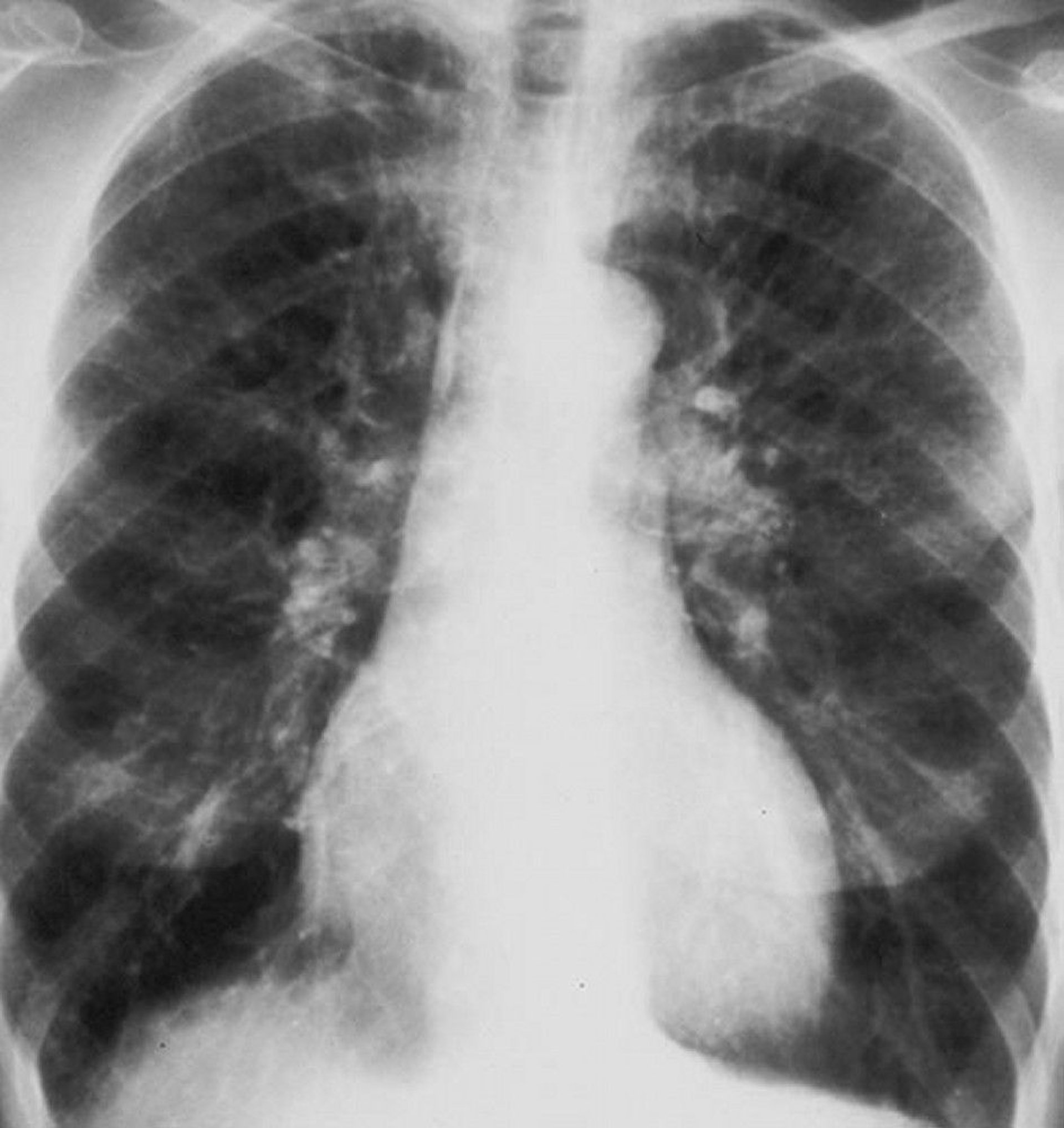
By permission of the publisher. From Barnes P. In Bone's Atlas of Pulmonary and Critical Care Medicine . Edited by J Crapo. Philadelphia, Current Medicine, 2005.
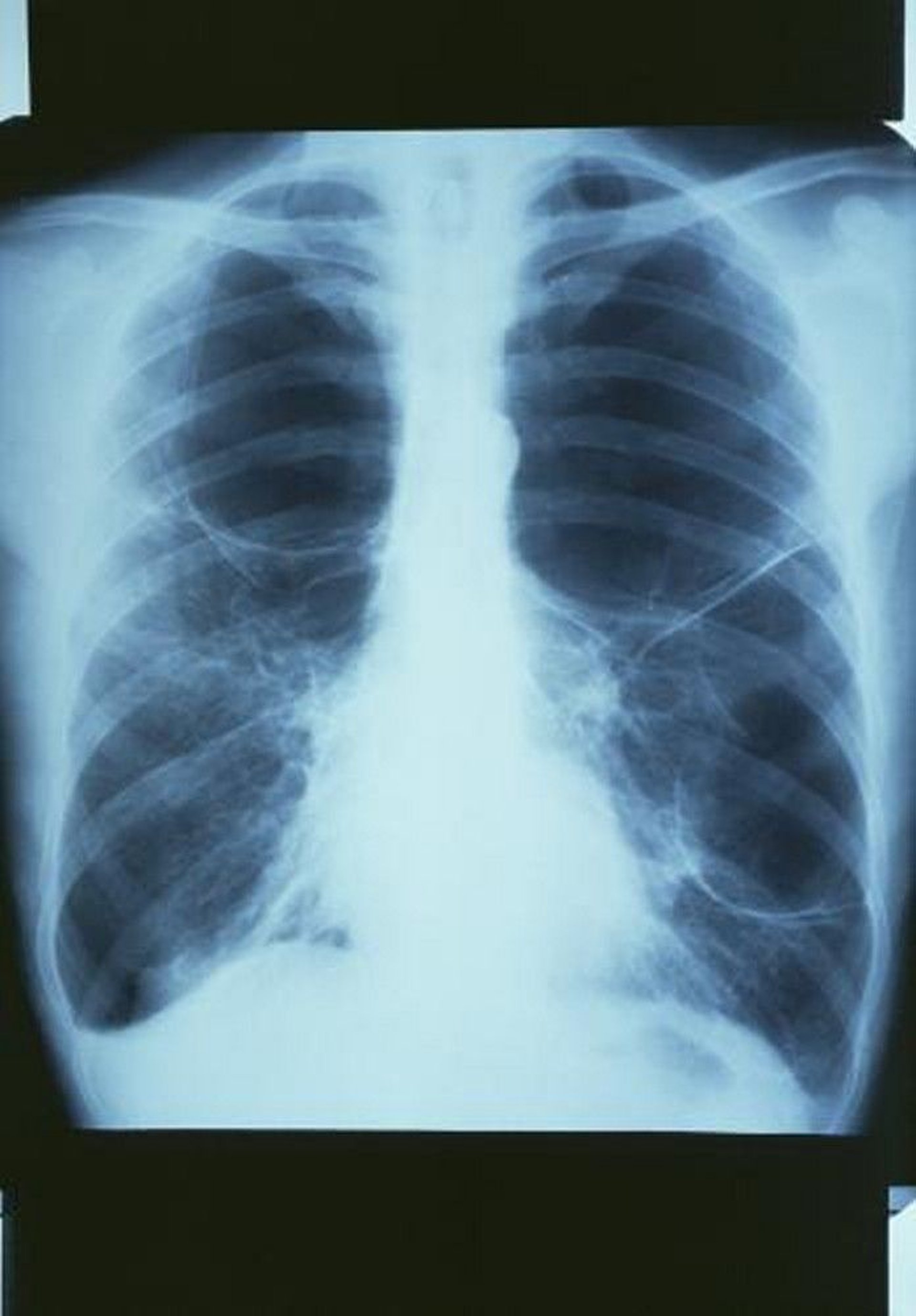
GJLP/CNRI/SCIENCE PHOTO LIBRARY
Prominent hila suggest large central pulmonary arteries that may signify pulmonary hypertension . Right ventricular enlargement that occurs in cor pulmonale may be masked by lung hyperinflation or may manifest as encroachment of the heart shadow on the retrosternal space or by widening of the transverse cardiac shadow in comparison with previous chest radiographs.
Chest CT may reveal abnormalities that are not apparent on the chest radiograph and may also suggest coexisting or complicating disorders, such as pneumonia , pneumoconiosis , or lung cancer . CT helps assess the extent and distribution of emphysema, estimated either by visual scoring or with analysis of the distribution of lung density. Indications for obtaining CT in patients with COPD include evaluation for lung volume reduction procedures, suspicion of coexisting or complicating disorders that are not clearly evident or excluded by chest radiograph, suspicion of lung cancer, and screening for lung cancer . Enlargement of the pulmonary artery diameter greater than the ascending aorta diameter suggests pulmonary hypertension ( 4 ).
Adjunctive tests
Alpha-1 antitrypsin levels should be measured in patients < 50 years with symptomatic COPD and in patients of any age with COPD who do not smoke to detect alpha-1 antitrypsin deficiency . Other indications of possible alpha-1 antitrypsin deficiency include a family history of premature COPD or unexplained liver disease, lower-lobe distribution of emphysema, and COPD associated with antineutrophil cytoplasmic antibody (ANCA)-positive vasculitis. If levels of alpha-1 antitrypsin are low, the diagnosis should be confirmed by genetic testing to establish the alpha-1 antitrypsin phenotype.
ECG , often done to exclude cardiac causes of dyspnea, typically shows diffusely low QRS voltage with a vertical heart axis caused by lung hyperinflation and increased P-wave voltage or rightward shifts of the P-wave vector caused by right atrial enlargement in patients with advanced emphysema. Findings of right ventricular hypertrophy include an R or R ′ wave as tall as or taller than the S wave in lead V1; an R wave smaller than the S wave in lead V6; right-axis deviation > 110 ° without right bundle branch block; or some combination of these findings. Multifocal atrial tachycardia , an arrhythmia that can accompany COPD, manifests as a tachyarrhythmia with polymorphic P waves and variable PR intervals.
Echocardiography is occasionally useful for assessing right ventricular function and pulmonary hypertension, although air trapping makes it technically difficult in patients with COPD. Echocardiography is most often indicated when coexistent left ventricular or valvular heart disease is suspected.
Hemoglobin and hematocrit are of little diagnostic value in the evaluation of COPD but may show erythrocythemia (hematocrit > 48%) if the patient has chronic hypoxemia. Patients with anemia (for reasons other than COPD) have disproportionately severe dyspnea. The differential white blood cell count may be helpful. A growing body of evidence indicates that eosinophilia predicts response to inhaled corticosteroids.
Serum electrolytes are of little value but may show an elevated bicarbonate level if patients have chronic hypercapnia. Venous blood gases are useful for diagnosis of acute or chronic hypercapnia.
Evaluation of exacerbations
Patients with acute exacerbations usually have combinations of increased cough, sputum, dyspnea, and work of breathing, as well as low oxygen saturation determined with pulse oximetry, diaphoresis, tachycardia, anxiety, and cyanosis. Patients with exacerbations accompanied by retention of carbon dioxide may be lethargic or somnolent, a very different appearance.
All patients requiring hospitalization for an acute exacerbation should undergo testing to quantify hypoxemia and hypercapnia. Hypercapnia may exist without hypoxemia.
Findings of partial pressure of arterial oxygen (PaO2) < 50 mm Hg, or partial pressure of carbon dioxide in arterial blood (PaCO2) > 50 mm Hg, or partial pressure of carbon dioxide in venous blood (PvCO2) > 55 mm Hg in patients with respiratory acidemia (pH < 7.35) define acute respiratory failure . Some patients chronically manifest such levels of PaO2 and PaCO2 in the absence of acute respiratory failure.
A chest radiograph is often done to check for pneumonia or pneumothorax . Point of care ultrasound (POCUS) may prove to be a useful adjunctive procedure for rapid diagnosis of pneumonia or pneumothorax. In patients with acute onset of symptoms, chest CT angiogram is done to check for pulmonary embolism . Very rarely, among patients receiving chronic systemic corticosteroids, infiltrates may represent Aspergillus pneumonia .
Yellow or green sputum is a reliable indicator of neutrophils in the sputum and suggests bacterial colonization or infection.
Sputum culture is usually done in hospitalized patients but is not usually necessary in outpatients. In samples from outpatients, Gram stain usually shows neutrophils with a mixture of organisms, often gram-positive diplococci ( Streptococcus pneumoniae ), gram-negative bacilli ( H. influenzae ), or both. However, culture and microscopic examination of sputum is usually not necessary for outpatients. Other oropharyngeal commensal organisms, such as Moraxella catarrhalis (formerly known as Branhamella catarrhalis ), occasionally cause exacerbations. In hospitalized patients, resistant gram-negative organisms (eg, Pseudomonas ) or, rarely, Staphylococcus may be identified in culture specimens.
During influenza season, a rapid influenza test will guide treatment with anti-influenza agents, and a respiratory viral panel for respiratory syncytial virus (RSV), rhinovirus, and metapneumovirus may allow tailoring of antimicrobial therapy. Testing for COVID-19 and consideration of COVID-19–specific therapies is also indicated.
Serum C-reactive protein (CRP) is helpful in guiding the use of antibiotics during exacerbations. In clinical trials, use of antibiotics can be decreased in patients with low CRP without evidence of harm ( 5, 6 ).
Diagnosis references
1. Lange P, Celli B, Agusti A, et al : Lung-function trajectories leading to chronic obstructive pulmonary disease. N Engl J Med 373(2):111–122, 2015.
2. Bhakta NR, Bime C, Kaminsky DA, et al : Race and ethnicity in pulmonary function test interpretation: An Official American Thoracic Society Statement. Am J Respir Crit Care Med 207(8):978–995, 2023. doi:10.1164/rccm.202302-0310ST
3. Global Initiative for Chronic Obstructive Lung Disease (GOLD) : Diagnosis and assessment. Global Strategy for the Prevention, Diagnosis, and Management of COPD: 2024 report.
4. Iyer AS, Wells JM, Vishin S, et al : CT scan-measured pulmonary artery to aorta ratio and echocardiography for detecting pulmonary hypertension in severe COPD. Chest 145(4):824–832, 2014.
5. Butler CC, Gillespie D, White P, et al : C-Reactive protein testing to guide antibiotic prescribing for COPD exacerbations. N Engl J Med 381(2):111–120, 2019. doi: 10.1056/NEJMoa1803185
6. Prins HJ, Duijkers R, van der Valk P, et al : CRP-guided antibiotic treatment in acute exacerbations of COPD in hospital admissions. Eur Respir J 53(5):1802014, 2019. doi: 10.1183/13993003.02014-2018
Treatment of COPD
(See also Treatment of Stable COPD and Treatment of Acute COPD Exacerbation .)
Smoking cessation
Inhaled bronchodilators, corticosteroids, or both
Supportive care (eg, oxygen therapy, pulmonary rehabilitation)
COPD management involves treatment of chronic stable disease and prevention and treatment of exacerbations . Treatment of cor pulmonale , a common complication of long-standing, severe COPD, is discussed elsewhere.
Smoking cessation is critical in treatment of COPD.
Treatment of chronic stable COPD aims to prevent exacerbations and improve lung and physical function. Treatment relieves symptoms rapidly with primarily short-acting beta-adrenergic medications and decreases exacerbations with inhaled corticosteroids, long-acting beta-adrenergic medications, long-acting anticholinergic medications, or a combination (see table Pharmacotherapy of COPD ).
Pulmonary rehabilitation includes structured and supervised exercise training, nutrition counseling, and self-management education.
Oxygen therapy is indicated for selected patients.
Treatment of exacerbations ensures adequate oxygenation and near-normal blood pH, reverses airway obstruction, and treats any cause.
Immunization against influenza , pneumococcus , COVID-19 , and respiratory syncytial virus (RSV —in patients age 60 years and older) should also be recommended as a preventive measure.
End-of-life care
In patients with very severe disease, exercise is unwarranted and activities of daily living are arranged to minimize energy expenditure. For example, patients may arrange to live on one floor of the house, have several small meals rather than fewer large meals, and avoid wearing shoes that must be tied. End-of-life care should be discussed, including whether to pursue mechanical ventilation, the use of palliative sedation, and appointment of a surrogate medical decision-maker in the event of the patient’s incapacitation.
Prognosis for COPD
Severity of airway obstruction predicts survival in patients with COPD. For an FEV1 35 to 55% predicted, the 5-year mortality is 40% ( 1 ). For an FEV1less than 35% predicted, the 5-year mortality is 55% ( 1 ).
More accurate prediction of risk of death is possible by simultaneously measuring body mass index ( B ), the degree of airflow obstruction ( O , which is the FEV1), dyspnea ( D , which is measured using the Modified British Medical Research Council (mMRC) Questionnaire ), and exercise capacity ( E , which is measured with a 6-minute walk test ); this is the BODE index. Also, older age and the presence of heart disease, anemia, resting tachycardia, hypercapnia, or hypoxemia predict decreased survival, whereas a significant response to bronchodilators predicts improved survival. Risk factors for death in patients with acute exacerbation requiring hospitalization include older age, higher PaCO2, and use of maintenance oral corticosteroids.
Patients at high risk of imminent death are those with progressive unexplained weight loss or severe functional decline (eg, those who experience dyspnea with self-care, such as dressing, bathing, or eating).
Mortality in COPD may result from comorbidities (eg, cardiovascular disease, cancer) or intercurrent illnesses rather than from progression of the underlying disorder in patients who have stopped smoking. Death is generally caused by acute respiratory failure, pneumonia, lung cancer, heart disease, or pulmonary embolism.
Prognosis reference
1. Almagro P, Martinez-Camblor P, Soriano JB, et al : Finding the best thresholds of FEV1 and dyspnea to predict 5-year survival in COPD patients: the COCOMICS study. PLoS One 9(2):e89866, 2014. doi: 10.1371/journal.pone.0089866
Cigarette smoking in susceptible people is the major cause of chronic obstructive pulmonary disease (COPD) in the developed world.
Diagnose COPD and differentiate it from disorders that have similar characteristics (eg, asthma, heart failure) primarily by routine clinical information, such as symptoms (particularly time course), age at onset, risk factors, and results of routine tests (eg, chest radiograph, pulmonary function tests).
Reductions of FEV1, FVC, and the ratio of FEV1/FVC post-bronchodilator are characteristic findings.
Categorize patients based on symptoms and exacerbation risk into one of 3 groups and use that category to guide treatment.
Relieve symptoms rapidly with primarily short-acting beta-adrenergic medications and decrease exacerbations with inhaled corticosteroids, long-acting beta-adrenergic medications, long-acting anticholinergic medications, or a combination.
Encourage smoking cessation using multiple interventions.
Immunization against influenza, pneumococcus, COVID-19, and respiratory syncytial virus ([RSV] in patients age ≥ 60 years) should also be done as a preventive measure.

Copyright © 2024 Merck & Co., Inc., Rahway, NJ, USA and its affiliates. All rights reserved.
- Cookie Preferences

Global Initiative for Chronic Obstructive Lung Disease - GOLD
GOLD Teaching Slide Set
Updated November 2024. PowerPoint slide set summarizing GOLD’s objectives, documents, and management recommendations from the 2024 update of the GOLD Report, with background information about COPD and the burden of this disease.
Ohio State nav bar
The Ohio State University
- BuckeyeLink
- Find People
- Search Ohio State
Pathophysiology and Clinical Presentation
The lungs are the main organ of the respiratory system. Their main function is to assist in the exchange of oxygen and carbon dioxide using the air that we inhale (McCance & Huether, 2019). The right lung has three lobes and the left lung has two lobes. The pulmonary artery brings deoxygenated blood to the capillaries that form respiratory membranes with the alveoli (McCance & Huether, 2019). The alveoli will perform gas exchange, and then the pulmonary veins will return the now oxygenated blood back to the heart so that it can be sent throughout the body (McCance & Huether, 2019). Around the lungs is the pleura which is made up of two layers, the visceral and parietal pleural layers. Between these two layers there is a small amount of pleural fluid that works as a lubricant to prevent any friction, as well as an adhesive to bring the lungs to the thoracic wall so that it can assist in the movement of lungs with every breath (McCance & Huether, 2019). With normal lung function, the alveoli in the lungs have strong elastic walls that allow air to expand and contract the little sacs. The bronchioles are nice and clear and allow air to flow in and out of them smoothly (McCance & Huether, 2019). This is normal lung function.
COPD is Chronic Obstructive Pulmonary Disease. This is a lung disease that is obstructive in nature, irreversible, and can get worse over time (McCance & Huether, 2019). COPD is a common disease that is preventable. There are two main conditions that cause COPD. One is emphysema , and the other is chronic bronchitis . In some situations, you may find a genetic susceptibility such as in the case of alpha-1 antitrypsin deficiency (McCance & Huether, 2019). COPD is the third leading cause of death in the United States and the sixth leading cause of death worldwide (McCance & Huether, 2019).
COPD happens when the lungs are exposed to harmful particles and gases which cause the lungs to have an abnormal inflammatory response (McCance & Huether, 2019). The most common harmful cause is cigarette smoking. COPD can also occur from exposure to occupational dusts and chemicals, indoor air pollution (such as fuels used for cooking and heating), outdoor air pollution, any factor involved in lung growth during gestation and childhood, and genetic susceptibilities such as a mutation in the alpha-1 antitrypsin gene (McCance & Huether, 2019). In both chronic bronchitis and emphysema you will see involvement of neutrophils, macrophages, and lymphocytes to the lungs, which will lead to inflammation, oxidative stress, extracellular matrix proteolysis, and apoptotic and autophagic cell death, all of which cause progressive damage (McCance & Huether, 2019).
Chronic bronchitis is one type of COPD. In chronic bronchitis, patients exhibit a chronic productive cough and experience excess mucus build up that leads to irritation and mucus throughout the large and small airways of the lungs (McCance & Huether, 2019). The lining within the airways becomes swollen and irritated and the cilia function becomes impaired, making it harder to breathe. This happens for at least three months of the year and for at least two years in a row. These patients will end up with a ventilation-perfusion mismatch with hypoxemia (Department of Pulmonary Rehab, 2009).
Imagine retrieved from mayoclinic.org
Emphysema is a second type of COPD. It is a disease of the alveoli. In emphysema, there is irritation to the alveoli in the lungs which eventually leads to damage and a reduction of air exchange in the lungs (McCance & Huether, 2019). This makes it hard for the patient to be able to move oxygen into the blood or take carbon dioxide out of the blood. Patients with emphysema will have permanent enlargement of the gas-exchange airways as well as damage to the walls of the alveoli (McCance & Huether, 2019). They lose their normal elasticity that allowed them to expand and contract, letting air in and out as with normal, healthy alveoli (Department of Pulmonary Rehab, 2009).
Clinical Presentation:
Often with COPD, patients you will see some combination of both presentations seen in chronic bronchitis and emphysema.
In review, COPD causes the flow of air out of the lungs to be blocked. The air is therefore trapped in the lungs, making it hard for the lungs to send the right amount of oxygen to the rest of the body (McCance & Huether, 2019). Patients can breathe air in, but getting air out is a challenge. Often, these patients will present with coughing (which can be productive or nonproductive), wheezing, shortness of breath that gets worse with exertion, and feelings of tightness in the chest (Department of Pulmonary Rehab, 2009).
The main causes of COPD are smoking, exposure to secondhand smoke, and working in environments where you are breathing in toxic dusts, fumes or gases (McCance & Huether, 2019).
Patients with COPD need to understand that this disease is chronic, obstructive in nature, and progressive over time. This means that they cannot reverse the disease, but they can stop it in its tracks and keep it from getting worse. One of the best ways to do this is to stop smoking if the patient is a smoker (Department of Pulmonary Rehab, 2009).
An official website of the United States government
Here’s how you know
Official websites use .gov A .gov website belongs to an official government organization in the United States.
Secure .gov websites use HTTPS A lock ( Lock Locked padlock icon ) or https:// means you’ve safely connected to the .gov website. Share sensitive information only on official, secure websites.

- Health Topics
- Drugs & Supplements
- Medical Tests
- Medical Encyclopedia
- About MedlinePlus
- Customer Support
Diagnosis and Tests
Treatments and therapies, living with, related issues, see, play and learn, test your knowledge, statistics and research, clinical trials.
- Journal Articles
Reference Desk
Find an expert, older adults, patient handouts, what is copd (chronic obstructive pulmonary disease).
COPD (chronic obstructive pulmonary disease) is a group of lung diseases that make it hard to breathe and get worse over time.
Normally, the airways and air sacs in your lungs are elastic or stretchy. When you breathe in, the airways bring air to the air sacs. The air sacs fill up with air, like a small balloon. When you breathe out, the air sacs deflate, and the air goes out. If you have COPD, less air flows in and out of your airways because of one or more problems:
- The airways and air sacs in your lungs become less elastic
- The walls between many of the air sacs are destroyed
- The walls of the airways become thick and inflamed
- The airways make more mucus than usual and can become clogged
What are the types of COPD (chronic obstructive pulmonary disease)?
COPD includes two main types:
- Emphysema affects the air sacs in your lungs, as well as the walls between them. They become damaged and are less elastic.
- Chronic bronchitis , in which the lining of your airways is constantly irritated and inflamed. This causes the lining to swell and make mucus.
Most people with COPD have both emphysema and chronic bronchitis, but how severe each type is can be different from person to person.
What causes COPD (chronic obstructive pulmonary disease)?
The cause of COPD is usually long-term exposure to irritants that damage your lungs and airways. In the United States, cigarette smoke is the main cause. Pipe, cigar, and other types of tobacco smoke can also cause COPD, especially if you inhale them.
Exposure to other inhaled irritants can contribute to COPD. These include secondhand smoke , air pollution , and chemical fumes or dusts from the environment or workplace.
Rarely, a genetic condition called alpha-1 antitrypsin deficiency can play a role in causing COPD.
Who is at risk for COPD (chronic obstructive pulmonary disease)?
The risk factors for COPD include:
- Smoking. This the main risk factor. Up to 75% of people who have COPD smoke or used to smoke.
- Long-term exposure to other lung irritants , such as secondhand smoke, air pollution, and chemical fumes and dusts from the environment or workplace
- Age. Most people who have COPD are at least 40 years old when their symptoms begin.
- Genetics. This includes alpha-1 antitrypsin deficiency, which is a genetic condition. Also, smokers who get COPD are more likely to get it if they have a family history of COPD.
- Asthma. People who have asthma have more risk of developing COPD than people who don't have asthma. But most people with asthma will not get COPD.
What are the symptoms of COPD (chronic obstructive pulmonary disease)?
At first, you may have no symptoms or only mild symptoms. As the disease gets worse, your symptoms usually become more severe. They can include:
- Frequent coughing or a cough that produces a lot mucus
- A whistling or squeaky sound when you breathe
- Shortness of breath, especially with physical activity
- Tightness in your chest
Some people with COPD get frequent respiratory infections such as colds and the flu . In severe cases, COPD can cause weight loss, weakness in your lower muscles, and swelling in your ankles, feet, or legs.
How is COPD (chronic obstructive pulmonary disease) diagnosed?
Your health care provider may use many tools to make a diagnosis:
- A medical history, which includes asking about your symptoms
- A family history
- Various tests, such as lung function tests , a chest x-ray or CT scan , and blood tests
Your doctor will diagnose COPD based on your signs and symptoms, your medical and family histories, and test results.
What are the treatments for COPD (chronic obstructive pulmonary disease)?
There is no cure for COPD. However, treatments can help with symptoms, slow the progress of the disease, and improve your ability to stay active. There are also treatments to prevent or treat complications of the disease. Treatments include:
- Quitting smoking if you are a smoker. This is the most important step you can take to treat COPD.
- Avoiding secondhand smoke and places where you might breathe in other lung irritants
- Ask your health care provider for an eating plan that will meet your nutritional needs. Also ask about how much physical activity you can do. Physical activity can strengthen the muscles that help you breathe and improve your overall wellness.
- Bronchodilators, which relax the muscles around your airways. This helps open your airways and makes breathing easier. Most bronchodilators are taken through an inhaler. In more severe cases, the inhaler may also contain steroids to reduce inflammation.
- Vaccines for the flu and pneumococcal pneumonia, since people with COPD are at higher risk for serious problems from these diseases
- Antibiotics if you get a bacterial or viral lung infection
- Oxygen therapy , if you have severe COPD and low levels of oxygen in your blood. Oxygen therapy can help you breathe better. You may need extra oxygen all the time or only at certain times.
- An exercise program
- Disease management training
- Nutritional counseling
- Psychological counseling
- Remove damaged lung tissue
- Remove large air spaces (bullae) that can form when air sacs are destroyed. The bullae can interfere with breathing.
- For severe COPD, some people may need lung transplant
If you have COPD, it's important to know when and where to get help for your symptoms. You should get emergency care if you have severe symptoms, such as trouble catching your breath or talking. Call your health care provider if your symptoms are getting worse or if you have signs of an infection, such as a fever .
Can COPD (chronic obstructive pulmonary disease) be prevented?
Since smoking causes most cases of COPD, the best way to prevent it is to not smoke. It's also important to try to avoid lung irritants such as secondhand smoke, air pollution, chemical fumes, and dusts.
NIH: National Heart, Lung, and Blood Institute
- Chronic Obstructive Pulmonary Disease (COPD) (Centers for Disease Control and Prevention)
- Chronic Obstructive Pulmonary Disease (COPD) (American Thoracic Society) - PDF
- Breathlessness (American Thoracic Society) - PDF
- Spirometry (American Lung Association)
- Spirometry (Mayo Foundation for Medical Education and Research) Also in Spanish
- COPD Medications (National Jewish Health)
- Medicines for COPD (American Thoracic Society) - PDF
- Surgery for COPD (American Lung Association)
- Nutrition and COPD (American Lung Association)
- Oxygen Therapy (American Lung Association)
- Asthma and COPD: Differences and Similarities (American Academy of Allergy, Asthma, and Immunology)
- Five Steps for Successful Flying with Oxygen (American Lung Association)
- Sleep Problems in Asthma and COPD (American Thoracic Society) - PDF Also in Spanish
- Traveling with Portable Oxygen (American College of Chest Physicians) - PDF
- Metered dose inhaler use - Series (Medical Encyclopedia) Also in Spanish
- Nebulizer use - series (Medical Encyclopedia) Also in Spanish
- Peak flow meter use - Series (Medical Encyclopedia) Also in Spanish
- Spacer use - Series (Medical Encyclopedia) Also in Spanish
- Managing COPD Quiz (Medical Encyclopedia) Also in Spanish
- Smoking Myths & Facts Quiz (Medical Encyclopedia) Also in Spanish
- Test Your Knowledge of COPD Triggers (Medical Encyclopedia) Also in Spanish
- FastStats: Chronic Obstructive Pulmonary Disease (COPD) Includes: Chronic Bronchitis and Emphysema (National Center for Health Statistics)
Journal Articles References and abstracts from MEDLINE/PubMed (National Library of Medicine)
- Article: Effects of scapulothoracic exercises on chest mobility, respiratory muscle strength, and...
- Article: Effectiveness and safety of acupuncture as an adjunctive therapy for chronic...
- Article: The effect of nebulized N-acetylcysteine on the phlegm of chronic obstructive...
- COPD -- see more articles
- American Lung Association
- Chronic Obstructive Pulmonary Disease COPD (Department of Health and Human Services, Office on Women's Health)
- COPD: Unique to Older Adults (AGS Health in Aging Foundation)
- Blood gases (Medical Encyclopedia) Also in Spanish
- Chronic obstructive pulmonary disease (COPD) (Medical Encyclopedia) Also in Spanish
- Chronic obstructive pulmonary disease - adults - discharge (Medical Encyclopedia) Also in Spanish
- COPD - control drugs (Medical Encyclopedia) Also in Spanish
- COPD - managing stress and your mood (Medical Encyclopedia) Also in Spanish
- COPD - quick-relief drugs (Medical Encyclopedia) Also in Spanish
- COPD and other health problems (Medical Encyclopedia) Also in Spanish
- COPD flare-ups (Medical Encyclopedia) Also in Spanish
- Day to day with COPD (Medical Encyclopedia) Also in Spanish
- How to breathe when you are short of breath (Medical Encyclopedia) Also in Spanish
- Pulmonary function tests (Medical Encyclopedia) Also in Spanish
- Smoking and COPD (Medical Encyclopedia) Also in Spanish
- Using oxygen at home (Medical Encyclopedia) Also in Spanish
The information on this site should not be used as a substitute for professional medical care or advice. Contact a health care provider if you have questions about your health.
- News and Features
- Conferences
- Clinical Tools
- Special Collections
Chronic Obstructive Pulmonary Disease (COPD)
Chronic obstructive pulmonary disease (COPD) refers to a group of conditions that include emphysema and chronic bronchitis and is characterized by progressive airflow limitation and tissue destruction. 1 It is associated with structural changes in the lungs due to chronic inflammation, typically caused by prolonged exposure to noxious gasses or particles. This chronic inflammation, most notably derived from cigarette smoke, causes airway narrowing and decreased lung recoil. 2
COPD is the third most common cause of morbidity and death globally 2 and was the fourth leading cause of mortality in the U.S. in 2018 in women. 1 The prognosis of COPD is largely variable based on significant comorbidities and the patient’s adherence to treatment, especially stopping smoking and pulmonary rehabilitation. 2
History of Chronic Obstructive Pulmonary Disease
The term “chronic bronchitis and emphysema” (CB&E) was used for several decades to describe what is now known as COPD. 3 The first clear description of acute and chronic bronchitis was published in 1808 by Charles Badham, who later became Professor of Practice of Physic at the University of Glasgow. René Laennec, who invented the monaural stethoscope in 1815, went on to publish the first clinical descriptions of emphysema.
Some features of the disease he described were:
- The presence of peripheral airway obstruction
- Loss of lung recoil
- Collateral ventilation
- Chronic bronchitis
- Right ventricular hypertrophy in advanced disease 3
In time, advancements in spirometry technology and knowledge of respiratory physiology led to the development of effective oxygen treatment methods in some academic hospitals. Despite this, in 1923, E.L. Collis, Professor of Preventive Medicine in Cardiff, presented a more realistic view of the typical medical management of bronchitis in Britain, concluding that smoke abatement in industrial districts and great towns and an improvement in the socioeconomic status of the lower grades of society could help prevent the condition. 3
In the 1930s, and again in the early years after World War II, there were major advances in the development of lung function tests and an increase in gathering systematic data about public health. This led to considerable developments in the assessment of chronic bronchitis and emphysema, and by the end of the 1950s, a new emphasis was placed on the effects of cigarette smoking and urban pollution. 3 By the early 1960s, smoking was determined to be the primary cause of CB&E, as is still the case today, although, at the time, the precise mechanisms of lung damage were unknown. Throughout the 1960s and 1970s, COPD gradually replaced CB&E as the designated term for the disease. 3
Epidemiology of Chronic Obstructive Pulmonary Disease
In 2018, 1 chronic lower respiratory disease, primarily chronic obstructive pulmonary disease, was the fourth leading cause of death in the United States, but by 2020 had dropped to sixth. 4 COPD is the third most common cause of morbidity and death across the globe. 2 Smokers are roughly 12 to 13 times more likely to die from chronic obstructive pulmonary disease than those who have never smoked. 5 Around 15.7 million Americans report having been diagnosed with COPD, but the prevalence is likely to be underestimated due to underdiagnosis — earlier surveys have shown that more than 50% of adults with low pulmonary function were unaware that they had the disease. 1
COPD most commonly presents in smokers and those over 40 years of age. 2 Historically, chronic obstructive pulmonary disease was thought of as a predominantly male-oriented disease, but since 2000 more women than men have died from COPD in the United States, and there are currently more women living with chronic obstructive pulmonary disease than men. 1 In 2013, the following groups were more likely to report a COPD diagnosis:
- Current or former smokers
- People aged 65 or older
- American Indians/Alaska Natives and multiracial non-Hispanics
- People with a history of asthma
- People who were divorced, widowed, or separated
- People who were unemployed, retired, or unable to work
- People with less than a high school education
Etiology and Risk Factors
COPD is caused by prolonged exposure to harmful particles or gasses, usually from cigarette smoke. 1,2,8
Smoking is the most common etiology of COPD worldwide, 2 causing approximately 85 to 90% of all chronic obstructive pulmonary disease cases. 5 Overall, 10% to 15% of smokers develop COPD. It should be noted that symptoms depend on the intensity of smoking, years of exposure, and baseline lung function. The more a person smokes, both in terms of years and volume of consumption (overall number of cigarettes), the greater the person’s risk of COPD. This risk is not limited to cigarette smokers but applies to pipe smokers, cigar smokers, and marijuana smokers. 7
Other possible causes of chronic obstructive pulmonary disease include:
- Secondhand smoke 2,5,8
- Environmental and occupational exposures to air pollutants and chemicals — chronic exposure to vapors, chemical fumes, and dusts can irritate and inflame the lungs 2,7,8
- Alpha-1 antitrypsin deficiency (AATD) — a genetic disorder that reduces the protective protein in the lungs 2,7,8
Additional risk factors that may raise the risk of developing COPD include:
- Repeat or severe respiratory infections, or a history of childhood respiratory infections 1,5,8
- Allergies 6
- Asthma — a chronic inflammatory airway disease that may be a risk factor, especially when combined with smoking. About 1 in 5 people with COPD have asthma. 8
- Genetic factors — factors beyond Alpha-1-antitrypsin deficiency are likely to increase the susceptibility of COPD in certain smokers. 7
COPD Pathophysiology
COPD is an inflammatory condition that affects the pulmonary vasculature, lung parenchyma, and airways. The pathophysiology of chronic obstructive pulmonary disease is characterized by progressive airflow limitation and tissue destruction, with chronic inflammation causing airway narrowing and decreased lung recoil. 2 One of the structural changes observed in COPD is emphysema, in which an irritant (smoking, air pollution, etc.) causes an inflammatory response, which, in turn, causes alveolar air sacs to be destroyed, leading to obstructive physiology.
Elastin is also destroyed, resulting in loss of elastic recoil, which causes airway collapse while exhaling. Altogether, this results in a decrease in the forced expiratory volume (FEV1), airflow limitation and impaired gas exchange. Imaging studies often show hyperinflation of the lungs due to air trapping from airway collapse during exhalation. Increases in CO2 levels stem from an inability to exhale fully and progressively impaired gas exchange, and CO2 retention can cause pulmonary hypertension due to diffuse vasoconstriction from hypoxemia. 2
Chronic Obstructive Pulmonary Disease Prognosis
There is no cure for chronic obstructive pulmonary disease (COPD). 9 The prognosis for those diagnosed with the disease varies based on adherence to treatment, especially regarding smoking cessation and pulmonary rehabilitation. 2 Airflow limitation and dyspnea associated with chronic obstructive pulmonary disease are typically progressive, 2 but lifestyle changes and treatments can improve symptoms and slow disease progression. 9 A poorer prognosis is associated with patients who exhibit other comorbidities, such as:
- Lung cancer
- Sleep-related breathing disorders
- Cognitive dysfunction/psychiatric illness
- Cardiovascular disease
- Pulmonary hypertension 2
- Metabolic syndrome
- Bronchiectasis 6
Also, those with asthma and chronic obstructive pulmonary disease tend to have a poorer quality of life with higher mortality rates. The BODE (body-mass index, airflow obstruction, dyspnea, and exercise capacity) index can be used to determine the mortality risk for patients with COPD. Scored on a point scale of 1 to 10, the BODE index provides a cumulative score that correlates with four-year survival, based on these criteria: 2
- Six-minute walk distance
- Modified Medical Research Council dyspnea scale
- FEV1 % of predicted
- BMI (body-mass index)
Other prognostic indicators of disease burden and mortality include: 6
- DLCO (diffusion capacity for carbon monoxide)
- Blood gas measurements
- Clinical state
- Radiographic severity
Presentation and Diagnosis of Chronic Obstructive Pulmonary Disease
COPD usually presents in adulthood, as it is a chronic and progressive disease, and commonly during the winter. Patients typically complain of chronic and progressive dyspnea, sputum production, and cough. They may also exhibit wheezing and chest tightness. 2 A full list of signs and symptoms of chronic obstructive pulmonary disease may include: 7
- Shortness of breath (dyspnea), especially during physical activities
- Frequent respiratory infections
- A chronic cough that may include sputum (sputum coloration may be clear, white, yellow, or greenish.)
- Chest tightness
- Unintended weight loss (in later stages)
- Lack of energy
- Swelling in ankles, feet, or legs
Symptoms such as coughing, wheezing, and shortness of breath typically become worse as the disease progresses. In later stages, significant weight loss can derive from systemic inflammation and the increased energy demands of breathing. 6
Diagnosis of COPD is confirmed by pulmonary function testing (PFT), particularly spirometry. 6 Patients exhibiting relative symptoms and risk factors (especially a history of smoking) should be evaluated for COPD. 2 An FEV1/FVC of less than 0.7 confirms the diagnosis of COPD. Radiography is not required for diagnosis but is usually one of the first steps when COPD is suspected in order to rule out other causes. 6
Physical Examination Findings
In the early stages of chronic obstructive pulmonary disease, the physical examination may be normal. Most patients present with the nonspecific symptoms of chronic shortness of breath and cough. The cough may or may not come with sputum production. 6 Patient history is critical in assisting with disease diagnosis, as there is a history of smoking in most cases. 2
The smell of smoke and signs of nicotine staining on the hands and fingernails may identify current smokers. 6 During auscultation, the clinician may hear:
- Distant breath sounds
- Crackles at the lung bases
- Distant heart sounds 6
Acute exacerbations of chronic obstructive pulmonary disease often present with productive cough, wheezing, and increased dyspnea. 2 Some other physical examination findings that may be observed are as follows: 2
- Significant respiratory distress
- Muscle wasting
- Use of accessory respiratory muscles
- Prolonged expiration
- Breathing through pursed lips
- Increase in the diameter of the anterior-posterior chest wall (barrel chest)
- Central cyanosis (when arterial oxygenation is low)
- Lower extremity edema in right heart failure
- Digital clubbing
Chronic Obstructive Pulmonary Disease Diagnostic Workup
Patient history.
The clinician must obtain a thorough patient history to help with an accurate diagnosis. Patients should be questioned on:
- History of smoking
- Exposure to secondhand smoke
- Occupational and environmental exposures
- Family history 2
If they do smoke, it is essential to identify the age at which they started smoking or stopped smoking (if relevant) and the total pack years. If the patient smoked previously but has quit, the clinician needs to know how many years have passed since they last smoked.6 The clinician needs to be made aware of any prior history of other diseases such as:
- Childhood respiratory infections
Patients with a family history of emphysema, basilar emphysema, and liver disease should raise suspicion for Alpha-1 antitrypsin deficiency (AATD),2 and any younger patient with emphysema symptoms and no or minimal smoking history should be tested for AATD.6
Pulmonary Function Testing
Pulmonary function testing (PFT) is crucial for accurate diagnosis, staging, and monitoring of chronic obstructive pulmonary disease. Spirometry should be performed before and after the administration of an inhaled bronchodilator. Inhaled bronchodilators may be a short-acting anticholinergic, a short-acting beta2-agonist (SABA), or both combined. An FEV1/FVC less than 0.07 is confirmation of a COPD diagnosis. Patients who exhibit signs of dyspnea and a significantly reduced FEV1 should be evaluated for oxygenation with arterial blood gas analysis or pulse oximetry.
The Global Initiative for Chronic Obstructive Lung Disease (GOLD) is a program that was instituted by the World Health Organization alongside the National Heart, Lung, and Blood Institute. GOLD provides updated reports on the recommendations for the diagnosis and treatment of chronic obstructive pulmonary disease. These recommendations are often used to determine disease severity and decide on the choice of therapy. 2 Guidelines for determining the stages of COPD are published by GOLD based on the severity of airflow limitation. The COPD GOLD criteria are as follows: 6
- Mild with FEV1 greater or equal to 80% predicted
- Moderate with FEV1 less than 80% predicted
- Severe with FEV1 less than 50% predicted
- Very severe with FEV1 less than 30% predicted
6-Minute Walk Test
A 6-minute walk test is a commonly performed assessment used to determine a patient’s submaximal functional capacity. This test should be performed indoors on a straight, flat surface, typically in a hallway with a length of 100 feet. The patient has six minutes to cover as much distance as possible while walking. 2
Laboratory Testing
A complete blood count to assess for anemia, infection, and polycythemia should be completed as part of the laboratory testing. Alpha-1 antitrypsin levels should also be checked to determine if there are other potential causes of chronic obstructive pulmonary disease. 2
Radiographic Imaging
Radiographic imaging includes computed tomography (CT) and a chest X-ray. These are not required for diagnosis but help to rule out other causes of symptoms, such as bronchiectasis, pleural disease, lung cancer with airway obstruction, or heart failure . Chest X-rays can show flattening of the diaphragm, hyperinflation, and increased anterior-posterior diameter. Bronchial-wall thickening may be present in cases of chronic bronchitis. CT imaging can be of use in patients with malignancy, bronchiectasis, or if planning surgical procedures, and a CT scan of the chest in patients diagnosed with COPD will be significant for centrilobular emphysema. The subpleural regions may contain bullae. 2
The diagnosis of chronic obstructive pulmonary disease does not require a biopsy; however, histopathologic findings include an increase in inflammatory cells, lymphoid follicles, and structural changes. 2
An acute worsening of respiratory symptoms constitutes an acute exacerbation of COPD. Mild exacerbations are defined by the presence of one of these symptoms — worsening dyspnea, purulence, and sputum volume — in addition to one of the following:
- Increased cough
- Increased wheezing
- Upper respiratory infection within five days
- Fever without another cause
- Increase in the patient’s baseline heart rate or respiratory rate
Moderate exacerbations are defined by the presence of two of the symptoms, while severe exacerbations are indicated by the presence of three. Patients may have acute respiratory failure along with physical findings of hypercapnia and hypoxemia. 2
Mild to moderate chronic obstructive pulmonary disease does not typically require arterial blood gas analysis, but when an assessment of hypercapnia is needed in severe airflow obstruction or oxygen saturation goes below 92%, an arterial blood gas analysis is indicated. 6
Differential Diagnosis of Chronic Obstructive Pulmonary Disease
Chronic obstructive pulmonary disease (COPD) presents with nonspecific symptoms and, therefore, has broad differential diagnoses. A list of some differential diagnoses for COPD is as follows:
- Asthma-COPD overlap syndrome 2
- Bronchiectasis 2
- Bronchiolitis obliterans 2
- Bronchopulmonary mycosis 6
- Central airway obstruction 6
- Chronic bronchitis with normal spirometry 6
- Chronic obstructive asthma 6
- Chronic respiratory failure 6
- Constrictive bronchiolitis 6
- Cor pulmonale 6
- Cystic fibrosis 2
- Diffuse panbronchiolitis 2
- Heart failure 2
- Interstitial lung disease 2
- Lymphangioleiomyomatosis 2
- Malignancy 2
- Pulmonary hypertension 6
- Spontaneous pneumothorax 6
- Thromboembolic disease 2
- Tuberculosis 2
Chronic Obstructive Pulmonary Disease Management
There is currently no cure for chronic obstructive pulmonary disease, but modifying risk factors and managing symptoms have shown to be effective in slowing disease progression and optimizing the quality of living. 6 The main goals of treatment are to:
- Improve the quality of life
- Control symptoms
- Reduce exacerbations and mortality 2
Nonpharmacological Therapy
Nonpharmacological interventions revolve around smoking cessation and pulmonary rehabilitation. Treatment choices will vary and should be based on individual patient needs.
All patients with COPD should receive an annual influenza vaccination. Those who are 65 years of age or older should receive the 13-valent pneumococcal conjugate vaccine (PCV13) and the 23-valent pneumococcal polysaccharide vaccine (PPSV23) at a minimum of one year apart. For patients with significant comorbidities (e.g., chronic heart disease, chronic lung disease, diabetes mellitus) who are 64 years of age or younger, the PPSV23 is recommended. 2
Pharmacological Therapy
Commonly used medications for COPD management include:
- Antibiotics. 2
- Bronchodilators (beta2-agonists, antimuscarinics, methylxanthines)
- Inhaled corticosteroids (ICS)
- Phosphodiesterase-4 (PDE4) inhibitors
- Systemic glucocorticoids
These are all considered to fall under medical therapy. 6
Bronchodilators
The primary mechanisms of action for bronchodilators can be split into two categories: anticholinergic medications and beta2 agonists. These are considered first-line drugs for chronic obstructive pulmonary disease and are delivered through inhalation. These bronchodilators improve FEV1 by altering the smooth muscle tone of the airways, therefore improving exercise tolerance. Bronchodilators are typically given on a regular basis to prevent and reduce:
- Exacerbations
- Hospitalizations 6
Short-acting antimuscarinic antagonists (SAMA) and short-acting beta2 agonists (SABA) should be prescribed as needed in order to manage intermittent dyspnea, typically for immediate relief. Long-acting antimuscarinic antagonists (LAMA) and long-acting beta2 agonists (LABA) are utilized for maintenance therapy in cases of more than occasional dyspnea or in increasing dyspnea; however, GOLD criteria advises against their use during exacerbations.
Another bronchodilator should be added if the symptoms persist while on a single bronchodilator. 6 Methylxanthines, which work by relaxing the smooth muscle in the airways that causes mild bronchodilation, are generally not recommended, as they have shown decreased efficacy and can cause a significant amount of nausea and vomiting along with increased frequency of tremors, palpitations, and arrhythmias. N-acetylcysteine, magnesium, chest physiotherapy, and mucoactive actions are also not recommended due to lack of documented efficacy.
SABA is the mainstay in COPD exacerbation and can be used with or without anticholinergics. LABA (which, again, is not recommended during exacerbations) includes, among others:
- Indacaterol
Arrhythmias, hypokalemia, and tremors are all potential side effects. Tachycardia may precipitate heart failure , so caution should be taken for patients with a prior history of heart disease.
Read more: Lidocaine Antiarrhythmic Uses
Acetyl-choline-induced bronchoconstriction is inhibited by the use of anticholinergics. SAMA includes oxitropium and ipratropium. LAMA, such as tiotropium, can be administered once a day. 6
Inhaled Corticosteroids
ICS is used in step-up therapy as an add-on therapy to bronchodilators. ICS includes budesonide, beclomethasone, fluticasone, etc. 6 Combining ICS and LABA has proven to be more beneficial than either of the drugs when used alone. 2 Common side effects of these drugs include:
- Local infection
All patients with acute chronic obstructive pulmonary disease exacerbation should be given oral systemic corticosteroids, but these should be avoided in stable patients due to more adverse effects. 6
Oral Phosphodiesterase-4 Inhibitors
These drugs are fast-acting and work by reducing inflammation. PDE4 inhibitors can be added if there is severe airflow obstruction and no improvement has been seen with the use of the bronchodilators or ICS. 6
Triple Inhaled Therapy
This therapy entails an inhaled combination of LABA, LAMA, and ICS. It is taken only once per day. 6
Antibiotics
COPD symptoms can be aggravated by respiratory infections. Antibiotics, such as azithromycin, can help treat episodes of worsening COPD, but they should not be used for prevention, as this could lead to antibiotic resistance. 12
Supportive Therapy
Supportive therapy includes:
- Pulmonary rehabilitation
- Oxygen therapy
- Ventilatory support
- Palliative care 6
Pulmonary rehabilitation is indicated in all stages of COPD 2 and can reduce dyspnea and hospitalizations for patients with severe symptoms and multiple exacerbations. 6 The purpose of pulmonary rehabilitation is to improve a patient’s physical function and psychological condition, and it typically involves therapies such as:
- Exercise training
- Behavioral changes. 2
In stable patients, routine supplemental oxygen has not been shown to improve clinical outcomes or the quality of life. Patients with COPD with PaO2 less than 55 mmHg (or oxygen saturation less than 88%) or PaO2 less than 59 mm Hg in case of cor pulmonale should be put on continuous long-term (longer than 15 hours) supplemental oxygen. Survival rates of these patients with severe resting hypoxemia are increased by oxygen therapy. Intermittent oxygen will help those who desaturate with exercise. Oxygen saturation greater than 90% is the goal for any patient. 6
Noninvasive positive pressure ventilation (NPPV) should be tried as the first mode of ventilation in patients with COPD exacerbation with respiratory failure, as long as there is no contraindication. NPPV decreases morbidity and mortality in patients with acute respiratory failure. This supportive therapy improves:
- Gas exchange
- Reduces work of breathing
- Improves VQ matching
- Decreases hospitalization duration
- Improves survival
If NPPV fails to work in a chronic obstructive pulmonary disease patient, the patient should be intubated and put on a ventilator. 6
Palliative care is typically recommended for GOLD stage D, although it is available from the time of diagnosis. This is simply additional care to the patient’s ongoing treatment plan with the goal of providing the best quality of life possible. Palliative care can include:
- Patient education
- Determination of the goal of care
- Discussions about advance care planning
- Patient reassurance
- Depression and anxiety management
- Exploration of end-of-life care plans 6
Interventional Therapy
Severe cases of COPD may require surgical intervention, including:
- Lung volume reduction surgery (improves elastic recoil and reduces hyperinflation 6 )
- Lung transplantation (typically implemented when FEV1 and/or DLCO are less than 20%, 6 it can improve the patient’s ability to breathe and to be active but can result in organ rejection and will require lifelong use of immune-suppressing medications 12 )
- Bullectomy 2 (removes bullae from the lungs, which can improve airflow 12 )
Severe conditions where symptoms are not controlled with medical therapy alone can indicate the need for surgical intervention. 2
Monitoring Side Effects, Adverse Events, Drug-Drug Interactions
Health care optimization in patients with COPD involves the collaboration of an interprofessional team. Comprehensive patient-tailored treatment plans should be implemented and monitored by:
- Primary care providers
- Pharmacists
- Respiratory therapists
- Rehabilitation specialists
- Consultants 2
Pharmacological intervention should be carefully monitored to avoid potential drug-drug interactions, especially in patients with comorbidities. There are adverse effects associated with some of the medications used to treat COPD.
- Arrhythmias
- Hypokalemia
- Local infection 6
- Hypertension
- Bone fracture
- Metabolic issues (e.g., hyperglycemia, type 2 diabetes, weight gain) 13
- Fast heartbeat
- Weight loss 12
Chronic Obstructive Pulmonary Disease Complications
Chronic obstructive pulmonary disease (COPD) patients are prone to develop many different long-term complications. These complications can include:
- Acute exacerbation of COPD 2
- Acute or chronic respiratory failure 2
- Adverse reactions to glucocorticoids 2
- Bacterial infections 2
- Chronic atelectasis 6
- Depression 12
- Heart problems 12
- Interstitial emphysema 6
- Lung cancer 12
- Pneumonia 6
- Pneumothorax 6
- Recurrent respiratory tract infections 6
- Respiratory acidosis, hypoxia, and coma 6
- Respiratory insufficiency or failure 6
- Weight loss 2
COPD Guidelines
Multiple U.S.-based guidelines are available, covering everything from COPD nursing diagnosis to pocket guides for diagnosis, management, and prevention. The GOLD guidelines are considered the standard for COPD diagnosis and treatment recommendations.
- Global Initiative for Chronic Obstructive Lung Disease (GOLD) — GLOBAL STRATEGY FOR PREVENTION, DIAGNOSIS AND MANAGEMENT OF COPD: 2022 Report
- American Thoracic Society — Pharmacologic Management of COPD: An Official ATS Clinical Practice Guideline 2020
COPD Factoids
- COPD is a chronic inflammatory lung disease that causes tissue destruction and irreversible airflow limitation. 2 Emphysema and chronic bronchitis are two types of COPD. 1
- Smoking is the most common risk factor worldwide, causing approximately 85 to 90% of all COPD cases. 5
- COPD most commonly affects adults over 40 years of age. 2
- Diagnosis is made by spirometry with a post-bronchodilator FEV1/FVC ratio <0.7. 2
- Patients with emphysema, especially younger ones, should be screened for AATD. 2
- Patients should obtain annual influenza vaccination. 2
- Patients should cease smoking and avoid secondhand smoke or other harmful exposures. 2
- Patients 65 years or older should receive PCV13 and PPSV23 at least one year apart. 2
- PPSV23 is recommended in COPD patients younger than 65 years who have significant comorbid conditions. 2
- Pharmacologic treatment is based on disease severity and usually involves the administration of bronchodilators and inhaled corticosteroids. 2
- Long-term oral corticosteroids are not recommended due to adverse effects. 6
- Common complications include acute exacerbations, bacterial pneumonia, and pulmonary hypertension. 2
- Current smoker or quit within the past 15 years
- 20+ pack-year smoking history
- Lung transplantation can improve functional capacity but may not increase the prognosis 2
- Depression and anxiety are very common in end-stage lung disease. Medication can be used accordingly for treatment. 6
Read more: Lung Cancer Statistics
- Centers for Disease Control and Prevention. Basics about COPD . Updated June 9, 2021. Accessed August 1, 2022.
- Agarwal AK, Raja A, Brown BD. Chronic obstructive pulmonary disease (COPD) . In: StatPearls. NCBI Bookshelf version. StatPearls Publishing: 2022. Accessed August 1, 2022.
- Watson RA, Pride NB. Early history of chronic obstructive pulmonary disease 1808–1980 . COPD: Journal of Chronic Obstructive Pulmonary Disease . 2015;13(2):262-273. doi:10.3109/15412555.2015.1043521
- Centers for Disease Control and Prevention. FastStats – Deaths and mortality . Updated January 13, 2022. Accessed August 1, 2022.
- American Lung Association. COPD causes and risk factors | American Lung Association . Updated March 5, 2021. Accessed August 1, 2022.
- Parul P, Akshay A, Sandeep S. Emphysema . StatPearls . January, 2022. Accessed August 1, 2022.
- Mayo Clinic. COPD – Symptoms and causes . Published April 15, 2020. Accessed August 1, 2022.
- National Heart, Lung, and Blood Institute. COPD – Causes and risk factors . Updated March 24, 2022. Accessed August 1, 2022.
- National Heart, Lung, and Blood Institute. COPD – Living with . Updated March 24, 2022. Accessed August 1, 2022.
- Agarwal AK, Raja A, Brown BD. [Figure, Table 4. BODE index for predicting COPD survival. Contributed by Anuj Agarwal, MD] . In: StatPearls. NCBI Bookshelf version. StatPearls Publishing: 2022. Accessed August 1, 2022.
- Agarwal AK, Raja A, Brown BD. [Figure, Figure 1. COPD assessment adapted from GOLD 2020 report. Contributed by Anuj Agarwal, MD] . In: StatPearls. NCBI Bookshelf version. StatPearls Publishing: 2022. Accessed August 1, 2022.
- Mayo Clinic. COPD – Diagnosis and treatment . Published April 15, 2020. Accessed August 1, 2022.
- Rice JB, White AG, Scarpati LM, Wan G, Nelson WW. Long-term systemic corticosteroid exposure: A systematic literature review . Clinical Therapeutics . 2017;39(11):2216-2229. doi:10.1016/j.clinthera.2017.09.011
Jonathan Poole is a freelance writer and copyeditor with a BSc in Exercise Science living in West Lafayette, IN. When not writing, he owns and operates a fitness training company, Unstoppable Athletes. More information regarding his training business can be found here: https://www.unstoppableathletes.com
Picked For You
Latest News
Want to read more?
Please login or register first to view this content.
Login Register
Got any suggestions?
We want to hear from you! Send us a message and help improve Slidesgo
Top searches
Trending searches

hispanic heritage month
21 templates

mexican independence
67 templates

49 templates

indigenous canada
47 templates

independencia de mexico
14 templates

22 templates
Chronic Obstructive Pulmonary Disease (COPD)
It seems that you like this template, chronic obstructive pulmonary disease (copd) presentation, free google slides theme, powerpoint template, and canva presentation template.
Chronic obstructive pulmonary disease, also known as COPD, encompasses a group of diseases that cause problems with breathing. In the United States alone it affects about 16 million people. If you are preparing a presentation about it you can use this Slidesgo proposal. It has a simple style, with a white background and light blue waves and lines, which convey elegance and serenity. In addition, we have included a multitude of resources that you can edit to convey your information, such as graphics, map, infographics, etc.
Features of this template
- 100% editable and easy to modify
- 26 different slides to impress your audience
- Contains easy-to-edit graphics such as graphs, maps, tables, timelines and mockups
- Includes 500+ icons and Flaticon’s extension for customizing your slides
- Designed to be used in Google Slides, Canva, and Microsoft PowerPoint
- 16:9 widescreen format suitable for all types of screens
- Includes information about fonts, colors, and credits of the free resources used
How can I use the template?
Am I free to use the templates?
How to attribute?
Attribution required If you are a free user, you must attribute Slidesgo by keeping the slide where the credits appear. How to attribute?

Register for free and start downloading now
Related posts on our blog.

How to Add, Duplicate, Move, Delete or Hide Slides in Google Slides

How to Change Layouts in PowerPoint

How to Change the Slide Size in Google Slides
Related presentations.

Create your presentation Create personalized presentation content
Writing tone, number of slides.

Premium template
Unlock this template and gain unlimited access

Register for free and start editing online

An official website of the United States government
The .gov means it’s official. Federal government websites often end in .gov or .mil. Before sharing sensitive information, make sure you’re on a federal government site.
The site is secure. The https:// ensures that you are connecting to the official website and that any information you provide is encrypted and transmitted securely.
- Publications
- Account settings
The PMC website is updating on October 15, 2024. Learn More or Try it out now .
- Advanced Search
- Journal List
- v.36(1); Jan-Feb 2019
Physical signs in patients with chronic obstructive pulmonary disease
Malay sarkar.
Department of Pulmonary Medicine, Indira Gandhi Medical College, Shimla, Himachal Pradesh, India
Rajeev Bhardwaz
1 Department of Cardiology, Indira Gandhi Medical College, Shimla, Himachal Pradesh, India
Irappa Madabhavi
2 Department of Medical and Pediatric Oncology, GCRI, Ahmedabad, Gujarat, India
3 Department of Pathology, University of Pennsylvania, Philadelphia, PA, USA
We reviewed the various physical signs of chronic obstructive pulmonary disease, their pathogenesis, and clinical importance. We searched PubMed, EMBASE, and the CINAHL from inception to March 2018. We used the following search terms: chronic obstructive pulmonary disease, physical examination, purse-lip breathing, breath sound intensity, forced expiratory time, abdominal paradox, Hoover's sign, barrel-shaped chest, accessory muscle use, etc. All types of studies were chosen. Globally, history taking and clinical examination of the patients is on the wane. One reason can be a significant development in the field of medical technology, resulting in overreliance on sophisticated diagnostic machines, investigative procedures, and medical tests as first-line modalities of patient's management. In resource-constrained countries, detailed history taking and physical examination should be emphasized as one of the important modalities in patient's diagnosis and management. Declining bedside skills and clinical aptitude among the physician is indeed a concern nowadays. Physical diagnosis of chronic obstructive pulmonary disease (COPD) is the quickest and reliable modalities that can lead to early diagnosis and management of COPD patients. Bedside elicitation of physical signs should always be the starting point for any diagnosis and therapeutic approach.
INTRODUCTION
Chronic obstructive pulmonary disease (COPD) is a common, preventable, and treatable disease characterized by persistent respiratory symptoms and airflow limitation that is due to airway and/or alveolar abnormalities usually caused by significant exposure to noxious particles or gases.[ 1 ] COPD is a major global public health issue because of its high prevalence, morbidity, and mortality.[ 2 , 3 ] The socioeconomic impact of COPD is also substantial.[ 4 ] Adeloye et al. [ 2 ] in a systematic review and meta-analysis reported a global prevalence of spirometry-defined COPD of 11.7%. According to the World Health Organization report, more than 3 million people died of COPD in 2012 and majority of the deaths occurred in developing countries.[ 3 ] The mortality due to COPD is rising, and it is expected to become the third leading cause of death globally by 2030.[ 5 ] Underdiagnosis of COPD is a global phenomenon. Lamprecht et al. [ 6 ] had shown that 81.4% of (spirometrically defined) COPD cases remain undiagnosed. Solution to the problem of underdiagnosis lies with proper planning and implementation of strategies focused on an early and accurate diagnosis of COPD. Among the various diagnostic modalities, physical diagnosis is rapid, cost-effective, and can lead to the earlier institution of various preventive and treatment strategies. The initial clinical examination (history and physical) helps in developing a rapport with the patients, identifying the severity of symptoms, determining prognosis, and monitoring therapy.[ 7 ] Clinical examination also helps in developing a pretest probability of the disease. It is highly relevant to establish the efficacy of clinical diagnosis, as an early diagnosis by clinical criteria will increase the number of patients for spirometric confirmation of diagnosis. There should be judicious use of various diagnostic modalities such as chest radiograph, pulmonary function tests, or other laboratory tests along with the evaluation of detailed history and physical signs. Interpretations of diagnostic tests without clinical findings will lose its importance. In this review, we will discuss various physical signs of COPD, their pathogenesis, and clinical importance.
PATHOPHYSIOLOGICAL CHANGES IN CHRONIC OBSTRUCTIVE PULMONARY DISEASE
COPD patients often develop hyperinflation. Hyperinflation occurs due to expiratory flow limitation caused by reduced lungs’ elastic recoil and increased airway resistance. Hyperinflation increases during exercise and acute exacerbation. Hyperinflation has a significant negative impact on respiratory muscles, particularly the diaphragm. Studies done in animals, healthy humans, and COPD patients have shown that hyperinflation increases the contribution of rib cage and neck muscles and decreases relative contribution of the diaphragm.[ 8 ] The effect of hyperinflation on diaphragmatic length is the main mechanism by which it affects the force-generating capacity of the diaphragm.[ 8 , 9 ] The diaphragm becomes flattened and shorter in emphysema. According to Laplace's law, flattened diaphragm because of its increased radius of curvature has reduced force-generating capacity.[ 10 ] Moreover, increase in resistive and elastic load and hypoxemia also causes inspiratory muscle fatigue.[ 11 ] The impact of hyperinflation on inspiratory intercostal muscles is substantially less compared to that of diaphragm.[ 12 ] Dysfunction of the diaphragm may lead to the development of various clinical signs such as abdominal paradox, Hoover's sign, and tripod position.[ 13 ]
ACCESSORY MUSCLE ACTIVITY
Accessory muscles of respiration include the sternocleidomastoid, scalene, trapezius, internal intercostal, and abdominal muscles. Dynamic hyperinflation and air trapping in COPD patients place the diaphragm and intercostal muscles in a mechanically disadvantageous position. Because of this, the diaphragm and intercostals are unable to provide adequate ventilation, leading to the recruitment of accessory muscles. Two important accessory muscles of inspiration are the scalene and sternocleidomastoid. Accessory muscle use is one of the earliest signs of airway obstruction. Use of accessory muscles indicates severe disease and signifies that the forced expiratory volume in 1 s (FEV 1 ) is decreased to 30% of the normal or less. More than 90% patients with acute exacerbations of COPD exhibit use of accessory respiratory muscles.[ 14 ] The activity of these muscles is best judged by palpation. The contraction of scalenes is felt by pressing the fingertips into the floor of the posterior triangle of the neck gently. The sternomastoids are examined by drawing them backward with the thumb and first finger to feel the contraction.
Chronic use of sternocleidomastoids may lead to the development of noticeable hypertrophy, and they may become thicker than patient's own thumb. The sternocleidomastoid activity can be seen in respiratory distress due to any cause, but their activity is mainly seen in patients with COPD.[ 15 ] Sternocleidomastoid activity leads to elevation of the clavicles, and more than 5 mm upward movement of the clavicle is a valuable sign of severe obstruction, correlating with FEV 1 of 0.6 L.[ 16 ] Magendie[ 17 ] described the cyclical inspiratory contraction of the scalene muscles as “respiratory pulse.” The scalene muscle is recruited earlier than the sternomastoid muscles. De Troyer et al. [ 18 ] had shown that most patients of COPD use the scalenes and not the sternocleidomastoids and trapezii during resting condition, suggesting that both the sternocleidomastoids and trapezii in humans have a very high threshold of activation. Sternomastoids are usually recruited at very high lung volumes and during periods of high levels of ventilation as in exercise.[ 19 ] Gandevla et al. [ 20 ] similarly demonstrated that in stable COPD patients, the inspiratory discharge frequencies were significantly greater for both the parasternal and scalene muscles compared to the controls; however, the sternomastoid muscle is not activated. However, whether scalene should be considered as accessory muscle is a big question as the study had shown its activity even at rest in healthy individuals.[ 15 ] Stubbing et al. [ 8 ] had shown that only contraction of scalene muscles but not the sternomastoid muscles is correlated with the degree of obstruction and the duration of symptoms. Accessory expiratory muscles are the abdominal respiratory muscles (rectus abdominis, transverse abdominis, and external and internal obliques). They augment the passive recoil of the lungs during expiration and also help in inspiration. Their contraction cause lengthening of the diaphragm, thereby diminishing its radius of curvature, which helps in generating greater inspiratory pressure by the diaphragm.[ 21 , 22 ] Dodd et al. [ 22 ] postulated that abdominal muscle recruitment can store elastic and gravitational energy within the diaphragm which when released during inspiration augments inspiratory pressure generation. Ninane et al. [ 23 ] demonstrated that many stable patients of severe COPD contract their abdominal muscles during expiration and it is confined to the transverse muscles. Abdominal muscles are active in expiration when the minute ventilation exceeds 40 L.[ 24 ] The accessory muscle recruitment has a likelihood ratio (LRs) of 4.75 (2.29–9.82; P < 0.0001) for the diagnosis of COPD.[ 25 ]
PURSE-LIP BREATHING
Patients with COPD frequently adopt purse-lip breathing (PLB) pattern either spontaneously or as a part of the pulmonary rehabilitation programs. In PLB, patients tend to exhale through pursed lips. PLB reduces the respiratory rate (both at rest and during exercise), reduces carbon dioxide level, and improves ventilation and oxygenation.[ 26 ] PLB decreases the respiratory rate by lengthening the period of expiration. During the PLB maneuver, resistance to expiratory airflow is increased, resulting in the development of a positive expiratory pressure in the airways. This positive intraluminal pressure reduces airway collapse, airway resistance, and residual volume and improves ventilation.[ 27 ] Bianchi et al. [ 28 ] had shown that PLB causes a significant decrease in end-expiratory volume of the chest wall and this decrease is related to baseline FEV 1, but not to hyperinflation. PLB also increases the inspiratory muscle strength over time. Lesser inspiratory force is required for each breath which reduces the sensation of dyspnea.[ 29 , 30 ] Due to different time constants, there is disproportionately less delivery of tidal ventilation in lung segments with a higher resistance. Moreover, increase in respiratory rate in COPD patients will further accentuate the uneven distribution. PLB by slowing the respiratory rate improves ventilation into these subdivisions.[ 27 ] Overall, PLB significantly reduces the PaCO 2 level and respiratory rate, increases the tidal volume, and improves the distribution of ventilation. PLB by slowing the respiratory rate reduces the work of breathing and subsequently carbon dioxide production. PLB decreases the PaCO 2 level by 5% compared to patients with normal breathing pattern. An appreciable drop in PaCO 2 level occurs after 10 min of PLB maneuver.[ 27 ]
Breslin[ 31 ] demonstrated a shift in the recruitment pattern of inspiratory and expiratory muscles with PLB maneuver. There is a greater activity in the rib cage and accessory muscles compared with diaphragmatic activity. The diaphragmatic tension-time index is reduced. Therefore, PLB by altering pattern of recruitment reduces the possibility of diaphragmatic fatigue and ameliorates dyspnea. PLB also increases recruitment of abdominal expiratory muscle. Expiratory muscle recruitment does not only facilitate expiration but also helps in inspiration by improving the length–tension relationship of the inspiratory muscles. The presence of PLB increases the probability of COPD with an LR of 5.05. The kappa score is 0.45 (moderate interobserver agreement).[ 25 ]
HOOVER'S CHEST SIGN
COPD patients with hyperinflation may show various abnormalities of chest wall motion, but the most common abnormality is the paradoxical inspiratory indrawing of the lateral rib cage (costal margin) popularly known as Hoover's sign.[ 32 , 33 ] Normally, the costal margin moves very little during quiet respiration. However, if it moves at all, the direction is outward and upward. In some healthy individuals, at the end of maximum inspiration, they may move slightly inward. However, in COPD patients, there is a gross exaggeration of this inward movement. The indrawing of the lateral rib cage may occur at the end of inspiration or throughout inspiration.[ 34 ] It occurs both at rest and during exercise. Gilmartin and Gibson[ 32 ] described the following types of paradoxical movement: late inspiratory paradox, a combination of late inspiratory paradox at the upper level and early inspiratory paradox at the lower level or intermittent paradoxical movement. The Hoover's sign is best appreciated by placing the first and second fingers on the costal margin near the anterior axillary line.[ 35 ] Occasionally, a biphasic Hoover's sign is seen when the costal margin moves out initially, then in, and then moves out again with the onset of expiration.[ 35 ] Paradoxical lateral rib cage movement is seen in both upper and lower rib cages, but it is greater at the lower rib cage level. COPD patients may also show inspiratory indrawing of the lower sternum known as anteroposterior ribcage paradox. It occurs typically in early inspiration and usually occurs along with lateral paradox.
Gilmartin and Gibson[ 32 ] demonstrated Hoover's sign in 77% of patients with chronic airflow obstruction. Garcia-Pachon and Padilla-Navas[ 36 ] reported the presence of Hoover's sign in 45% of stable COPD patients. The frequency of the sign increases with the severity of airflow obstruction. It was demonstrated in 36%, 43%, and 76% of patients with moderate, severe, and very severe COPD, respectively. Hoover's sign has a good interobserver agreement with kappa statistics of 0.74, which is better compared to other physical signs (wheezes, rhonchi, and reduced breath sounds) and clinical impression. The sensitivity and specificity of Hoover's sign for the detection of airway obstruction is 58% and 86%, respectively, and the positive LR is 4.16.[ 37 ] Hoover's sign is associated with a higher level of dyspnea (both at rest and after exercise) and higher use of health-care resources in COPD patients, including hospitalizations, and this finding is independent of the FEV 1 and the body mass index.[ 38 ] There is a conflicting report in the literature regarding the relationship between lateral ribcage paradox and FEV 1 . Gilmartin and Gibson[ 32 ] found a weak correlation, but Stubbing et al. [ 8 ] observed a correlation between paradoxical rib cage movement with FEV 1 and age.
Hoover's chest sign develops due to the inward pulling of the lateral rib cage by the flattened diaphragm.[ 39 ] Hyperinflation leads to the loss of zone of apposition, and the diaphragmatic fibers adopt horizontal orientation. When the horizontally orientated fibers contract, they pull the costal margin inward. However, Gorman et al. [ 40 ] in an ultrasonography-based study had shown that the zone of apposition length is reduced by 50% at residual volume, but not completely absent. Therefore, the conventional theory that Hoover's sign is the result of an inward pull of the lower ribs by radially oriented diaphragm muscle fibers is probably not correct. Troyer and Wilson[ 41 ] proposed the three-compartment model to explain the chest wall mechanics. During diaphragmatic contraction, pleural pressure (Ppl) falls which exert a caudal and inward force on the entire rib cage. However, via the “insertional force” and “appositional force,” the diaphragm exerts forces in the cranial and outward direction on the lower ribs. In the presence of hyperinflation, the zone of apposition is decreased, and pleural pressure becomes the dominant force on the lower ribs and rib displacement is reversed in caudal-inward direction.
BARREL-SHAPED CHEST
The normal chest is oval shaped, with its anteroposterior diameter less than its lateral diameter. The thoracic ratio, thoracic index, or chest index[ 42 ] is the ratio of the anteroposterior to lateral diameter and is normally approximately 0.70–0.75 in adults. The upper normal limit is approximately 0.9.[ 43 ] In a barrel-shaped chest, the anteroposterior diameter is equal to or greater than its lateral diameter and the thoracic ratio becomes >0.9. The ribs become more horizontal and dorsal kyphosis is present in the majority of cases.[ 34 , 44 ] Other findings are prominent sternum, elevated clavicles, shortened neck, and widened intercostal spaces.[ 43 ] Barrel-shaped chest is usually seen in advanced emphysema. Aging can also produce barrel-shaped deformity of the chest without any lung disease.
However, the increased anteroposterior chest diameter may be an illusory finding as Kilburn and Asmundsson[ 45 ] demonstrated that the anteroposterior diameter was not different significantly between the three groups: 25 patients with emphysema, 22 patients with other diseases, and 16 normal individuals. Hence, the authors hypothesized that the decreased abdominal diameters due to weight loss seen in COPD may be responsible for an illusory increase in the anteroposterior diameters of the chest. Walsh et al. [ 46 ] examined the structural changes of the thorax in hyperinflated individuals with COPD and compared it with age-matched normal individuals. They reported no difference in rib cage dimensions between the COPD patients and the controls. The barrel-shaped chest has an LR (95% confidence intervals) of 2.58 (1.45–4.57; P < 0.001) for the diagnosis of COPD.[ 25 ] The interobserver agreement for barrel chest is good with the kappa score of 0.62.[ 25 ] Patients without a barrel-shaped chest are significantly less likely to have airflow limitation.[ 47 , 48 ]
INSPIRATORY RECESSION OF SUPRACLAVICULAR FOSSA AND INTERCOSTAL SPACES
Some patients of COPD may show recession or indrawing of the supraclavicular fossa. It is attributed to a phase lag between the generation of a large negative inspiratory Ppl and a resultant change in lung volume. The phase leg is related to increased airway resistance and reduced FEV 1 level.[ 49 ] The same mechanism is also responsible for retraction of intercostal spaces. Godfrey et al. reported a correlation between airway obstruction and recession of the supraclavicular fossa.[ 50 ]
DYSPNEA-RELIEVING POSTURE
COPD patients often adopt instinctively during episodes of respiratory distress dyspnea-relieving position such as tripod position. In tripod position, the patients are in sitting and leaning forward posture with their outstretched hands on their knees. The forward-leaning position improves dyspnea by several mechanisms. The arm support in tripod position fixes and lifts the shoulder girdle and improves the length–tension relationship of other accessory muscles (pectoralis major and minor) that are attached between the ribs and the upper limb or shoulder girdle.[ 51 ] The tripod position by compressing abdominal contents and pushing the short, flattened diaphragm upward helps in restoring the normal dome-shaped appearance of the diaphragm.[ 52 , 53 ] It optimizes the length–tension relationship of the diaphragm and improves its functions. The tripod position also decreases the recruitment of sternocleidomastoid and scalene muscles.[ 53 ] The tripod position also improves thoracoabdominal movement.[ 54 ] O’Neill and McCarthy had shown that of six different positions, the seated leaning-forward position is the optimum position for the patients to generate maximum inspiratory pressures and to obtain greatest subjective relief of dyspnea.[ 14 ]
PERIPHERAL EDEMA
Pedal edema in patients with COPD may indicate right-sided heart failure or cor pulmonale. Right heart failure develops as a result of pulmonary hypertension.[ 55 ] However, renal and hormonal abnormalities, manifesting as edema or hyponatremia, are also commonly encountered in patients with COPD.[ 56 ] Both hypercapnia and hypoxemia can cause edema, but hypercapnia appears to have more prominent roles than hypoxemia.[ 57 ] Kilburn and Dowell had shown that in healthy individuals, moderately acute hypoxemia increases renal blood flow and only severe hypoxemia (PaO 2 <40 mmHg) reduces renal blood flow.[ 58 ] Reduction in renal blood flow leads to activation of the renin–angiotensin–aldosterone system, arginine–vasopressin and the sympathetic nervous system, and edema formation.[ 56 , 59 , 60 ]
INSPIRATORY MUSCLE FATIGUE
Cohen et al. [ 61 ] had shown the following sequence in the development of inspiratory muscle fatigue: electromyographic evidence of fatigue, tachypnea, respiratory alternans, abdominal paradox, and finally an increase in PaCO 2 , associated with a fall in minute ventilation and respiratory rate and worsening of respiratory acidemia. Diaphragmatic failure occurs within 45 min in a healthy person when the target transdiaphragmatic pressure (Pdi) is >40% of maximal transdiaphragmatic pressure.[ 62 ] Both the abdominal paradox and respiratory alternans are reliable clinical signs of inspiratory muscle fatigue.[ 61 ] Patients with asynchronous breathing have a poor prognosis. They show a significantly higher mortality and a significantly higher requirement of assisted ventilation. They also have significantly lower values for forced vital capacity (VC) and FEV 1 compared to patients with synchronous breathing.[ 63 ]
ABDOMINAL PARADOX OR RESPIRATORY PARADOX
The abdominal or respiratory paradox is defined by indrawing of the abdominal wall when the rib cage moves outward. Normally, during inspiration, the abdominal and thoracic wall move synchronously, both expanding in inspiration and contracting in exhalation. The diaphragm is attached to lower ribs via the zone of apposition, and its fibers are directed upward, parallel to the rib cage.[ 64 ] During inspiration, the descent of the diaphragm causes outward bulging of the abdominal wall and increase in abdominal pressure (Pab). The increased abdominal pressure causes displacement of the lower rib cage via the zone of apposition. COPD patients may develop diaphragmatic fatigue due to a mechanically disadvantageous position of the diaphragm and overwork. The pressure gradient (Pdi) produced by the diaphragm is zero. Since Pdi is the difference between Pab and Ppl,[ 21 ] in this condition, Pab equals Ppl. Therefore, with each inspiration, the fall in Ppl caused by the contraction of intercostal muscles sucks upward the fatigued diaphragm and abdomen moves inward. This is called abdominal or respiratory paradox. The best way to demonstrate abdominal paradox is bimanual palpation with one hand over the patient's chest and one over the abdomen. Palpation of the abdomen also helps in differentiating abdominal paradox from abdominal muscle contraction, which is present in many stable COPD patients.[ 23 ] The paradoxical movement may not be apparent in upright posture if the expiratory muscles contract and push the diaphragm upward during expiration, as during subsequent inspiration, the diaphragm returns to its resting position passively. However, in the supine position, paradoxical movement becomes obvious. The asynchronous rib cage and abdominal movement are more common in patients with severe COPD. Braun and Rochester[ 65 ] had shown that moderate and severe COPD patients with inspiratory muscle weakness do not retain carbon dioxide (CO 2 ) when the maximum inspiratory mouth pressure (PImax) is >−50 cm H 2 0. Tobin et al. [ 66 , 67 ] on the other hand proposed that abdominal paradox develops due to increase in respiratory load, rather than muscle fatigue.
RESPIRATORY ALTERNANS
Another sign of respiratory muscle fatigue is respiratory alternans. Patients with respiratory alternans exhibit alternate use of either the diaphragm or chest wall cyclically, so that most of the respiratory movements are abdominal for a few breaths, followed by another series of breaths that occur due to the displacement of the rib cage.[ 68 ] In some patients, it is seen only in the erect posture. When they lean forward, the increased intra-abdominal pressure can restore the dome of the flattened diaphragm. This may improve diaphragmatic efficiency and respiratory alternans disappears.[ 13 ] Both abdominal paradox and respiratory alternans are associated with or followed by the rise in PaCO 2 level, but the development of severe respiratory acidemia occurs late.[ 61 ]
JUGULAR VENOUS DISTENSION DURING EXPIRATION
Neck veins are inspected for estimation of the jugular venous pressure and an analysis of the venous pulse. Jugular venous distension during expiration indicates that the intrathoracic pressure has become excessively positive due to airway obstruction.[ 35 ] Due to the large swings in the intrathoracic pressure, the jugular venous pressure is often difficult to assess in COPD patients.
LOSS OF BUCKET-HANDLE MOVEMENTS OF THE CHEST
During inspiration, ribs undergo pump-handle and bucket-handle movement. Due to hyperinflation and elevation of the sternum in COPD patients, there is a loss of the bucket-handle movement about the vertebrosternal axis with retention or even exaggeration of the pump-handle movement. The bucket-handle movement of the lower rib cage is lost due to two factors: loss of zone of apposition and medial orientation of the diaphragm fibers. However, Godfrey et al. demonstrated a lack of correlation between the loss of bucket-handle movement with the degree of obstruction.[ 50 ]
LARYNGEAL HEIGHT OR TRACHEAL LENGTH
This is the distance between the top of the thyroid cartilage and suprasternal notch. The positive LR of laryngeal height is 5.21, and when it is combined with lung function questionnaire, the positive LR becomes 29.06.[ 69 ] In the Straus et al. series, maximum laryngeal height of ≤4 cm has an LR of 3.6 for the diagnosis of COPD.[ 70 ] The laryngeal height is shorter in COPD due to two reasons: clavicles and sternum are placed at a higher level due to hyperinflation. Second, the forceful diaphragmatic contraction may pull the trachea abnormally downward. Laryngeal descent is the difference between maximum and minimum laryngeal heights. Maximum laryngeal height is measured at the end of expiration and minimum laryngeal height is measured at the end of inspiration. Laryngeal descent was not found to be useful in ruling in or out obstructive airway disease. COPD patients may also develop distortion of tracheal shape. The ratio of the short to the long radius of trachea is a better parameter than tracheal index in detecting distortion.[ 71 ]
TRACHEAL DESCENT WITH INSPIRATION
Patients with chronic airflow obstruction may show downward displacement of trachea during inspiration. This sign is called Campbell sign and it is different from tracheal tug seen in patients with an aortic aneurysm (pulsation of aorta palpable through the trachea). Campbell sign is best felt by placing the tip of the index finger on the thyroid cartilage.[ 72 ] Campbell sign is probably produced by the downward pull of the depressed diaphragm.[ 35 ] Godfrey et al. [ 50 ] had demonstrated that tracheal descent was correlated significantly with FEV 1 and specific airway conductance. However, this sign is not specific for chronic airways obstruction and can be present in respiratory distress of any cause.
ASSESSMENT OF CARDIAC POSITION
Patients with COPD may present with an absent apical impulse and an impaired cardiac dullness. The cardiac apex beat in COPD may not be present at the usual location and may be shifted to the subxiphoid area.[ 73 ] Both these signs are related to the degree of airflow obstruction (FEV 1 ) and hyperinflation.[ 8 ] Shifting of apex beat to the subxiphoid area suggests a FEV1 of <50%.[ 74 ] The subxiphoid region should also be included routinely for the palpation of cardiac impulse in emphysema. Badgett et al. [ 73 ] noticed the presence of subxiphoid apical impulse in only six patients, and the sensitivity and specificity for moderate COPD are 27% and 98%, respectively. Absent cardiac dullness had a sensitivity of 16%, but specificity for moderate COPD is 99%. The kappa statistic was 0.49 (moderate interobserver agreement). The positive and negative LR of absent cardiac dullness is 16 and 0.8 for diagnosing COPD in patients with a history of smoking or self-reported COPD, respectively. A systolic heave in the left parasternal region indicates right ventricular hypertrophy. Hyperinflation may modify this finding.
Chest hyperresonance
The chest percussion should be done routinely in COPD patients to determine the type of percussion sounds. The percussion sound is hyperresonant, if the sound is more hollow than normal.[ 75 ] The characteristic finding in COPD is a generalized and symmetrical hyperresonance note. Oshaug et al. [ 76 ] showed that hyperresonance to percussion is the strongest predictor of COPD, with a sensitivity of 20.8%, a specificity of 97.8%, and an LR of 9.5.
DIAPHRAGMATIC EXCURSIONS
The diaphragmatic position and its range of movement can be demonstrated by percussion. Diaphragmatic excursion actually measures the movement of the dome as the dome moves more than the peripheral part.[ 77 ] The normal diaphragmatic excursion is 4–5 cm, and it is reduced in emphysema patients. However, a normal diaphragmatic movement is less likely useful in decreasing the likelihood of airflow limitation.[ 73 ]
DECREASED BREATH SOUND INTENSITY
A reduction in breath sound intensity (BSI) is often seen in patients with COPD. Pardee et al. [ 78 ] developed a scoring system for BSI. According to this system, the clinician listens sequentially over six locations on the patient's chest: bilaterally over the upper anterior portion of the chest, in the midaxillae, and at the posterior bases. At each site, the clinician grades the inspiratory sound as absent (0 points), barely audible (1 point), faint but definitely heard (2 points), normal (3 points), or louder than normal (4 points). The patient's total score may range from 0 (absent breath sounds) to 24 (very loud breath sounds). A BSI score of 9 or less greatly increases the probability of chronic airflow obstruction (LR = 10.2), whereas a score of 16 or more decreases the probability (LR = 0.1).[ 78 , 79 ] Badgett et al. [ 73 ] showed that the presence of diminished breath sounds is the best variable for diagnosing moderate COPD. Best strategy would be a combination of history and physical examination. Badgett et al. [ 73 ] proposed the following combined model: history of smoking more than 70 pack-years, history of chronic bronchitis or emphysema, and diminished breath sounds intensity. The positive LR of COPD diagnosis is 33.5, if answering yes to two of these questions. The kappa score for BSI determination is 0.96 (very good).
BREATH SOUNDS AT MOUTH
Breath sound at mouth is acoustically different from the sounds heard at chest wall. Breath sounds at mouth contain frequencies distributed widely from 200 to 2000 Hz, whereas breath sounds heard at chest wall do not contain frequencies above 200 Hz as they are filtered off by the alveolar air and chest wall.[ 80 ] In patients with chronic bronchitis and asthma, breath sound is easily audible even at a distance, and the intensity of the breath sounds at the mouth directly correlates with increased airway resistance, reduced FEV 1 , and peak expiratory flow rate (PEFR). In contrast, emphysema patients have quiet breathing at the mouth. This is because emphysema does not cause direct bronchial narrowing. Emphysema patients develop small airway obstruction due to the loss of elastic recoil of the lung.[ 81 ] The decreased intensity of breath sounds can be either due to poor sound production or poor sound transmission by emphysematous lung parenchyma.[ 82 ] The lung parenchymal tissue is an important conduit for sound transmission. Therefore, alveolar destruction and air-trapping decreases sound transmission. Ploysongsang et al. [ 83 ] had demonstrated that regional breath sound intensity in emphysema varies from breath to breath and is correlated with regional ventilation, which suggests that an airflow-dependent reduction in sound generation can explain the decreased intensity. Schreur et al. similarly suggest that diminished lung sounds in emphysema are predominantly due to concurrent airflow limitation.[ 84 ]
EARLY INSPIRATORY CRACKLES
Early inspiratory crackles appear at the beginning of inspiration and end before mid-inspiration. It is classically seen in COPD. Crackles are usually due to airway secretions within large airway and disappear on coughing. These crackles are scanty, gravity-independent, usually audible at the mouth, and strongly associated with severe airway obstruction. Nath and Capel had shown that among patients with known obstructive lung disease, early inspiratory crackles imply a severe disease (i.e., mean FEV 1 /VC 31%).[ 85 ] The positive LR of early inspiratory crackles is 14.6.[ 86 , 87 , 88 ] The positive LR for detecting severe chronic airflow obstruction is 20.8.[ 85 ]
Wheezes are produced by the vibration of the narrowed walls of airway. The presence of unforced wheezing has an LR of 2.6 for COPD diagnosis.
OTHER AUSCULTATORY SIGNS
The clinical examination to detect the signs of cor pulmonale in COPD is insensitive due to the hyperinflation of the chest.[ 89 , 90 ] Splitting of the 2 nd heart sound with an accentuated pulmonic component and occasionally the murmur of pulmonary valvular insufficiency indicates pulmonary hypertension, but is not a sensitive indicator of pulmonary hypertension in patients with COPD.[ 60 ] The presence of a right ventricular gallop sound intensified by a deep inspiration is a reliable index to the onset of right ventricular failure. Tricuspid regurgitation may also develop in patients with right ventricular dysfunction. The murmur of tricuspid regurgitation is holosystolic, best heard in the left fourth intercostal space in the parasternal area. The intensity of the murmur increases during inspiration and is known as Carvallo's sign.[ 91 ]
FORCED EXPIRATORY TIME
The forced expiratory time (FET) is a simple, inexpensive, reproducible bedside test to detect airflow obstruction.[ 92 , 93 ] It is the time taken by an individual to complete a forceful exhalation after maximal inspiration. The patient is instructed to take a full breath and then exhale as fast and complete as possible with the mouth wide open. The bell of the stethoscope is placed over the trachea in the suprasternal notch. The duration of audible expiration is measured to the nearest half second with the help of a stopwatch. In normal healthy individuals, 70%–80% of the VC is expelled in the first second of expiratory maneuver and remaining 20%–30% is expelled in further 2–3 s.[ 94 , 95 ] However, in COPD patients, exhalation takes longer time due to airway obstruction. A FET of <5 s indicated FEV 1 : VC of more than 60%; whereas, a FET more than 6 s indicates an FEV 1 : VC ratio of <50%.[ 96 ] The positive LR in patients with the age of 60 years or older is 0.42 for a cutoff of 4–6 s and 4.08 for a cutoff of >8 s. The interobserver agreement is good with kappa score of 0.70.[ 92 ] There is a weak correlation with the severity of obstruction as FET depends on FVC and air trapping may reduce FVC in some patients.[ 1 ]
MATCH TEST (SNIDER TEST)
It is a bedside test to detect airflow obstruction. The patient is first instructed to inspire maximally and is then asked to expire rapidly and forcefully with their mouths wide open to extinguish the standard cardboard match placed at a distance of 6 inch (15 cm). The ability to blow out the match depends on the velocity of air flow which is affected by airway obstruction.[ 97 ] The Snider test correlates with FEV 1 and maximum breathing capacity. In the Snider's series, 80% of the patients with maximum breathing capacity above 60 L/min and 85% of the patients with FEV 1 >1.60 L could extinguish the match. The test is positive if the patient fails to extinguish the match. This test is a simple screening test, and if positive, further pulmonary function test should be performed. The Snider's test can be positive in both obstructive and restrictive lung diseases. This test should not be done in patients receiving supplemental oxygen therapy.
ANCILLARY TESTS
Pulsus paradoxus.
Normally, there is an inspiratory fall in systolic blood pressure, but the magnitude is <10 mmHg. Pulsus paradoxus (PP) is defined by an inspiratory fall in systolic blood pressure of >10 mmHg. It is an exaggeration of normal physiologic fall in systolic blood pressure, so the term paradoxical is a misnomer. PP is classically detected in cardiac tamponade, acute asthma, and acute exacerbation of COPD.[ 98 ] PP is measured by the following sphygmomanometer-based method: initially, the sphygmomanometer cuff is inflated 20 mmHg above the systolic blood pressure level. The cuff pressure is slowly released at a rate of 2 mmHg/s until the first Korotkoff sound is heard only during expiration, and this value of systolic blood pressure is then noted. The cuff pressure is further reduced until the Korotkoff sounds become audible in both phases of the respiratory cycle. The difference between these two levels quantifies PP. PP of at least 15 mmHg indicates that the FEV 1 level is probably 25% or lower.[ 75 , 99 ]
Clubbing of the digits is not typical in COPD and when present should raise the possibilities of comorbidities such as lung cancer, interstitial lung disease, or bronchiectasis.
The presence of certain physical signs in individuals with chronic airflow obstruction is related to the degree of airflow obstruction, secondary effects of the airflow obstruction (e.g., hyperinflation), age of the patient, and duration of the disease. Stubbing et al. [ 8 ] found following physical signs to be significantly correlated with FEV 1 : tracheal descent, scalene muscle (but not sternomastoid) contraction, costal margin movement, and cardiac position (a combination of the impalpable apex beat and impaired cardiac dullness at percussion). The cardiac position was found to be correlated not only with FEV 1 but also with the degree of hyperinflation as assessed by FRC. They did not find any correlation between supraclavicular fossae recession, intercostal recession, upper rib cage movement, and tracheal length with FEV 1 or FRC.[ 8 ] Godfrey et al. [ 50 ] similarly found a significant correlation between specific conductance (an index of airway obstruction) and tracheal descent on inspiration, accessory muscle activities, recession of supraclavicular fossae on inspiration, increased resonance on percussion, and FET. There was no correlation between wheezing and airway obstruction and wheezing may be absent in patients with severe obstruction. The costal paradox and tracheal length were more closely related to age or duration of symptoms than to the narrowing of the air passages.
Badgett et al. [ 73 ] also evaluated the role of history and physical examination in the diagnosis of clinically significant COPD. They also reported the predictive value of various pulmonary signs and symptoms in the diagnosis of COPD. In multivariate analysis, a history of smoking and reduced breath sounds were the only parameters significantly associated with COPD. The sensitivity and specificity of the combination were 67% and 98%, respectively. The positive and negative predictive values for the FET were 57 and 85%, respectively. The sensitivity and specificity for displacement of cardiac impulse were 71% and 87%, respectively, and for reduced breath sound, it was 77% and 93%, respectively.
Holleman and Simel[ 100 ] in a prospective observational study reported that the number of years the patient had smoked cigarettes, patient-reported wheezing, and auscultated wheezing were independent predictors of airflow obstruction. FET and PEFR were additional independent predictors of airflow obstruction. However, in this study, other physical signs of airway obstruction were overlooked. The authors subsequently proposed a nomogram using the following criteria: patient-reported wheezing, number of years the patient had smoked, and auscultated wheezing or PEFR. It was validated for the bedside prediction of obstructive airway disease. In a case–control study, Mattos et al. [ 25 ] evaluated the accuracy of nine clinical variables in the diagnosis of COPD. Majority of patients had severe COPD. All the clinical signs showed high LR for COPD diagnosis: accessory muscle recruitment (LR, 4.75), PLB (LR, 5.05), barrel chest (LR, 2.58), and reduced breath sounds (LR, 7.17). Straus et al. [ 70 ] demonstrated that only four criteria of the history and physical signs are significantly associated with the diagnosis of airway obstruction in multivariate analysis. The four criteria are the followings: smoking for more than 40 pack-years (LR, 8.3), self-reported history of chronic obstructive airway disease (LR, 7.3), maximum laryngeal height of at least 4 cm (LR, 2.8), and age at least 45 years (LR, 1.3). Patients with all the four criteria have an LR of 220 for obstructive airway disease. Oshaug et al. [ 76 ] in a cross-sectional study evaluated the role of chest signs along respiratory symptoms and a history of smoking in the diagnosis of COPD. At least one chest sign was observed in 38.7% of the patients. They found hyperresonance to percussion, diminished breath sounds (odds ratio = 5.0), and wheezes as independent predictors of COPD in multivariate logistic regression analysis. Hyperresonance to percussion was the strongest predictor of COPD, with an LR of 9.5. Along with shortness of breath and pack-years, these three chest signs provided significant diagnostic information.
van Schayck et al. [ 101 ] studied the relationship between the physical signs of the chest and the degree of airflow obstruction in patients with asthma and COPD. They demonstrated following signs to be correlated with the degree of airflow obstruction: a prolonged expiration, low-standing diaphragm, decreased expiratory breath sounds, noisy inspiratory sounds, and decreased diaphragmatic excursions. They also reported a fair correlation between the number of physical signs and the degree of airflow obstruction. Although the sensitivity of the individual sign is low, it was increased with a combination of signs.
However, there are certain limitations of the physical signs. Findings from physical examination had high specificity (>90%), but a low sensitivity.[ 102 ] Physical findings are usually normal unless the FEV 1 is <50% predicted.[ 8 ] The interobserver agreement about respiratory signs is often highly variable. We definitely need larger and better-designed study related to the role of physical signs in the diagnosis of COPD in future.
Legendary physician Sir William Osler once said that “Medicine is learned by the bedside and not in the classroom.” Patients will get maximum benefit when a careful history and physical examination is combined with tailored laboratory investigations. The physical examination helps the physician to develop a rapport with the patients, reduces the risk of unnecessary investigations, and renders the physician more autonomous in his or her diagnostic skills. Physical diagnosis should not be considered a cabalistic rite;[ 103 ] it will remain as an important armamentarium for the diagnosis of the patients for years to come.
Financial support and sponsorship
Conflicts of interest.
There are no conflicts of interest.
IMAGES
VIDEO
COMMENTS
This limited airflow is known as obstruction. Symptoms include trouble breathing, a daily cough that brings up mucus and a tight, whistling sound in the lungs called wheezing. COPD is most often caused by long-term exposure to irritating smoke, fumes, dust or chemicals. The most common cause is cigarette smoke.
Chronic obstructive pulmonary disease (COPD) is a common and treatable disease characterized by progressive airflow limitation and tissue destruction. It is associated with structural lung changes due to chronic inflammation from prolonged exposure to noxious particles or gases most commonly cigarette smoke. Chronic inflammation causes airway narrowing and decreased lung recoil.
COPD 101 is an overview of key clinical concepts for COPD, including risk factors, epidemiology, screening and diagnostics, and treatment strategies. This presentation is the starting point for anyone new to COPD or seeking to improve overall care for their COPD population. Patient education materials, created specifically to assist clear ...
Chronic obstructive pulmonary disease (COPD) is a common respiratory condition characterized by cough, dyspnea, and airflow limitation [1]. Approximately 10 percent of individuals aged 40 years or older have COPD, although the prevalence varies between countries and increases with age [1-4]. COPD is consistently ranked among the top causes of ...
Chronic obstructive pulmonary disease (COPD) is estimated to affect 32 million persons in the United States and is the third leading cause of death in this country. Patients typically have symptoms of chronic bronchitis and emphysema, but the classic triad also includes asthma (see the image below).
Overview. Chronic obstructive pulmonary disease (COPD) is a common lung disease causing restricted airflow and breathing problems. It is sometimes called emphysema or chronic bronchitis. In people with COPD, the lungs can get damaged or clogged with phlegm. Symptoms include cough, sometimes with phlegm, difficulty breathing, wheezing and tiredness.
In the United States, approximately 24 million people have airflow limitation, of whom approximately 16 million have a diagnosis of COPD (1).COPD is a leading cause of death, resulting in approximately 140,000 deaths each year in the United States (2).Prevalence, incidence, and mortality rates increase with age.
Introduction. Chronic obstructive pulmonary disease (COPD) is a leading cause of morbidity and mortality. COPD is the seventh leading cause of years of life lost globally, and lower respiratory disease is the fourth leading cause of death in the United States. 1, 2 In the United States, the age-standardized COPD-related death rate was 39.1 deaths per 100,000 in 2014 (44.3 in men and 35.6 in ...
GOLD Teaching Slide Set. Updated November 2024. PowerPoint slide set summarizing GOLD's objectives, documents, and management recommendations from the 2024 update of the GOLD Report, with background information about COPD and the burden of this disease. Download GOLD Teaching Slide Set.
This is normal lung function. COPD is Chronic Obstructive Pulmonary Disease. This is a lung disease that is obstructive in nature, irreversible, and can get worse over time (McCance & Huether, 2019). COPD is a common disease that is preventable. There are two main conditions that cause COPD. One is emphysema, and the other is chronic bronchitis.
COPD (chronic obstructive pulmonary disease) is a group of lung diseases that make it hard to breathe and get worse over time. Normally, the airways and air sacs in your lungs are elastic or stretchy. When you breathe in, the airways bring air to the air sacs. The air sacs fill up with air, like a small balloon.
This generally includes an acute change in one or more of the following cardinal symptoms: The clinical manifestations and evaluation of patients with exacerbations of COPD are discussed in detail here. A table to assist with emergency management of severe acute exacerbations of COPD is provided (table 1). The diagnosis and treatment of stable ...
Presentation and Diagnosis of Chronic Obstructive Pulmonary Disease. COPD usually presents in adulthood, as it is a chronic and progressive disease, and commonly during the winter. Patients typically complain of chronic and progressive dyspnea, sputum production, and cough.
COPD National Action Plan Goals. Empower people with COPD, their families, and caregivers to recognize and reduce the burden of COPD. Improve the prevention, diagnosis, treatment, and management of COPD by improving the quality of care delivered across the health care continuum. Collect, analyze, report, and disseminate COPD-related public ...
A major challenge is the heterogeneous nature of the clinical presentation, and alternative causes for acute deterioration, such as heart failure, pneumothorax, pulmonary emboli, or anxiety, must be considered. ... The relationship between COPD exacerbations and health-related quality of life was first reported by Seemungal and colleagues, 4 ...
Free Google Slides theme, PowerPoint template, and Canva presentation template. Chronic obstructive pulmonary disease, also known as COPD, encompasses a group of diseases that cause problems with breathing. In the United States alone it affects about 16 million people. If you are preparing a presentation about it you can use this Slidesgo proposal.
Physical diagnosis of chronic obstructive pulmonary disease (COPD) is the quickest and reliable modalities that can lead to early diagnosis and management of COPD patients. Bedside elicitation of physical signs should always be the starting point for any diagnosis and therapeutic approach. KEY WORDS: Accessory muscle use, barrel-shaped chest ...
CASE PRESENTATION: 68-year-old patient admitted for inpatient evaluation with hypertensive emergency and a precipitous decline in renal function. Prior pulmonary history is notable for cryptogenic organizing pneumonia diagnosed and treated with steroids twenty years ago with sparse pulmonary follow-up.
Presentation Exertional dyspnea and chronic dry cough for a long duration and fever with productive cough for the last 2 weeks. ... It is manifested by advanced pulmonary fibrosis with widespread honeycombing, interlobular and intralobular septal thickening, and traction bronchiectasis. Our case shows superimposed secondary bacterial infection ...