- How it works


Useful Links
How much will your dissertation cost?
Have an expert academic write your dissertation paper!
Dissertation Services

Get unlimited topic ideas and a dissertation plan for just £45.00
Order topics and plan

Get 1 free topic in your area of study with aim and justification
Yes I want the free topic

Radiology Dissertation topics – Based on The Latest Study and Research
Published by Ellie Cross at December 29th, 2022 , Revised On May 16, 2024
A dissertation is an essential part of the radiology curriculum for an MD, DNB, or DMRD degree programme. Dissertations in radiology can be very tricky and challenging due to the complexity of the subject.
Students must conduct thorough research to develop a first-class dissertation that makes a valuable contribution to the file of radiology. The first step is to choose a well-defined and clear research topic for the dissertation.
We have provided some interesting and focused ideas to help you get started. Choose one that motivates you so you don’t lose your interest in the research work halfway through the process.
Other Subject Links:
- Evidence-based Practice Nursing Dissertation Topics
- Child Health Nursing Dissertation Topics
- Adult Nursing Dissertation Topics
- Critical Care Nursing Dissertation Topics
- Palliative Care Nursing Dissertation Topics
- Mental Health Nursing Dissertation Topics
- Nursing Dissertation Topics
- Coronavirus (COVID-19) Nursing Dissertation Topics
List of Radiology Dissertation Topics
- The use of computed tomography and positron emission tomography in the diagnosis of thyroid cancer
- MRI diffusion tensor imaging is used to evaluate traumatic spinal injury
- Analysing digital colour and subtraction in comparison patients with occlusive arterial disorders and Doppler
- Functional magnetic resonance imaging is essential for ensuring the security of brain tumour surgery
- Doppler uterine artery preeclampsia prediction
- Utilising greyscale and Doppler ultrasonography to assess newborn cholestasis
- MRI’s reliability in detecting congenital anorectal anomalies
- Multivessel research on intrauterine growth restriction (arterial, venous) Doppler speed
- Perfusion computed tomography is used to evaluate cerebral blood flow, blood volume, and vascular permeability for brain neoplasms
- In post-radiotherapy treated gliomas, compare perfusion magnetic resonance imaging with magnetic resonance spectroscopy to identify recurrence
- Using multidetector computed tomography, pediatric retroperitoneal masses are evaluated. Tomography
- Female factor infertility: the role of three-dimensional multidetector CT hysterosalpingography
- Combining triphasic computed tomography with son elastography allows for assessing localised liver lesions
- Analysing the effects of magnetic resonance imaging and transperineally ultrasonography on female urinary stress incontinence
- Using dynamic contrast-enhanced and diffusion-weighted magnetic resonance imaging, evaluate endometrial lesions
- For the early diagnosis of breast lesions, digital breast tomosynthesis and contrast-enhanced digital mammography are also available
- Using magnetic resonance imaging and colour Doppler flow, assess portal hypertension
- Magnesium resonance imaging enables the assessment of musculoskeletal issues
- Diffusion magnetic resonance imaging is a crucial diagnostic technique for neoplastic or inflammatory brain lesions
- Children with chest ailments that are HIV-infected and have a radiological spectrum high-resolution ultrasound for childhood neck lumps
- Ultrasonography is useful when determining the causes of pelvic discomfort in the first trimester
- Magnetic resonance imaging is used to evaluate diseases of the aorta or its branches. Angiography’s function
- Children’s pulmonary nodules can be distinguished between benign and malignant using high-resolution CT
- Research on multidetector computed urography for treating diseases of the urinary tract
- The evaluation of the ulnar nerve in leprosy patients involves significantly high-resolution sonography
- Using computed tomography and magnetic resonance imaging, radiologists evaluate musculoskeletal tumours that are malignant and locally aggressive before surgery
- The function of MRI and ultrasonography in acute pelvic inflammatory disorders
- Ultrasonography is more efficient than computed tomographic arthrography for evaluating shoulder discomfort
- For patients with blunt abdominal trauma, multidetector computed tomography is a crucial tool
- Compound imaging and expanded field-of-view sonography in the evaluation of breast lesions
- Focused pancreatic lesions are assessed using multidetector CT and perfusion CT
- Ct virtual laryngoscopy is used to evaluate laryngeal masses
- In the liver masses, triple-phase multidetector computed tomography
- The effect of increasing the volume of brain tumours on patient survival
- Colonic lesions can be diagnosed using perfusion computed tomography
- A role for proton MRI spectroscopy in the diagnosis and management of temporal lobe epilepsy
- Functions of multidetector CT and Doppler ultrasonography in assessing peripheral arterial disease
- There is a function for multidetector computed tomography in paranasal sinus illness
- In neonates with an anorectal malformation, transperineal ultrasound
- Using multidetector CT, comprehensive imaging of an acute ischemic stroke is performed
- The diagnosis of intrauterine neurological congenital disorders requires the use of fetal MRI
- Children with chest masses may benefit from multidetector computed angiography
- Multimodal imaging for the evaluation of palpable and non-palpable breast lesions
- As measured by sonography and in relation to fetal outcome, fetal nasal bone length at 11–28 gestational days
- Relationship between bone mineral density, diffusion-weighted MRI imaging, and vertebral marrow fat in postmenopausal women
- A comparison of the traditional catheter and CT coronary imaging angiogram of the heart
- Evaluation of the descending colon’s length and diameter using ultrasound in normal and intrauterine-restricted fetuses
- Investigation of the hepatic vein waveform in liver cirrhosis prospectively. A connection to Child Pugh’s categorisation
- Functional assessment of coronary artery bypass graft patency in symptomatic patients using CT angiography
- MRI and MRI arthrography evaluation of the labour-ligamentous complex lesion in the shoulder
- The evaluation of soft tissue vascular abnormalities involves imaging
- Colour Doppler ultrasound and high-resolution ultrasound for scrotal lesions
- Comparison of low-dose computed tomography and ultrasonography with colour Doppler for diagnosing salivary gland disorders
- The use of multidetector CT to diagnose lesions of the salivary glands
- Low dose CT venogram and sonography comparison for evaluating varicose veins: a pilot study
- Comparison of dynamic contrast-enhanced MRI and triple phase CT in patients with liver cirrhosis
- Carotid intima-media thickness and coronary artery disease are examined in individuals with coronary angiography for suspected CAD
- Unenhanced computed tomography assessment of hepatic fat levels in fatty liver disease
- Bone mineral density in postmenopausal women and vertebral marrow fat on spectroscopic and diffusion-weighted MRI images are correlated
- Evaluation of CT coronary angiography against traditional catheter coronary angiography in comparison
- High-frequency ultrasonography and colour Doppler evaluation of the median nerve in carpal tunnel syndrome in contrast to nerve conduction tests
- Role of MR urethrography in the surgical therapy of obliterative urethral stricture compared to conventional urethrography
- High-resolution computed tomography evaluation of the temporal bone in cholesteatoma patients.
- Ultrasonographic assessment of sore shoulders and linkage of clinical examination and rotator cuff diseases
- A Study to Evaluate the Performance of Magnetisation Transfer Ratio in Distinguishing Neurocysticercosis from Tuberculoma
- Deep learning applications in radiology diagnostics.
- Radiomics for personalised cancer therapy.
- AI-driven image enhancement techniques in radiology.
- Role of virtual reality in radiology education.
- Nanotechnology advancements in radiology imaging.
- Radiogenomics for predicting treatment response.
- IoT-enabled devices for remote radiology consultations.
- Biomarker discovery through radiological imaging.
- 3D printing in pre-surgical planning for radiology.
- Radiological imaging for early detection of Alzheimer’s disease.
- Applications of machine learning in radiology workflow optimization.
- Radiological imaging modalities for sports injuries assessment.
- Role of radiology in assessing COVID-19 complications.
- Interventional radiology techniques for stroke management.
- Automated reporting systems in radiology.
- Radiology-guided minimally invasive surgeries.
- Quantitative imaging for assessing tumour heterogeneity.
- Big data analytics in radiology for population health.
- Augmented reality for intraoperative radiological guidance.
- Radiological imaging in assessing cardiovascular risks.
- Radiology applications in detecting rare diseases.
- Role of radiology in precision medicine.
- Artificial intelligence for improving mammography accuracy.
- Radiological imaging is used to monitor Parkinson’s disease progression.
- Tele-radiology applications in resource-limited settings.
- Radiological imaging in pediatric orthopaedics.
- Artificial intelligence for improving CT image reconstruction.
- Role of radiology in assessing infectious diseases.
- Radiological imaging for assessing lung fibrosis.
- 3D visualization techniques in radiology reporting.
- Radiology applications in evaluating renal disorders.
- Imaging biomarkers for predicting dementia risk.
- Radiomics for predicting treatment response in prostate cancer.
Hire an Expert Writer
Orders completed by our expert writers are
- Formally drafted in an academic style
- Free Amendments and 100% Plagiarism Free – or your money back!
- 100% Confidential and Timely Delivery!
- Free anti-plagiarism report
- Appreciated by thousands of clients. Check client reviews

Final Words
You can use or get inspired by our selection of the best radiology diss. You can also check our list of critical care nursing dissertation topics and biology dissertation topics because these areas also relate to the discipline of medical sciences.
Choosing an impactful radiology dissertation topic is a daunting task. There is a lot of patience, time and effort that goes into the whole process. However, we have tried to simplify it for you by providing a list of amazing and unique radiology dissertation topics for you. We hope you find this blog helpful.
Also learn about our dissertation services here .
Free Dissertation Topic
Phone Number
Academic Level Select Academic Level Undergraduate Graduate PHD
Academic Subject
Area of Research
Frequently Asked Questions
How to find radiology dissertation topics.
For radiology dissertation topics:
- Research recent advancements.
- Identify unexplored areas.
- Consult experts and journals.
- Focus on patient care or tech.
- Consider ethical or practical issues.
- Select a topic resonating with your passion and career objectives.
You May Also Like
Feminist dissertation topics focus on the people who believe that women should have equal chances and rights as men. Feminism is a historical, social, and political movement founded by women to achieve gender equality and remove injustice.
Need interesting and manageable Marketing dissertation topics or thesis? Here are the trending Operations Marketing dissertation titles so you can choose the most suitable one.
A child’s behavior is affected by his or her experiences and behaviors. Language, IQ, and motor abilities are just some of the domains in which change is studied. Theories of development give different weights to experience.
USEFUL LINKS
LEARNING RESOURCES

COMPANY DETAILS

- How It Works
Radiology Research Paper Topics

Radiology research paper topics encompass a wide range of fascinating areas within the field of medical imaging. This page aims to provide students studying health sciences with a comprehensive collection of radiology research paper topics to inspire and guide their research endeavors. By delving into various categories and exploring ten thought-provoking topics within each, students can gain insights into the diverse research possibilities in radiology. From advancements in imaging technology to the evaluation of diagnostic accuracy and the impact of radiological interventions, these topics offer a glimpse into the exciting world of radiology research. Additionally, expert advice is provided to help students choose the most suitable research topics and navigate the process of writing a research paper in radiology. By leveraging iResearchNet’s writing services, students can further enhance their research papers with professional assistance, ensuring the highest quality and adherence to academic standards. Explore the realm of radiology research paper topics and unleash your potential to contribute to the advancement of medical imaging and patient care.
100 Radiology Research Paper Topics
Radiology encompasses a broad spectrum of imaging techniques used to diagnose diseases, monitor treatment progress, and guide interventions. This comprehensive list of radiology research paper topics serves as a valuable resource for students in the field of health sciences who are seeking inspiration and guidance for their research endeavors. The following ten categories highlight different areas within radiology, each containing ten thought-provoking topics. Exploring these topics will provide students with a deeper understanding of the diverse research possibilities and current trends within the field of radiology.
Academic Writing, Editing, Proofreading, And Problem Solving Services
Get 10% off with 24start discount code.
Diagnostic Imaging Techniques
- Comparative analysis of imaging modalities: CT, MRI, and PET-CT.
- The role of artificial intelligence in radiological image interpretation.
- Advancements in digital mammography for breast cancer screening.
- Emerging techniques in nuclear medicine imaging.
- Image-guided biopsy: Enhancing accuracy and safety.
- Application of radiomics in predicting treatment response.
- Dual-energy CT: Expanding diagnostic capabilities.
- Radiological evaluation of traumatic brain injuries.
- Imaging techniques for evaluating cardiovascular diseases.
- Radiographic evaluation of pulmonary nodules: Challenges and advancements.
Interventional Radiology
- Minimally invasive treatments for liver tumors: Embolization techniques.
- Radiofrequency ablation in the management of renal cell carcinoma.
- Role of interventional radiology in the treatment of peripheral artery disease.
- Transarterial chemoembolization in hepatocellular carcinoma.
- Evaluation of uterine artery embolization for the treatment of fibroids.
- Percutaneous vertebroplasty and kyphoplasty: Efficacy and complications.
- Endovascular repair of abdominal aortic aneurysms: Long-term outcomes.
- Interventional radiology in the management of deep vein thrombosis.
- Transcatheter aortic valve replacement: Imaging considerations.
- Emerging techniques in interventional oncology.
Radiation Safety and Dose Optimization
- Strategies for reducing radiation dose in pediatric imaging.
- Imaging modalities with low radiation exposure: Current advancements.
- Effective use of dose monitoring systems in radiology departments.
- The impact of artificial intelligence on radiation dose optimization.
- Optimization of radiation therapy treatment plans: Balancing efficacy and safety.
- Radioprotective measures for patients and healthcare professionals.
- The role of radiology in addressing radiation-induced risks.
- Evaluating the long-term effects of radiation exposure in diagnostic imaging.
- Radiation dose tracking and reporting: Implementing best practices.
- Patient education and communication regarding radiation risks.
Radiology in Oncology
- Imaging techniques for early detection and staging of lung cancer.
- Quantitative imaging biomarkers for predicting treatment response in solid tumors.
- Radiogenomics: Linking imaging features to genetic profiles in cancer.
- The role of imaging in assessing tumor angiogenesis.
- Radiological evaluation of lymphoma: Challenges and advancements.
- Imaging-guided interventions in the treatment of hepatocellular carcinoma.
- Assessment of tumor heterogeneity using functional imaging techniques.
- Radiomics and machine learning in predicting treatment outcomes in cancer.
- Multimodal imaging in the evaluation of brain tumors.
- Imaging surveillance after cancer treatment: Optimizing follow-up protocols.
Radiology in Musculoskeletal Disorders
- Imaging modalities in the evaluation of sports-related injuries.
- The role of imaging in diagnosing and monitoring rheumatoid arthritis.
- Assessment of bone health using dual-energy X-ray absorptiometry (DXA).
- Imaging techniques for evaluating osteoarthritis progression.
- Imaging-guided interventions in the management of musculoskeletal tumors.
- Role of imaging in diagnosing and managing spinal disorders.
- Evaluation of traumatic injuries using radiography, CT, and MRI.
- Imaging of joint prostheses: Complications and assessment techniques.
- Imaging features and classifications of bone fractures.
- Musculoskeletal ultrasound in the diagnosis of soft tissue injuries.
Neuroradiology
- Advanced neuroimaging techniques for early detection of neurodegenerative diseases.
- Imaging evaluation of acute stroke: Current guidelines and advancements.
- Role of functional MRI in mapping brain functions.
- Imaging of brain tumors: Classification and treatment planning.
- Diffusion tensor imaging in assessing white matter integrity.
- Neuroimaging in the evaluation of multiple sclerosis.
- Imaging techniques for the assessment of epilepsy.
- Radiological evaluation of neurovascular diseases.
- Imaging of cranial nerve disorders: Diagnosis and management.
- Radiological assessment of developmental brain abnormalities.
Pediatric Radiology
- Radiation dose reduction strategies in pediatric imaging.
- Imaging evaluation of congenital heart diseases in children.
- Role of imaging in the diagnosis and management of pediatric oncology.
- Imaging of pediatric gastrointestinal disorders.
- Evaluation of developmental hip dysplasia using ultrasound and radiography.
- Imaging features and management of pediatric musculoskeletal infections.
- Neuroimaging in the assessment of pediatric neurodevelopmental disorders.
- Radiological evaluation of pediatric respiratory conditions.
- Imaging techniques for the evaluation of pediatric abdominal emergencies.
- Imaging-guided interventions in pediatric patients.
Breast Imaging
- Advances in digital mammography for early breast cancer detection.
- The role of tomosynthesis in breast imaging.
- Imaging evaluation of breast implants: Complications and assessment.
- Radiogenomic analysis of breast cancer subtypes.
- Contrast-enhanced mammography: Diagnostic benefits and challenges.
- Emerging techniques in breast MRI for high-risk populations.
- Evaluation of breast density and its implications for cancer risk.
- Role of molecular breast imaging in dense breast tissue evaluation.
- Radiological evaluation of male breast disorders.
- The impact of artificial intelligence on breast cancer screening.
Cardiac Imaging
- Imaging evaluation of coronary artery disease: Current techniques and challenges.
- Role of cardiac CT angiography in the assessment of structural heart diseases.
- Imaging of cardiac tumors: Diagnosis and treatment considerations.
- Advanced imaging techniques for assessing myocardial viability.
- Evaluation of valvular heart diseases using echocardiography and MRI.
- Cardiac magnetic resonance imaging in the evaluation of cardiomyopathies.
- Role of nuclear cardiology in the assessment of cardiac function.
- Imaging evaluation of congenital heart diseases in adults.
- Radiological assessment of cardiac arrhythmias.
- Imaging-guided interventions in structural heart diseases.
Abdominal and Pelvic Imaging
- Evaluation of hepatobiliary diseases using imaging techniques.
- Imaging features and classification of renal masses.
- Radiological assessment of gastrointestinal bleeding.
- Imaging evaluation of pancreatic diseases: Challenges and advancements.
- Evaluation of pelvic floor disorders using MRI and ultrasound.
- Role of imaging in diagnosing and staging gynecological cancers.
- Imaging of abdominal and pelvic trauma: Current guidelines and techniques.
- Radiological evaluation of genitourinary disorders.
- Imaging features of abdominal and pelvic infections.
- Assessment of abdominal and pelvic vascular diseases using imaging techniques.
This comprehensive list of radiology research paper topics highlights the vast range of research possibilities within the field of medical imaging. Each category offers unique insights and avenues for exploration, enabling students to delve into various aspects of radiology. By choosing a topic of interest and relevance, students can contribute to the advancement of medical imaging and patient care. The provided topics serve as a starting point for students to engage in in-depth research and produce high-quality research papers.
Radiology: Exploring the Range of Research Paper Topics
Introduction: Radiology plays a crucial role in modern healthcare, providing valuable insights into the diagnosis, treatment, and monitoring of various medical conditions. As a dynamic and rapidly evolving field, radiology offers a wide range of research opportunities for students in the health sciences. This article aims to explore the diverse spectrum of research paper topics within radiology, shedding light on the current trends, innovations, and challenges in the field.
Radiology in Diagnostic Imaging : Diagnostic imaging is one of the core areas of radiology, encompassing various modalities such as X-ray, computed tomography (CT), magnetic resonance imaging (MRI), ultrasound, and nuclear medicine. Research topics in this domain may include advancements in imaging techniques, comparative analysis of modalities, radiomics, and the integration of artificial intelligence in image interpretation. Students can explore how these technological advancements enhance diagnostic accuracy, improve patient outcomes, and optimize radiation exposure.
Interventional Radiology : Interventional radiology focuses on minimally invasive procedures performed under image guidance. Research topics in this area can cover a wide range of interventions, such as angioplasty, embolization, radiofrequency ablation, and image-guided biopsies. Students can delve into the latest techniques, outcomes, and complications associated with interventional procedures, as well as explore the emerging role of interventional radiology in managing various conditions, including vascular diseases, cancer, and pain management.
Radiation Safety and Dose Optimization : Radiation safety is a critical aspect of radiology practice. Research in this field aims to minimize radiation exposure to patients and healthcare professionals while maintaining optimal diagnostic image quality. Topics may include strategies for reducing radiation dose in pediatric imaging, dose monitoring systems, the impact of artificial intelligence on radiation dose optimization, and radioprotective measures. Students can investigate how to strike a balance between effective imaging and patient safety, exploring advancements in dose reduction techniques and the implementation of best practices.
Radiology in Oncology : Radiology plays a vital role in the diagnosis, staging, and treatment response assessment in cancer patients. Research topics in this area can encompass the use of imaging techniques for early detection, tumor characterization, response prediction, and treatment planning. Students can explore the integration of radiomics, machine learning, and molecular imaging in oncology research, as well as advancements in functional imaging and image-guided interventions.
Radiology in Neuroimaging : Neuroimaging is a specialized field within radiology that focuses on imaging the brain and central nervous system. Research topics in neuroimaging can cover areas such as stroke imaging, neurodegenerative diseases, brain tumors, neurovascular disorders, and functional imaging for mapping brain functions. Students can explore the latest imaging techniques, image analysis tools, and their clinical applications in understanding and diagnosing various neurological conditions.
Radiology in Musculoskeletal Imaging : Musculoskeletal imaging involves the evaluation of bone, joint, and soft tissue disorders. Research topics in this area can encompass imaging techniques for sports-related injuries, arthritis, musculoskeletal tumors, spinal disorders, and trauma. Students can explore the role of advanced imaging modalities such as MRI and ultrasound in diagnosing and managing musculoskeletal conditions, as well as the use of imaging-guided interventions for treatment.
Pediatric Radiology : Pediatric radiology focuses on imaging children, who have unique anatomical and physiological considerations. Research topics in this field may include radiation dose reduction strategies in pediatric imaging, imaging evaluation of congenital anomalies, pediatric oncology imaging, and imaging assessment of developmental disorders. Students can explore how to tailor imaging protocols for children, minimize radiation exposure, and improve diagnostic accuracy in pediatric patients.
Breast Imaging : Breast imaging is essential for the early detection and diagnosis of breast cancer. Research topics in this area can cover advancements in mammography, tomosynthesis, breast MRI, and molecular imaging. Students can explore topics related to breast density, imaging-guided biopsies, breast cancer screening, and the impact of artificial intelligence in breast imaging. Additionally, they can investigate the use of imaging techniques for evaluating breast implants and assessing high-risk populations.
Cardiac Imaging : Cardiac imaging focuses on the evaluation of heart structure and function. Research topics in this field may include imaging techniques for coronary artery disease, valvular heart diseases, cardiomyopathies, and cardiac tumors. Students can explore the role of cardiac CT, MRI, nuclear cardiology, and echocardiography in diagnosing and managing various cardiac conditions. Additionally, they can investigate the use of imaging in guiding interventional procedures and assessing treatment outcomes.
Abdominal and Pelvic Imaging : Abdominal and pelvic imaging involves the evaluation of organs and structures within the abdominal and pelvic cavities. Research topics in this area can encompass imaging of the liver, kidneys, gastrointestinal tract, pancreas, genitourinary system, and pelvic floor. Students can explore topics related to imaging techniques, evaluation of specific diseases or conditions, and the role of imaging in guiding interventions. Additionally, they can investigate emerging modalities such as elastography and diffusion-weighted imaging in abdominal and pelvic imaging.
Radiology offers a vast array of research opportunities for students in the field of health sciences. The topics discussed in this article provide a glimpse into the breadth and depth of research possibilities within radiology. By exploring these research areas, students can contribute to advancements in diagnostic accuracy, treatment planning, and patient care. With the rapid evolution of imaging technologies and the integration of artificial intelligence, the future of radiology research holds immense potential for improving healthcare outcomes.
Choosing Radiology Research Paper Topics
Introduction: Selecting a research topic is a crucial step in the journey of writing a radiology research paper. It determines the focus of your study and influences the impact your research can have in the field. To help you make an informed choice, we have compiled expert advice on selecting radiology research paper topics. By following these tips, you can identify a relevant and engaging research topic that aligns with your interests and contributes to the advancement of radiology knowledge.
- Identify Your Interests : Start by reflecting on your own interests within the field of radiology. Consider which subspecialties or areas of radiology intrigue you the most. Are you interested in diagnostic imaging, interventional radiology, radiation safety, oncology imaging, or any other specific area? Identifying your interests will guide you in selecting a topic that excites you and keeps you motivated throughout the research process.
- Stay Updated on Current Trends : Keep yourself updated on the latest advancements, breakthroughs, and emerging trends in radiology. Read scientific journals, attend conferences, and engage in discussions with experts in the field. By staying informed, you can identify gaps in knowledge or areas that require further investigation, providing you with potential research topics that are timely and relevant.
- Consult with Faculty or Mentors : Seek guidance from your faculty members or mentors who are experienced in the field of radiology. They can provide valuable insights into potential research areas, ongoing projects, and research gaps. Discuss your research interests with them and ask for their suggestions and recommendations. Their expertise and guidance can help you narrow down your research topic and refine your research question.
- Conduct a Literature Review : Conducting a thorough literature review is an essential step in choosing a research topic. It allows you to familiarize yourself with the existing body of knowledge, identify research gaps, and build a strong foundation for your study. Analyze recent research papers, systematic reviews, and meta-analyses related to radiology to identify areas that need further investigation or where controversies exist.
- Brainstorm Research Questions : Once you have gained an understanding of the current state of research in radiology, brainstorm potential research questions. Consider the gaps or controversies you identified during your literature review. Develop research questions that address these gaps and contribute to the existing knowledge. Ensure that your research questions are clear, focused, and answerable within the scope of your study.
- Consider the Practicality and Feasibility : When selecting a research topic, consider the practicality and feasibility of conducting the study. Evaluate the availability of resources, access to data, research facilities, and ethical considerations. Assess the time frame and potential constraints that may impact your research. Choosing a topic that is feasible within your given resources and time frame will ensure a successful and manageable research experience.
- Collaborate with Peers : Consider collaborating with your peers or forming a research group to enhance your research experience. Collaborative research allows for a sharing of ideas, resources, and expertise, fostering a supportive environment. By working together, you can explore more complex research topics, conduct multicenter studies, and generate more impactful findings.
- Seek Multidisciplinary Perspectives : Radiology intersects with various other medical disciplines. Consider exploring interdisciplinary research topics that integrate radiology with fields such as oncology, cardiology, neurology, or orthopedics. By incorporating multidisciplinary perspectives, you can address complex healthcare challenges and contribute to a broader understanding of patient care.
- Choose a Topic with Clinical Relevance : Select a research topic that has direct clinical relevance. Focus on topics that can potentially influence patient outcomes, improve diagnostic accuracy, optimize treatment strategies, or enhance patient safety. By choosing a clinically relevant topic, you can contribute to the advancement of radiology practice and have a positive impact on patient care.
- Seek Ethical Considerations : Ensure that your research topic adheres to ethical considerations in radiology research. Patient privacy, confidentiality, and informed consent should be prioritized when conducting studies involving human subjects. Familiarize yourself with the ethical guidelines and regulations specific to radiology research and ensure that your study design and data collection methods are in line with these principles.
Choosing a radiology research paper topic requires careful consideration and alignment with your interests, expertise, and the current trends in the field. By following the expert advice provided in this section, you can select a research topic that is engaging, relevant, and contributes to the advancement of radiology knowledge. Remember to consult with mentors, conduct a thorough literature review, and consider practicality and feasibility. With a well-chosen research topic, you can embark on an exciting journey of exploration, innovation, and contribution to the field of radiology.
How to Write a Radiology Research Paper
Introduction: Writing a radiology research paper requires a systematic approach and attention to detail. It is essential to effectively communicate your research findings, methodology, and conclusions to contribute to the body of knowledge in the field. In this section, we will provide you with valuable tips on how to write a successful radiology research paper. By following these guidelines, you can ensure that your paper is well-structured, informative, and impactful.
- Define the Research Question : Start by clearly defining your research question or objective. It serves as the foundation of your research paper and guides your entire study. Ensure that your research question is specific, focused, and relevant to the field of radiology. Clearly articulate the purpose of your study and its potential implications.
- Conduct a Thorough Literature Review : Before diving into writing, conduct a comprehensive literature review to familiarize yourself with the existing body of knowledge in your research area. Identify key studies, seminal papers, and relevant research articles that will support your research. Analyze and synthesize the literature to identify gaps, controversies, or areas for further investigation.
- Develop a Well-Structured Outline : Create a clear and well-structured outline for your research paper. An outline serves as a roadmap and helps you organize your thoughts, arguments, and evidence. Divide your paper into logical sections such as introduction, literature review, methodology, results, discussion, and conclusion. Ensure a logical flow of ideas and information throughout the paper.
- Write an Engaging Introduction : The introduction is the opening section of your research paper and should capture the reader’s attention. Start with a compelling hook that introduces the importance of the research topic. Provide background information, context, and the rationale for your study. Clearly state the research question or objective and outline the structure of your paper.
- Conduct Rigorous Methodology : Describe your research methodology in detail, ensuring transparency and reproducibility. Explain your study design, data collection methods, sample size, inclusion/exclusion criteria, and statistical analyses. Clearly outline the steps you took to ensure scientific rigor and address potential biases. Include any ethical considerations and institutional review board approvals, if applicable.
- Present Clear and Concise Results : Present your research findings in a clear, concise, and organized manner. Use tables, figures, and charts to visually represent your data. Provide accurate and relevant statistical analyses to support your results. Explain the significance and implications of your findings and their alignment with your research question.
- Analyze and Interpret Results : In the discussion section, analyze and interpret your research results in the context of existing literature. Compare and contrast your findings with previous studies, highlighting similarities, differences, and potential explanations. Discuss any limitations or challenges encountered during the study and propose areas for future research.
- Ensure Clear and Coherent Writing : Maintain clarity, coherence, and precision in your writing. Use concise and straightforward language to convey your ideas effectively. Avoid jargon or excessive technical terms that may hinder understanding. Clearly define any acronyms or abbreviations used in your paper. Ensure that each paragraph has a clear topic sentence and flows smoothly into the next.
- Citations and References : Properly cite all the sources used in your research paper. Follow the citation style recommended by your institution or the journal you intend to submit to (e.g., APA, MLA, or Chicago). Include in-text citations for direct quotes, paraphrased information, or any borrowed ideas. Create a comprehensive reference list at the end of your paper, following the formatting guidelines.
- Revise and Edit : Take the time to revise and edit your research paper before final submission. Review the content, structure, and organization of your paper. Check for grammatical errors, spelling mistakes, and typos. Ensure that your paper adheres to the specified word count and formatting guidelines. Seek feedback from colleagues or mentors to gain valuable insights and suggestions for improvement.
Conclusion: Writing a radiology research paper requires careful planning, attention to detail, and effective communication. By following the tips provided in this section, you can write a well-structured and impactful research paper in the field of radiology. Define a clear research question, conduct a thorough literature review, develop a strong outline, and present your findings with clarity. Remember to adhere to proper citation guidelines and revise your paper before submission. With these guidelines in mind, you can contribute to the advancement of radiology knowledge and make a meaningful impact in the field.
iResearchNet’s Writing Services
Introduction: At iResearchNet, we understand the challenges faced by students in the field of health sciences when it comes to writing research papers, including those in radiology. Our writing services are designed to provide you with expert assistance and support throughout your research paper journey. With our team of experienced writers, in-depth research capabilities, and commitment to excellence, we offer a range of services that will help you achieve your academic goals and ensure the success of your radiology research papers.
- Expert Degree-Holding Writers : Our team consists of expert writers who hold advanced degrees in various fields, including radiology and health sciences. They possess extensive knowledge and expertise in their respective areas, allowing them to deliver high-quality and well-researched papers.
- Custom Written Works : We understand that each research paper is unique, and we tailor our services to meet your specific requirements. Our writers craft custom-written research papers that align with your research objectives, ensuring originality and authenticity in every piece.
- In-Depth Research : Research is at the core of any high-quality paper. Our writers conduct comprehensive and in-depth research to gather relevant literature, scientific articles, and other credible sources to support your research paper. They have access to reputable databases and libraries to ensure that your paper is backed by the latest and most reliable information.
- Custom Formatting : Formatting your research paper according to the specified guidelines can be a challenging task. Our writers are well-versed in various formatting styles, including APA, MLA, Chicago/Turabian, and Harvard. They ensure that your paper adheres to the required formatting standards, including citations, references, and overall document structure.
- Top Quality : We prioritize delivering top-quality research papers that meet the highest academic standards. Our writers pay attention to detail, ensuring accurate information, logical flow, and coherence in your paper. We conduct thorough editing and proofreading to eliminate any errors and improve the overall quality of your work.
- Customized Solutions : We understand that every student has unique research requirements. Our services are tailored to provide customized solutions that address your specific needs. Whether you need assistance with topic selection, literature review, methodology, data analysis, or any other aspect of your research paper, we are here to support you at every step.
- Flexible Pricing : We strive to make our services affordable and accessible to students. Our pricing structure is flexible, allowing you to choose the package that suits your budget and requirements. We offer competitive rates without compromising on the quality of our work.
- Short Deadlines : We recognize the importance of meeting deadlines. Our team is equipped to handle urgent orders with short turnaround times. Whether you have a tight deadline or need assistance in a time-sensitive situation, we can deliver high-quality research papers within as little as three hours.
- Timely Delivery : Punctuality is a priority for us. We understand the significance of submitting your research papers on time. Our writers work diligently to ensure that your paper is delivered within the agreed-upon timeframe, allowing you ample time for review and submission.
- 24/7 Support : We provide round-the-clock support to address any queries or concerns you may have. Our customer support team is available 24/7 to assist you with any questions related to our services, order status, or any other inquiries you may have.
- Absolute Privacy : We prioritize your privacy and confidentiality. Rest assured that all your personal information and research paper details are handled with the utmost discretion. We adhere to strict privacy policies to protect your identity and ensure confidentiality throughout the process.
- Easy Order Tracking : We provide a user-friendly platform that allows you to easily track the progress of your order. You can stay updated on the status of your research paper, communicate with your assigned writer, and receive notifications regarding the completion and delivery of your paper.
- Money Back Guarantee : We are committed to your satisfaction. In the rare event that you are not satisfied with the delivered research paper, we offer a money back guarantee. Our aim is to ensure that you are fully content with the final product and receive the value you expect.
At iResearchNet, we understand the challenges students face when it comes to writing research papers in radiology and other health sciences. Our comprehensive range of writing services is designed to provide you with expert assistance, customized solutions, and top-quality research papers. With our team of experienced writers, in-depth research capabilities, and commitment to excellence, we are dedicated to helping you succeed in your academic endeavors. Place your order with iResearchNet and experience the benefits of our professional writing services for your radiology research papers.
Unlock Your Research Potential with iResearchNet
Are you ready to take your radiology research papers to the next level? Look no further than iResearchNet. Our team of expert writers, in-depth research capabilities, and commitment to excellence make us the perfect partner for your academic success. With our range of comprehensive writing services, you can unlock your research potential and achieve outstanding results in your radiology studies.
Why settle for average when you can have exceptional? Our team of expert degree-holding writers is ready to work with you, providing custom-written research papers that meet your specific requirements. We delve deep into the world of radiology, conducting in-depth research and crafting well-structured papers that showcase your knowledge and expertise.
Don’t let the complexities of choosing a research topic hold you back. Our expert advice on selecting radiology research paper topics will guide you through the process, ensuring that you choose a topic that aligns with your interests and has the potential to make a meaningful contribution to the field of radiology.
It’s time to unleash your potential and achieve academic excellence in your radiology studies. Place your trust in iResearchNet and experience the exceptional quality and support that our writing services offer. Let us be your partner in success as you embark on your journey of writing remarkable radiology research papers.
Take the first step towards elevating your radiology research papers by contacting us today. Our dedicated support team is available 24/7 to assist you with any inquiries and guide you through the ordering process. Don’t settle for mediocrity when you can achieve greatness with iResearchNet. Unlock your research potential and exceed your academic expectations.
ORDER HIGH QUALITY CUSTOM PAPER

- Google Meet
- Mobile Dialer
Resent Search

Management Assignment Writing

Technical Assignment Writing

Finance Assignment Writing

Medical Nursing Writing

Resume Writing

Civil engineering writing

Mathematics and Statistics Projects

CV Writing Service

Essay Writing Service

Online Dissertation Help

Thesis Writing Help

RESEARCH PAPER WRITING SERVICE

Case Study Writing Service

Electrical Engineering Assignment Help

IT Assignment Help

Mechanical Engineering Assignment Help

Homework Writing Help

Science Assignment Writing

Arts Architecture Assignment Help

Chemical Engineering Assignment Help

Computer Network Assignment Help

Arts Assignment Help

Coursework Writing Help

Custom Paper Writing Services

Personal Statement Writing

Biotechnology Assignment Help

C Programming Assignment Help

MBA Assignment Help

English Essay Writing

MATLAB Assignment Help

Narrative Writing Help

Report Writing Help

Get Top Quality Assignment Assistance

Online Exam Help

Macroeconomics Homework Help

Change Management Assignment Help

Operation management Assignment Help

Strategy Assignment Help

Human Resource Management Assignment Help

Psychology Assignment Writing Help

Algebra Homework Help

Best Assignment Writing Tips

Statistics Homework Help

CDR Writing Services

TAFE Assignment Help

Auditing Assignment Help

Literature Essay Help

Online University Assignment Writing

Economics Assignment Help

Programming Language Assignment Help

Political Science Assignment Help

Marketing Assignment Help

Project Management Assignment Help

Geography Assignment Help

Do My Assignment For Me

Business Ethics Assignment Help

Pricing Strategy Assignment Help

The Best Taxation Assignment Help

Finance Planning Assignment Help

Solve My Accounting Paper Online

Market Analysis Assignment

4p Marketing Assignment Help

Corporate Strategy Assignment Help

Project Risk Management Assignment Help

Environmental Law Assignment Help

History Assignment Help

Geometry Assignment Help

Physics Assignment Help

Clinical Reasoning Cycle

Forex Assignment Help

Python Assignment Help

Behavioural Finance Assignment Help

PHP Assignment Help

Social Science Assignment Help

Capital Budgeting Assignment Help

Trigonometry Assignment Help

Java Programming Assignment Help

Corporate Finance Planning Help

Sports Science Assignment Help

Accounting For Financial Statements Assignment Help

Robotics Assignment Help

Cost Accounting Assignment Help

Business Accounting Assignment Help

Activity Based Accounting Assignment Help

Econometrics Assignment Help

Managerial Accounting Assignment Help

R Studio Assignment Help

Cookery Assignment Help

Solidworks assignment Help

UML Diagram Assignment Help

Data Flow Diagram Assignment Help

Employment Law Assignment Help

Calculus Assignment Help

Arithmetic Assignment Help

Write My Assignment

Business Intelligence Assignment Help

Database Assignment Help

Fluid Mechanics Assignment Help

Web Design Assignment Help

Student Assignment Help

Online CPM Homework Help

Chemistry Assignment Help

Biology Assignment Help

Corporate Governance Law Assignment Help

Auto CAD Assignment Help

Public Relations Assignment Help

Bioinformatics Assignment Help

Engineering Assignment Help

Computer Science Assignment Help

C++ Programming Assignment Help

Aerospace Engineering Assignment Help

Agroecology Assignment Help

Finance Assignment Help

Conflict Management Assignment Help

Paleontology Assignment Help

Commercial Law Assignment Help

Criminal Law Assignment Help

Anthropology Assignment Help

Biochemistry Assignment Help

Get the best cheap assignment Help

Online Pharmacology Course Help

Urgent Assignment Help

Paying For Assignment Help

HND Assignment Help

Legitimate Essay Writing Help

Best Online Proofreading Services

Need Help With Your Academic Assignment

Assignment Writing Help In Canada

Assignment Writing Help In UAE

Online Assignment Writing Help in the USA

Assignment Writing Help In Australia

Assignment Writing Help In the UK

Scholarship Essay Writing Help

University of Huddersfield Assignment Help

Ph.D. Assignment Writing Help

Law Assignment Writing Help

Website Design and Development Assignment Help

University of Greenwich Assignment Assistance in the UK

University of Warwick Assignment Writing Help

Academic Counselling Sessions

Academic Counselling and Sample Service

Professional Proofreading Services

Technical Assistance

Counseling Sample & Proofreading

Radiology Thesis Research Topics
A dissertation, or thesis, is an integral part the Radiology curriculum. It can be called MD, DNB, or DMRD. For your convenience, we have tried to collect radiology thesis topics from different sources. Writing a Radiology thesis is not for everyone. There is no way around it so accept it and get on with it. #PhilosophyGyan!). Get started on your thesis as soon as you can. You can finish your thesis before the exams to avoid stress. Your thesis may need to be edited many times so be ready for this and plan your time accordingly.
Here are some tips for choosing the right topic and thesis in Radiology research:
- Prospective studies are more effective than retrospective ones.
- For your radiology thesis, choose a topic that is simple.
- You can choose a new topic if you're really interested in research and have a mentor to guide you. After you're done, make sure you publish your research.
- It is a good idea to stick with a topic for your thesis that won't take too much of your time in most cases.
- This does not mean you should abandon your thesis or 'Ctrl L + CtrlV' it from someone from another university. Writing your thesis is the first step in research methodology. Please do it honestly.
- However, don't spend too much time writing/collecting data to support your thesis.
- Don't put off preparing your thesis. Once you have been given a guideline, begin researching the topics and writing the review.
- Do not rush to finish your thesis until a few months before the exam.
- Some people have been unable to appear on the exam due to not having submitted their thesis on time. Do not take your thesis lightly.
- I will reiterate once more: Do not choose the thesis topic of someone else. Learn about the types of cases your Hospital treats. A good thesis on a common topic is better than one that is poorly written on a more obscure one.
List of Radiology Thesis Topics
- The state of the art in MRI for the diagnosis of hepatic focal lesion
- Multimodality imaging evaluation for sacroiliitis in patients newly diagnosed with spondyloarthropathy
- Multidetector computed Tomography in Oesophageal Varices
- The role of positron emission imaging tomography and computed tomography for the diagnosis of thyroid cancer
- Ultrasound elastography is used to evaluate focal breast lesions
- Assessment of traumatic spinal injuries: role of MRI diffusion tensor imagery
- Sonographic imaging for male infertility
- Comparative analysis of digital subtraction and color Doppler in patients with occlusive arterial diseases
- CT urography and haematuria: What is its role?
- Functional magnetic resonance imaging plays a vital role in brain tumor surgery safety
- Prediction of preeclampsia by Doppler uterine artery
- Evaluation of neonatal Cholestasis: Role of Doppler ultrasonography and gray scale
- Validity of MRI for diagnosis of congenital anorectal abnormalities
- Assessment of clubfoot: Role of sonography
- Diffusion MRI plays a role in the preoperative evaluation for brain neoplasms
- Pre-anaesthetic evaluation and laryngeal conditions.
- Study of intrauterine growth restriction: multivessel (arterial, venous) Doppler velocity
- Multiparametric 3tesla-MRI for suspected prostatic malignancy
- Sonography is an important tool for identifying benign nodules in the thyroid.
- Multiple sclerosis: Role of advanced magnetic resonance imaging sequences
- Evaluation of jaw lesions: role of multidetector computed Tomography
- Ultrasound and MR Imaging are important in the evaluation of Musculotendinous Pathologies of Shoulder Joint
- Perfusion computed tomography plays a role in the assessment of cerebral blood flow, blood volume, and vascular permeability for cerebral neoplasms
- MRI flow quantification is used to assess the most common csf flow abnormalities
- Diffusion-weighted MRI is important in the evaluation of prostate lesions. It also helps to determine histopathological correlation.
- CT enterography for evaluation of small bowel problems
- To detect recurrence, compare perfusion magnetic resonance imaging and magnetic resonance spectroscopy in post-radiotherapy treated gliomas.
- Evaluation of paediatric retroperitoneal masses using multidetector computed Tomography
- Multidetector computed tmography plays a role in neck lesions
- Indian population estimates standard liver volume
Topics for a Radiology dissertation
- Multislice CT scan, barium swallow and their role in the estimation of the length of oesophageal tumors
- Malignant Lesions-A Prospective Study.
- Ultrasonography is an important tool for the diagnosis of acute abdominal disease in children.
- Role of three dimensional multidetector CT hysterosalpingography in female factor infertility
- Comparative evaluation of multidetector computedtomography (MDCT), virtual tracheobronchoscopy, and fiberoptic traceo-bronchoscopy for airway diseases
- The role of multidetector CT for small bowel obstruction evaluation
- Sonographic evaluation of adhesive capsulitis in the shoulder
- Utility of MR Urography Versus Other Techniques in Obstructive Uropathy
- An MRI of the postoperative knee
- 64-slice multi detector computed tomography plays an important role in the diagnosis of mesenteric and bowel injury after blunt abdominal trauma.
- In the evaluation of focal liver lesion, sonoelastography is combined with triphasic computed Tomography
- Evaluation of the role of transperineal ultrasound and magnetic resonance imaging in urinary stress incontinence in women
- Multidetector computed morphographic features of abdominal hernias
- Ultrasound elastography is used to evaluate lesions in major salivary glands
- Female urinary incontinence: Transvaginal ultrasound and Magnetic Resonance Imaging
- Evaluation of colonic lesions using MDCT colonography and double contrast barium enema
- Role of MRI for diagnosis and staging urinary bladder carcinoma
- Children with febrile neutropenia: Spectrum of imaging findings
- Children with chest tuberculosis: Spectrum of radiographic appearances
- Computerized tomography plays a role in the evaluation of mediastinal masses during paediatrics
- Diagnosis of renal artery stenosis by comparison of multimodality imaging in diabetics
- Multidetector CT virtual Hysteroscopy is an important tool in the diagnosis of female infertility.
- Evaluation of Crohn's Disease: The role of multislice computed Tomography
- CT quantification of airway and parenchymal parameters using 64-slice MDCT in patients with chronic obstructive lung disease
- Comparative evaluation of MDCT versus 3t MRI in radiographically diagnosed jaw lesions.
- Evaluation of the diagnostic accuracy of ultrasonography, colour-Doppler sonography, and low dose computed Tomography in acute appendicitis
- Ultrasonography , magnetic resonance cholangio-pancreatography (MRCP) in assessment of pediatric biliary lesions
- Multidetector computed Tomography in Hepatobiliary Lesions
- Assessment of peripheral nerve lesions using high resolution ultrasonography (HRU) and colour Doppler
- Multidetector computed Tomography in Pancreatic Lesions
Thesis topics in DNB radiology
- Magnetic resonance perfusion weighted imagery & spectroscopy are used to grade gliomas by correlating the perfusion parameter of the lesion and the final histopathological grade
- Magnetic resonance assessment of abdominal tuberculosis.
- Low dose spiral HRCT for diffuse lung disease is useful in diagnosing
- Evaluation of endometrial lesion evaluations using dynamic contrast enhanced and diffusion-weighted magnetic resonance imaging
- Digital breast tomosynthesis and contrast enhanced digital mammography are both available for early diagnosis of breast lesions.
- Assessment of Portal Hypertension using Colour Doppler flow and magnetic resonance imaging
- Magnetic resonance imaging allows for the evaluation of musculoskeletal problems
- Diffusion magnetic resonance imaging is an important tool in the diagnosis of brain lesions that are neoplastic or inflammatory.
- Radiological spectrum of HIV-infected children with chest diseases High resolution ultrasonography for neck masses in children
- With surgical findings
- Evaluation of spinal trauma: Role of MRI
- Type 2 diabetes mellitus: Sonographic evaluation of the peripheral nerves
- Perfusion computed tomography plays a role in the evaluation neck masses and correlation
- Ultrasonography plays a role in diagnosing knee joint problems
- Ultrasonography plays a role in the evaluation of different causes of pelvic pain during the first trimester.
- The Evaluation of Diseases of the Aorta or its Branches: Magnetic Resonance Angiography's Role
- MDCT fistulography for evaluation of fistulas in Ano
- Multislice CT plays a role in the diagnosis of small intestinal tumors
- High resolution CT plays a role in the differentiation of benign and malignant pulmonary nodules among children
- Multidetector computed urography in the treatment of urinary tract disorders: A study
- High resolution sonography plays an important role in the assessment of the ulnar nerve for patients suffering from leprosy.
- Radiological pre-operative evaluation of malignant and locally aggressive musculoskeletal tumors using magnetic resonance imaging and computed tomography.
- In acute pelvic inflammatory diseases, the role of MRI and ultrasound
- In the evaluation of shoulder pain, ultrasonography is more effective than computed tomographicarthrography
- Multidetector Computed Tomography is an important tool for patients suffering from blunt abdominal trauma.
- Evaluation of breast lesions: The role of extended field-of-view sonography and compound imaging
- Multidetector CT, perfusion CT are used to evaluate focal pancreatic lesion.
- Assessment of breast masses using sono-mammography or colour Doppler imaging
- Evaluation of laryngeal masses: role of CT virtual laryngoscopy
- Triple phase multi-detector computed tomography in the liver masses
Radiology thesis topics for reference
- Ultrasound elastography is used to evaluate hepatic dysfunction in chronic liver disease.
- Assessment of hydrocephalus in children: Role of MRI
- Sonoelastography is an important tool in the diagnosis of breast lesions
- Patients with intracranial tumors: The impact of volumetric tumor doubling on survival
- Perfusion computed tomography plays a role in the diagnosis of colonic lesions
- Proton MRI spectroscopy plays a role in the evaluation and treatment of temporal lobe epilepsy
- Evaluation of peripheral arterial disease: role of multidetector CT and Doppler ultrasound
- Multidetector computed Tomography plays a role in paranasal sinus disease
- Virtual endoscopy with MDCT is an effective tool for diagnosing and evaluating gastric problems
- High resolution 3 Tesla MRI for the assessment of hindfoot and ankle pain.
- Ultrasonography transperineal in infants suffering from anorectal malformation
- In order to detect varices in patients with cirrhotics, CT portography uses MDCT instead of color Doppler
- CT urography plays a role in the evaluation of a dilapid ureter
- Dynamic contrast-enhanced multidetector CT characterizes pulmonary nodules
- Comprehensive CT imaging of an acute ischemic stroke using multidetector CT
- Fetal MRI plays a vital role in diagnosing intrauterine neurological congenital abnormalities
- Multidetector computed angiography plays a role in pediatric chest mass
- Multimodality imaging for the assessment of breast lesions that are palpable or non-palpable.
- Sonographic Assessment of Fetal Nasal Bone Length at 11-28 Gestational Days and Its Relationship to Fetal Outcome.
- The Role Of Sonoelastography and Contrast-Enhanced Computed Tomography in Evaluation Of Lymph Node Metastasis in Head and Neck Cancers
- Unenhanced computed Tomography allows for assessment of the hepatic fat in fatty liver disease.
- Correlation between vertebral marrow fat and spectroscopy, diffusion-weighted MRI imaging, and bone mineral density in postmenopausal females
- Comparative assessment of CT coronary imaging with conventional catheter coronary angiography
- Ultrasound evaluation of the length and diameter of the descending colon in normal and intrauterine-restricted foetuses
- Prospective study of the hepatic vein waveform in liver cirrhosis. Correlation with Child Pugh's classification.
- CT angiography for evaluation of coronary artery bypass graft patency in symptomatic patients' functional assessment myocardium using cardiac MRI in patients suffering from myocardial injury
- MRI Evaluation of HIV Positive Patients with Central Nerv System Manifestations
- MDCT evaluation of mediastinal hilar masses
- Evaluation of labro-ligamentous complex lesion by MRI & MRI arthrography shoulder joint
- Imaging plays a role in the assessment of soft tissue vascular malformations
Thesis topics in MD radiology:
- The Role of CT Virtual Cystoscopy in Urinary Bladder Neoplasia Diagnosis
- Multislice CT is an essential diagnostic technique for small intestinal tumours.
- "Mri Flow Quantification in the Evaluation of the Most Common CSF Flow Anomals"
- "The Fetal Mri Role in the Diagnosis of Intrauterine Neurological CongenitalAnomalies"
- Transcranial Ultrasound in the Diagnosis of Neonatal Brain Insults
- "Interventional Imaging Procedures' Role in the Treatment of Specific Gynecological Disorders"
- The Role of Radiological Imaging in Endometrial Carcinoma Diagnosis
- "The Role of High Resolution CT in the Diagnosis of Benign and Malignant Pulmonary Nodules in Children"
- Ultrasonography is a valuable diagnostic technique for knee joint pathologies.
- "The Role of Diagnostic Imaging Modalities in Assessing Post-Liver Transplantation Recipient Complications"
- "In Diagnosis, Diffusion-Weighted Magnet Resonance Imaging
- Brain Tumor Characterization in Relation to Conventional Mri
- PET-CT and Hepatic Tumor Evaluation
- "The Role of CT in the Evaluation of Mediastinal Masses in Pediatric Patients"
- "Female Urinary Incontinence: Transvaginal Ultrasound and Magnetic Resonance Imaging
- Multidetector CT is an important tool in diagnosing urinary bladder cancer
- "The Role Of Transvaginal Ultrasound in Diagnosis and Treatment Of Female Infertility
- Role Of Diffusion-Weighted Mri Imaging In Evaluation Of Cancer Prostate
- "Role Of Emission Tomography With Computed Tomography In Diagnosis Of Cancer Thyroid"
- CT Urography in the Case of Haematuria: What Role Does It Play?
- "The Role of Ultrasonography in the Diagnosis of Acute Abdominal Disorders in Children"
- "The Role of Functional Magnetic Resonance Imaging in Increasing the Safety of Brain Tumor Surgery"
- The Role of Sonoelastography in the Characterization of Breast Lesions
- "Ultrasonography and Magnetic Resonance Cholangiopancreatography (MRCP) in Pediatric Biliary Lesions"
- "The Role of Ultrasound and Color Doppler Imaging in the Evaluation of Acute Abdominal Pain Caused by Female Genital Causes"
- "The Role of Multidetector CT Virtual Laryngoscopy in the Diagnosis of Laryngeal Mass Lesions"
- The Postoperative Knee MRI
- Mri's Role in Valvular Heart Disease Assessment
- Fetal Abdominal Abnormalities: The Role of 3D and 4D Ultrasonography
- State-of-the-Art Hispatic Focal Lesions
Get Trustworthy Thesis Assistance Offerd From AHECounselling
Take a deep breath, and think about the issues. What are your problems when writing a thesis. Why can't your thesis be transferred to our thesis-helpers? Are you looking for additional assistance in writing your thesis? These concerns can be frustrating. However, losing your marks is not something you want. Consider all the reasons you should visit AHECounselling to get dissertation help.
We are proud to offer our thesis writing services and assist scholars with these problems. You will find qualified thesis helpers at AHECounselling that can meet all your needs. We can answer all your questions about any topic. Professional thesis assistance is also available to help with the drafting, editing, and proofreading. It is now time to click the Order Now button. Place your order quickly before it is too late to submit your thesis!
Frequently asked questions
How do i choose a thesis for my radiology .
Select a straightforward subject for your radiology thesis. You can pick a unique topic if you have a competent mentor who will help you and are really engaged in research. Once you've completed that, be sure to publish your study as soon as it's finished.
What are the problems in radiology ?
The "invisible" radiologist, tissue characterization, and micro resolution are among the problems. Opportunities exist in interventional radiology and quantitative imaging. Radiological screening practices will alter due to in vitro diagnostics. Radiology may have varied effects from automation.
What are the 5 most common errors in radiology ?
In 2016, Johnson found that failure to consult earlier studies or reports, limitations in imaging technique (inappropriate or incomplete protocols), inaccurate or incomplete history, the lesion's location outside of the region of interest, and a failure to search were the most frequent causes of diagnostic errors.
What do radiology means ?
Imaging technology is used in the medical specialty of radiology to identify and treat illness. Diagnostic radiology and interventional radiology are two subfields of radiology. Radiologists are medical professionals with a focus on radiology.
What is an example of radiology ?
The most typical kinds of radiological diagnostic tests include: The term "computed tomography" (CT) is also used for CAT scans, which include CT angiography. upper gastrointestinal and barium enema fluoroscopy. MRI and MR angiography are terms for magnetic resonance imaging.
Does radiologist do surgery ?
A surgical operation, for instance, may be supported by medical imaging used by an interventional radiologist. With the use of this imaging, operations may be performed more safely and with a quicker recovery. Typically, interventional radiologists do keyhole surgery.
What does the future hold for radiology ?
Future phases of AI in radiology will build sophisticated deep learning algorithms, more complicated artificial neural networks, and intricate integration of several data systems (pathology and radiology) so that AI in medicine and radiology will continue to advance and become more potent.
Is AI going to replace radiologists ?
Radiologists cannot be replaced by AI. However, it can make radiologists' routine work easier. Early adopters of AI will therefore probably lead the radiology industry in the future. Some radiology medical students have changed their perspectives in response to this topic, which has raised concerns.
Which field is better nursing or radiology ?
Radiologic technologists made an average yearly pay of $56,450 as of 2012, according to the BLS. This is significantly greater than the average yearly salary of LPNs and certified vocational nurses, which was $42,400. But the majority of nurses make more money than radiologic technologists.
How do radiology techs make more money ?
You will be paid extra if you select a shift that starts later in the day. You will get paid extra if you pick shifts on the weekends. A radiologic technician who works the night shift gets paid much more per hour than one who works the day shift.
Do radiologists talk to patients ?
Direct patient interaction is already a common practice in several radiology subspecialties. Before, during, and after tests, sonologists, fluoroscopists, interventional radiologists, women's imagers, and pediatric radiologists frequently speak with their patients directly.
How long does it take to become a radiologist ?
You must complete a minimum of seven years of formal medical education. A master's in radiology follows a bachelor's in radiography with a biology and physics emphasis, similar to an MBBS or premedical degree.
Can radiologist do pain management ?
Numerous operations that our radiologists may carry out can aid in the pain reduction of suffering individuals. Many of those procedures can be very beneficial for people with joint pain, back pain, or chronic face discomfort.
List of Radiology Thesis Topics ?
- The role of positron emission imaging tomography and computed tomography in the diagnosis of thyroid cancer
Radiology thesis topics for reference ?

Top 10 Best Universities Ranking list in India 2022

Generic Conventions: Assignment Help Services

Research Paper Topics For Medical

Top 5 Resources for Writing Excellent Academic Assignments

How to Write a Literature Review for Academic Purposes

Tips for Writing a killer introduction to your assignment

How To Write A Compelling Conclusion For Your University Assignment

Research Papers Topics For Social Science

Best 150 New Research Paper Ideas For Students

7 Best Plagiarism Checkers for Students And Teachers in 2024
Enquiry form.
Get Instant Help From 5000+ Experts For
101 best radiology thesis topics and ideas.

Writing a thesis in radiology encompasses the exploration of cutting-edge imaging techniques, diagnostics, disease management, and advancements in the field of medical imaging. It involves extensive research, critical analysis, and the application of imaging technologies to contribute novel insights to the domain of radiology. Here’s an introductory overview of radiology thesis writing:
Introduction to Radiology Thesis Writing
A radiology thesis serves as a significant academic undertaking, focusing on the study, analysis, and advancement of medical imaging technologies and their applications in diagnosing, managing, and treating various medical conditions. It offers students an opportunity to explore innovative imaging techniques, interpret radiological data, and contribute to the evolution of radiological practices.
101 Radiology Thesis Topics
- The Role of Artificial Intelligence in Radiology Diagnosis
- Imaging Techniques for Early Detection of Alzheimer’s Disease
- Radiomics: Application in Oncological Imaging
- Comparative Analysis of MRI and CT Scans in Neurological Disorders
- Radiological Imaging in Pediatric Congenital Anomalies
- Emerging Trends in Breast Imaging and Cancer Detection
- Interventional Radiology Techniques in Vascular Conditions
- Imaging Modalities in Assessing Cardiovascular Risk Factors
- Radiological Evaluation of Sports Injuries
- Advances in Molecular Imaging and Nuclear Medicine
- Radiological Evaluation of Lung Diseases in COVID-19 Patients
- Role of Imaging in Precision Medicine
- Imaging Biomarkers for Prognosis in Traumatic Brain Injury
- Radiological Assessment of Gastrointestinal Tract Disorders
- Dual-Energy CT Imaging Applications in Musculoskeletal Disorders
- Ultrasound Imaging in Obstetrics and Gynecology
- Functional MRI (fMRI) in Psychiatric Disorders
- PET-CT Imaging in Oncology: Treatment Response Assessment
- Image-Guided Radiotherapy in Cancer Treatment
- Radiological Evaluation of Rheumatologic Conditions
- Imaging Techniques in Stroke Diagnosis and Management
- Applications of 3D Printing in Radiology
- Radiological Assessment of Liver Diseases
- Spectral Imaging in Abdominal Pathologies
- Imaging Modalities for Spinal Disorders
- Artificial Intelligence and Deep Learning in Chest X-rays
- Radiological Assessment of Renal Pathologies
- Imaging of Peripheral Arterial Disease
- Quantitative Imaging in Musculoskeletal Radiology
- Imaging Spectrum of Infectious Diseases
- Radiomics in Predicting Treatment Response in Oncology
- Role of Imaging in Neurodegenerative Diseases
- Imaging Evaluation of Endocrine Disorders
- Vascular Imaging Techniques in Atherosclerosis
- Contrast-Enhanced Ultrasound (CEUS) Applications
- Imaging Evaluation of Pancreatic Pathologies
- Interventional Radiology in Pain Management
- Imaging Assessment in Head and Neck Cancers
- PET-MRI Fusion Imaging: Applications and Challenges
- Imaging Evaluation of Bone Tumors
- AI-Assisted Reporting in Radiology: Opportunities and Challenges
- Fetal MRI: Applications in Prenatal Diagnosis
- Radiological Evaluation of Pituitary Disorders
- Imaging Techniques in Pediatric Musculoskeletal Trauma
- Interventional Radiology in Urological Conditions
- Virtual Colonoscopy: Advancements and Comparative Studies
- Imaging in Ophthalmology: Retinal Diseases
- AI-Based Automated Segmentation in Radiology
- Breast Tomosynthesis: Diagnostic Advantages
- Imaging Evaluation of Soft Tissue Pathologies
- Radiological Assessment of Lymphatic Disorders
- Multimodal Imaging Approaches in Brain Tumors
- Imaging Techniques for Assessing Arthritis
- CT Colonography: Current Practices and Future Directions
- Imaging Evaluation of Adrenal Pathologies
- AI-Based CAD Systems in Mammography
- Imaging of Acute Abdomen: Diagnostic Challenges
- Imaging in Dermatologic Conditions
- AI Applications in Image Reconstruction Techniques
- Imaging Evaluation of Skeletal Dysplasias
- Interventional Radiology in Pediatric Congenital Heart Diseases
- Advanced Imaging Techniques in Trauma Radiology
- Radiological Assessment of Laryngeal Disorders
- Imaging Modalities in Muscular Dystrophies
- MRI-Guided Interventions in Musculoskeletal Pathologies
- Dual-Energy CT in Abdominal Imaging
- Imaging of Neurovascular Disorders
- AI-Based Algorithms for Lung Nodule Detection
- Imaging Techniques in Thyroid Disorders
- Imaging Assessment in Vascular Malformations
- Radiological Evaluation of Hand and Wrist Pathologies
- PET Imaging in Neuro-Oncology
- Imaging Evaluation of Sinonasal Pathologies
- AI-Based Image Reconstruction in MRI
- Imaging Techniques for Evaluating Ankle Injuries
- Radiological Assessment of Biliary Pathologies
- Imaging in Pitfalls and Artifacts: Recognition and Correction
- AI in Quantitative Assessment of Lung Diseases
- Imaging Techniques in Skeletal Dysplasias
- Radiological Evaluation of Orbital Pathologies
- AI-Based Decision Support Systems in Radiology
- Imaging Assessment of Facial Trauma
- PET-MR Imaging in Pediatric Oncology
- Imaging Evaluation of Temporal Bone Disorders
- Radiological Assessment of Male and Female Pelvic Pathologies
- AI in Breast MRI Interpretation
- Imaging Techniques for Evaluating Elbow Injuries
- Interventional Radiology in Gastrointestinal Bleeding
- Imaging Evaluation of Splenic Pathologies
- 3D and 4D Imaging in Obstetrics
- AI in Automated Detection of Brain Lesions
- Imaging in Bone Marrow Disorders
- Radiological Assessment of Hepatic Pathologies
- Imaging Evaluation of Foot and Ankle Pathologies
- AI-Based Pattern Recognition in Radiology
- Imaging Techniques for Evaluating Shoulder Injuries
- Interventional Radiology in Portal Hypertension
- Imaging Evaluation of Renal Transplant Complications
- AI-Assisted Image Reconstruction in CT
- Imaging Techniques in Diagnosing Pelvic Floor Disorders
- Radiological Assessment of Placental Pathologies
These topics cover a broad spectrum of radiology, from various imaging modalities to applications in diverse medical conditions, diagnostics, and advancements in the field.
If you are stuck with your Radiology Thesis writing , send your requirement to [email protected] and get instant Radiology Thesis Help .
Book Your Assignment
Recent Posts

How To Prepare An Excellent Thesis Defense?
How to restate a thesis – a detailed guide, explanatory thesis: examples and guide for clear writing, how to write 3 types of thesis statements, how to effectively prepare for your thesis defense, popular categories, get assignment help from subject matter experts.
4.7/5 rating | 10,000+ happy students | Great tutors 24/7
ONLINE TO HELP YOU 24X7
OR GET MONEY BACK!
OUT OF 38983 REVIEWS
Radiology Research Topics
1. Revolutionizing Medical Imaging with Computed Tomography
Are you a medical imaging specialist looking to take your imaging capabilities to the next level? Look no further than high-precision computed tomography! Computed Tomography (CT) is an industry-leading medical imaging technology that provides clinicians with essential 3D images to diagnose potential illnesses as accurately as possible.
Using powerful x-ray beams and complex algorithms, CT scans create detailed internal images with far better resolution than most other diagnostic modalities, such as MRI or ultrasound. These highly intricate 3D depictions essentially act like a snapshot of the inner workings when scanning – making it easier for healthcare providers to detect problems related to cardiovascular diseases, cancer, trauma, infections, and soft tissue damage.
2. Gastro-Diagnostics: Taking an X-Ray of your Digestive System
This study will help you dive deep into the depths of your digestive system and take a good hard look at what’s happening inside you. The Gastro-Diagnostic system works safely and quickly to order special equipment for an endoscopy or colonoscopy procedure. This minimally invasive process involves only light anesthesia and is used for diagnostic purposes only — it does not establish any form of treatment.
Once complete, a radiologist will evaluate the results directly from the Imaging center via secure transfer to our facility. They are set up with full training and assistance in reading images securely online. The final diagnosis must be based upon a referral by physicians trained in this field of medical science
- Radiation Revolution: An Inside Look at Diagnostic Radiology
Are you curious to learn more about diagnostic radiology? Well, this is your chance! With this study, you’ll get all the necessary information.
Diagnostic radiology is an advanced imaging technology used in hospitals, clinics, and physician’s offices worldwide. It uses specialized equipment to produce cross-section images of body parts and identify problems that cannot be seen by just taking x-rays. These images are then used to diagnose and treat conditions like cancer, heart disease, stroke, neurodegenerative diseases, musculoskeletal ailments, and more!
Opting for diagnostic radiology instead of traditional x-ray procedure allows doctors to detect subtle changes related to or unrelated health issues much earlier. It enables them to plan suitable treatments accordingly. Moreover, this sophisticated imaging tool provides detailed information about bodily organs, often serving as a guide before undertaking minor or major surgeries.
- Magnifying Medical Miracles with MRI Technology
If you want to make medical miracles happen, it all starts with the right technology. Enter MRI technology – a powerful tool that gives doctors and physicians deep insight into human anatomy so they can effectively diagnose diseases and create successful treatment plans.
MRI stands for Magnetic Resonance Imaging, but we think of it as Major Resolution Imagery. Put simply; an MRI machine helps health care professionals locate problems ranging from fractures in bones to defects inside organs or arteries — something no other device on earth can do quite like this one! Plus, its cutting-edge imaging capabilities let them observe minute details without resorting to invasive surgery – true magnifying magic at work!
- Exploring Ultrasonography Medical Imaging
Ultrasonography is a medical imaging technology that creates images of inside organs and structures by using high-frequency sound waves. It is commonly used to assess the health of a fetus during pregnancy and diagnose and monitor conditions such as heart disease, cancer, and kidney stones. Examples include obstetric ultrasound for pregnant women and echocardiography for assessing heart health.
This cutting-edge medical imaging technology has revolutionized how medical professionals view the body’s inner workings. With ultrasonography, you can view organs, tissues, and even unborn babies with unparalleled clarity and detail.
- Role of RADS in Radiology
RADS stands for Radiology Assessment Database System. It is a system used by radiologists to store, manage, and analyze medical imaging data. Examples of popular RADS systems include PACS (Picture Archiving and Communication System) and RIS (Radiology Information System).
RADS also has powerful analytical tools that help you get the most out of your imaging datasets. It enables you to monitor patient outcomes, analyze diagnostic accuracy, and detect trends in image quality across your practice or institution. In addition, RADS includes a variety of reporting tools that let you generate custom reports and track results over time.
- Deciphering Exposure Indicators through Radiology
Exposure Indicators in Radiology are measurements used to determine the amount of radiation exposure a patient has received during a radiological procedure. Examples of popular exposure indicators include the dose-area product (DAP) and the computed tomography dose index (CTDI). The DAP is a measure of the total radiation dose delivered to a patient during an imaging procedure. At the same time, the CTDI is a measure of the radiation dose delivered to a specific region of the body.
These indicators are incredibly accurate and reliable, precisely measuring the radiation dose a patient receives during a radiological procedure. With this information, you can ensure your patients get the required dosage without exceeding it.
- Focal Spot/Area/Zone: Radiology
Do you want to get the most out of your radiology exams? This study will help you a lot!
Focal Spot/Area/Zone is a term used in radiology to refer to the area of the body that is being imaged. It is the area where the X-ray beam is focused and is usually the size of a pinhead. Popular examples include mammograms, which focus on the breast tissue, and CT scans, which focus on the head or chest.
Focal Spot/Area/Zone also provides safety benefits. With its pinpoint accuracy, radiation exposure time is limited and helps limit exposure to x-ray radiation. As a result, fewer images must be taken to get the desired results, reducing the risk to your patients.
- An Exploration of Contrast Medium
A contrast medium is a material that is used to improve the visibility of organs, vessels, and tissues during medical imaging procedures. The procedures include X-ray, computed tomography (CT), magnetic resonance imaging (MRI), and ultrasound. Popular examples of contrast media include barium sulfate for X-rays, gadolinium for MRI, and microbubbles for ultrasound.
Contrast medium helps in aiding quick diagnosis as it improves the accuracy and effectiveness of medical imaging procedures. The contrast medium lets your doctor get a detailed image for a great diagnosis. It also helps in warning about potential danger signs that may not be visible through standard imaging procedures.
Another advantage of using a contrast medium for medical imaging is its safety. It is FDA approved and noted to be safe for human usage.
10. A Clear Look at Mammography
A mammogram is a type of imaging test that uses low-dose X-rays to detect changes in the breast tissue. It is used to screen for and diagnose breast cancer and other conditions, such as cysts or benign tumors. Mammograms can also be used to monitor the progress of treatment for breast cancer.
Mammography involves squeezing the breasts between two plates and capturing an X-ray picture. This compression helps to spread out the breast tissue so that any abnormalities can be more easily seen on the X-ray image. The images are then sent to a radiologist, who will interpret them and report back with their findings.
11. A Guide to Abdominal Radiography
Abdominal radiography is an imaging technique used to view the internal organs and structures of the abdomen. It involves taking X-ray pictures of the abdomen, which can help diagnose various conditions such as gallstones, appendicitis, ulcers, hernias, and tumors. Abdominal radiography is also used to assess the abdominal organs’ health and monitor treatments such as chemotherapy or radiation therapy.
Whether you’re taking precautions or not sure what’s happening inside, abdominal radiography helps you and your doctor gain valuable insights into the health of your abdominal organs and provides an actual window into exactly what treatments — like chemotherapy or radiation therapy — are doing to make you feel better.
12. Marker Types – Nodules, Lesions, and Tumors:
Introducing the most comprehensive marker types – Nodules, Lesions, and Tumors! These markers provide a fast, easy and accurate way to identify different types of tissue changes with medical imaging and biopsy techniques.
Nodules are solid lumps that can form in any part of the body. They can be easily detected through CT, MRI, and ultrasounds. Lesions are an area of abnormal tissue caused by injury or disease. This can range from skin lesions such as moles and warts to brain lesions such as tumours. Finally, tumours are abnormal masses of tissue that can be either benign or malignant. Popular examples include breast cancer tumors and brain tumors
13. Exploring the Anatomy of Structures
Calling all curious learners who are interested in understanding the anatomy of structures! Explore the Skull, Chest Cavity, and Spine to satisfy your need for knowledge.
Learn the ins and outs of the Skeletal System by getting a closer look at these components. Start by delving into the Skull, the bony structure that houses and protects the brain – found in humans, cows, and other mammals. Then shift your focus to understanding the Chest Cavity and how it holds our most vital organs, like the heart and lungs. Finally, please take a look at the Spine, the column of bones that runs from head to toe and helps us stand and move.
- Exploring Necrosis and Its Effects
It is typically termed cell death which happens when cells are injured, infected, or otherwise destroyed. Necrotic tissue can be identified by its discolouration and the presence of an inflammatory response in the surrounding area. It is important to understand necrosis and its effects, as it can lead to serious health complications if not treated properly.
The process of necrosis begins with cellular damage, which may occur due to physical trauma, radiation exposure, extreme temperatures, toxic chemicals, or infectious agents such as bacteria and viruses. When this damage occurs on a cellular level, enzymes are released from lysosomes within the cell, which causes further destruction of the cell’s structure and membrane integrity.
- Understanding Inflammation and Its Impact
Inflammation is the body’s complicated biochemical response to injuries or illness. It is a natural process that aids in the body’s defence against external invaders such as germs and viruses while also mending damaged tissue. Inflammation can manifest itself in a variety of ways, ranging from modest redness and swelling to severe pain and fever.
While inflammation can be beneficial in some cases, it can also lead to chronic health problems if left unchecked. When inflammation becomes prolonged or excessive, it can damage healthy tissues and organs over time. This type of prolonged inflammation is known as chronic inflammation and may contribute to conditions like heart disease, diabetes, arthritis, asthma, and certain cancers.
- Embracing the Unconventional: Understanding Abnormality
In a world where conformity is often expected, it can be challenging to understand and accept those who are considered “abnormal.” But what does it mean to be abnormal? Abnormality is defined as any behavior or condition that deviates from the norm. This could include physical disabilities, mental health issues, social anxieties, religious beliefs and practices, or having different interests than those around you.
When we think of abnormality in society today, there is an inherent stigma associated with it. People may fear the unknown or feel uncomfortable when confronted with something unfamiliar; this can lead them to judge others without understanding why someone might act differently than they do. So don’t assume that just because someone acts differently than you do means they’re wrong or bad!
- Getting a Circular Look at Radial Angiography
Radial angiography is a medical imaging method that allows you to see the blood arteries in your body. It is commonly used to diagnose and treat coronary artery disease, aneurysms, and vascular malformations. Radial angiography utilizes X-ray images from different angles to create a circular view of the studied vessels. This allows doctors to get a better understanding of the anatomy and pathology of the vessels.
The process begins with an injection of contrast material into the patient’s bloodstream. This material helps to highlight any abnormalities or blockages that may be present in the vessels being studied. The patient is then placed in a special X-ray machine called a C-arm, which rotates around them while taking multiple images from different angles
18. Unlocking the Mysteries of a PET scan
Its full form is Positron Emission Tomography Scan. It is a powerful diagnostic tool used to detect and diagnose diseases in the body. It is a type of imaging test that uses a radioactive tracer to create detailed 3D images of the inside of the body. The tracer is injected into the patient’s bloodstream and then travels through the body. As it moves through organs and tissues, it emits signals detected by a special camera. This information is then used to create an image of the body’s internal structures.
PET scans help us diagnosing cancer, heart disease, brain disorders, and other conditions that affect organ function. They can also be used to monitor how well treatments for these conditions are working.
- An Inside Look at Fluoroscopy
Fluoroscopy in medical imaging employs X-rays to provide real-time pictures of the body. It is used to diagnose and treat a variety of conditions, including cancer, heart disease, and gastrointestinal disorders. Fluoroscopy can also be used to guide minimally invasive procedures such as biopsies and catheterizations.
During a fluoroscopy procedure, the patient lies on an examination table while an X-ray machine passes radiation through the body. A detector plate detects the radiation and displays a picture on a monitor in real time. This allows the doctor to observe the movement of organs or other structures within the body
- “The Not-so-Narrow Tunnel of Stenosis”
The study provides an in-depth look at the stenosis. Stenosis is a medical condition that occurs when a passageway or opening in the body narrows, such as the spinal canal or an artery. This narrowing can cause pressure on nerves and other structures, leading to pain and other symptoms. Many conditions, including age-related wear and tear of the spine, trauma, tumours, infection, and congenital abnormalities, can cause stenosis.
The most common type of stenosis is lumbar spinal stenosis (LSS). LSS occurs when the spinal canal narrows in the lower back area due to degenerative changes in the spine. This narrowing can pressure the nerves that travel through this area of the spine, causing pain and other symptoms.
- A Cross-Sectional Guide to Imaging Speak
Cross-sectional imaging creates a three-dimensional (3D) representation of the body by combining several images obtained from different angles. It diagnoses and monitors diseases, injuries, and other conditions. Cross-sectional imaging can be used to detect tumours, cysts, fractures, and other abnormalities in the body.
When performing cross-sectional imaging, doctors will often use contrast agents such as barium or iodine to help enhance the visibility of certain areas on the scan. Contrast agents are injected into the patient’s bloodstream before scanning so they can be seen more clearly on the scan.
- Bone Densitometry Classification System
Bone densitometry is a medical imaging technique used to measure the density of bones to diagnose and monitor bone diseases. The World Health Organization (WHO) Bone Densitometry Classification System is commonly used for classifying bone density. This approach was created in 1994 and has subsequently been recognized as the gold standard for measuring bone health by several nations.
The WHO Bone Densitometry Classification System uses a four-level scale to classify bone density. The first level, normal, indicates no signs of osteoporosis or other bone diseases. The second level, low-normal, suggests that there may be some signs of osteoporosis but not enough to warrant treatment. The third level, osteopenia, indicates an increased risk of developing osteoporosis and should be monitored closely. Finally, the fourth level, osteoporosis, indicates an advanced stage of bone loss and requires immediate treatment.
23. Unraveling the Mysteries of Computed Radiography
Computed radiography (CR) is a digital imaging technique that captures and stores X-ray images. It is an alternative to traditional film-based radiography, which uses photographic film to capture the image. CR technology has revolutionized the field of medical imaging, providing faster, more accurate results than ever before.
CR works by using a special phosphor plate that is exposed to X-rays. The plate absorbs the X-rays and stores them as an electrical charge. This charge is then scanned and turned into digital data, which may be displayed on a computer monitor or printed for further examination.
- Unlocking the Potential of Intraoperative Radiography
Intraoperative radiography (IORT) is a relatively new imaging technique that has the ability to alter how surgeons approach their profession. This technology allows for real-time imaging during surgery, providing surgeons with unprecedented accuracy and precision. IORT can be used to detect small tumours or other abnormalities that may not be visible to the naked eye, allowing for more precise surgical interventions.
The use of IORT in surgery has been steadily increasing over the past few years as its advantages have become more widely known. It is particularly useful in orthopedic surgeries, where it can help guide the placement of screws and other implants.
- Reimagining Radiography: The Power of Virtual Radiography
Virtual radiography (VR) uses computer-generated images to create detailed 3D models of the body. This allows doctors to quickly and accurately assess a patient’s condition without performing an invasive procedure or taking multiple X-rays. VR also eliminates the need for costly equipment, such as X-ray machines, which can be expensive to maintain and operate.
The use of virtual radiography has already been shown to improve accuracy and reduce costs in many areas of healthcare. For example, it has been used successfully in orthopedic surgery, where it can provide detailed images of bones and joints that are difficult to capture with traditional X-rays. It has also been used in cardiology, which can help identify blockages in arteries without requiring an invasive procedure.
- A Scintillating Look at Scintigraphy
Scintigraphy is a type of imaging technique used to diagnose and monitor various medical conditions. It involves using a radioactive tracer, injected into the body and then detected by a special camera. The camera produces images that can be used to identify areas of abnormal activity in the body, such as tumours or infections.
Scintigraphy has been used for decades to diagnose and monitor diseases such as cancer, heart disease, kidney disease, and thyroid disorders. It can also be used to detect bone fractures or other injuries. In addition, scintigraphy can be used to evaluate organ function and detect abnormalities in blood flow.
- The Science behind Doppler Flow Studies
Doppler flow studies are a type of medical imaging technique used to measure the speed and direction of blood flow in the body. This type of study is based on the Doppler Effect, which is an acoustic phenomenon that occurs when sound waves are reflected off moving objects. The Doppler Effect causes a change in the frequency of the sound waves, which can be detected by specialized equipment.
In medical imaging, Doppler flow studies use ultrasound technology to detect changes in blood flow. Ultrasound waves are sent into the body and bounce off red blood cells as they move through vessels. A transducer then picks up the reflected sound waves and converts them into electrical signals that a computer can analyse.
- Examining the Impact of Nuclear Medicine Studies
Nuclear medicine studies are a sort of medical imaging that employs small quantities of radioactive material to diagnose and cure disorders. Nuclear medicine studies can provide valuable information about the functioning of the body’s organs, bones, and other tissues. They are used to detect cancer, heart disease, kidney disease, and other conditions.
The use of nuclear medicine studies has increased significantly over the past few decades due to technological advances and an increased understanding of their potential benefits. However, there is still some debate about whether they should be used more widely.
- Take a Peek inside Apnea Imaging: A Visual Journey
Apnea imaging is a type of medical imaging that uses specialized techniques to visualize the airways and lungs. It is used to diagnose and monitor obstructive sleep apnea (OSA), a condition in which a person’s breathing stops and starts during sleep. Apnea imaging can be performed using X-rays, computed tomography (CT) scans, magnetic resonance imaging (MRI), or ultrasound.
X-Rays: X-rays are the most commonly used form of apnea imaging. They provide detailed images of the chest and lungs, allowing doctors to identify any blockages or abnormalities in the airway. X-rays are quick and easy to perform, but they provide less detail than other forms of apnea imaging.
- Anatomical Orientation: Coronal, Sagittal, Transverse
Anatomical orientation is a term used to describe the three-dimensional orientation of body structures, organs, and tissues. Medical professionals need to understand anatomical orientation to diagnose and treat patients accurately. The three main orientations are coronal, sagittal, and transverse.
The coronal orientation is referred to as a plane that divides the body into anterior (front) and posterior (back) parts. This plane runs from side to side, perpendicular to the body’s long axis. In this orientation, structures are viewed as if looking at them from the front or back.
Sagittal orientation describes a plane that divides the body into left and right halves. This plane runs from head to toe along the body’s long axis. In this orientation, structures are viewed as if looking at them from the side.
Transverse orientation describes a plane that divides the body into upper and lower sections. This plane runs across the body’s width, perpendicular to both coronal and sagittal planes. In this orientation, structures are viewed as if looking at them from above or below.
- Seeing Through the Mysteries of Radiopaque Materials
Radiopaque materials are substances that can be seen on X-ray imaging. These materials are used in a variety of medical and industrial applications, from diagnosing medical conditions to inspecting the integrity of pipelines. Radiopaque materials have unique properties that make them invaluable for these purposes, but what exactly makes them so special?
At its most basic level, radiopacity is the ability of a material to absorb X-rays and appear opaque on an X-ray image. The atomic structure of the material determines this property; some elements are naturally more radiopaque than others. For example, iodine is one of the most radiopaque elements, while carbon is relatively transparent to X-rays.
The most common type of radiopaque material used in medical imaging is barium sulfate. Barium sulfate has a high atomic number and therefore absorbs X-rays very well.
- Exploring Paracentric Radiation Therapy
Paracentric radiation therapy is a type of external beam radiation therapy used to treat cancer. It is a specialized form of radiotherapy that uses multiple beams of radiation from different angles to target the tumour while sparing surrounding healthy tissue. This technique has been used for many years in treating various types of cancer, including prostate, breast, lung, and head and neck cancers.
The paracentric approach utilizes several beams of radiation focused on the tumour from different angles. This allows for more precise tumour targeting while minimizing damage to nearby healthy tissue. The beams can be directed to varying depths within the body, allowing for more effective treatment of tumours located deep within the body.
- Achieving Optimal Clarity with Isotropic Resolution
Isotropic resolution refers to the ability of an imaging system to capture images with equal resolution in all directions. This means that the image will have the same level of detail regardless of the orientation or angle from which it is viewed.
The most common way to achieve isotropic resolution is through the use of multiple cameras, each capturing a different angle of view. By combining these images, a single image can be created that has equal detail in all directions. This technique is often used in medical imaging, allowing doctors tto understand better what they are looking at and make more accurate diagnoses.
- Taking a Closer Look at the Future of Tomosynthesis Scanning
Tomosynthesis scanning is a revolutionary imaging technique that has the potential to revolutionize medical diagnosis. This technology uses X-ray beams to create three-dimensional images of the body, allowing doctors to see more detail than ever before. Tomosynthesis scanning has already been used in mammography and is now being explored for use in other areas of medicine, such as orthopedics and cardiology.
Tomosynthesis scanning can also be used to detect diseases or conditions that may not appear on traditional X-rays. For example, tomosynthesis scans can detect small lesions or calcifications that may indicate breast cancer before they become visible on standard mammograms.
- Multiplanar Imaging: An Innovative Take on Diagnostics
Multiplanar imaging is an innovative approach to medical diagnostics that has revolutionized the way doctors and radiologists view and interpret images of the body. This technique combines multiple imaging modalities, such as MRI, CT, and ultrasound, to create a three-dimensional (3D) representation of the body’s anatomy. It allows for more accurate diagnosis and treatment planning by providing a comprehensive view of the patient’s condition.
The multiplanar imaging technique was first developed in the early 2000s to improve diagnostic accuracy and reduce radiation exposure. Multiplanar imaging is beneficial for diagnosing complex conditions such as cancer or heart disease. For example, it can help doctors determine if a tumour is malignant or benign by providing detailed information about its size, shape, and location within the body.
- Getting Radial: A Guide to Mastering Imaging Algorithms
Radial imaging algorithms are a powerful tool for medical professionals, allowing them to quickly and accurately diagnose a wide range of conditions. Radial imaging algorithms use mathematical equations to create images from data collected by medical devices such as MRI scanners or ultrasound machines. These images can then be used to diagnose diseases, detect abnormalities, and monitor the progress of treatments.
Radial imaging algorithms are based on the concept of “radial symmetry” – the idea that an object can be rotated around its center point without changing its shape or size. This allows medical professionals to take multiple images from different angles and combine them into one image that shows the entire object in detail. This is especially useful for diagnosing complex conditions such as tumors or heart defects, where multiple angles may be needed to get an accurate picture.
- Getting to the Core of Molecular Imaging
Molecular imaging is a rapidly growing field of medical science that has the potential to revolutionize the way we diagnose and treat diseases. Molecular imaging is a type of imaging technology that uses specialized techniques to visualize and measure molecular processes in living organisms. It is used to detect and monitor changes in biological systems at the molecular level, allowing for more accurate diagnosis and treatment of diseases.
Molecular imaging can study various biological processes, such as gene expression, protein synthesis, cell metabolism, and drug delivery. It can also be used to detect changes in tissue structure or function due to disease or injury. By providing detailed information about the underlying biology of a disease, molecular imaging can help physicians make more informed decisions about diagnosis and treatment.
- Exploring the Potential of Teleradiology Systems
Teleradiology systems are becoming increasingly popular in the medical field as they offer several advantages over traditional radiology services. Teleradiology is the practice of sending images and other medical data from one location to another via electronic means. This technology has revolutionized how radiologists can care for patients, allowing them to access imaging studies from any location with an internet connection.
Additionally, teleradiology systems allow for faster diagnosis and treatment decisions due to their ability to transmit images quickly between multiple locations. This can be especially beneficial in emergencies where time is of the essence.
- Computer Assisted Diagnosis (CAD) in Radiology
Computer Assisted Diagnosis (CAD) in radiology is a rapidly growing field of medical imaging technology. It involves using computer algorithms to analyze medical images and provide diagnostic information to radiologists. CAD systems are designed to detect abnormalities in medical images, such as tumours or lesions, and can be used to assist radiologists in making more accurate diagnoses.
Advances in computer technology and artificial intelligence have fueled the development of CAD systems (AI). AI algorithms are used to analyze medical images and identify patterns that may indicate an abnormality. These algorithms can also be trained on large datasets of medical images to improve their accuracy over time.
- Exploring New Radio-Pharmaceutical Drugs to Improve Care
The development of new radio-pharmaceutical drugs has been a major focus of medical research in recent years. Radio-pharmaceutical drugs are pharmaceuticals that contain radioactive elements, which allow them to be used for diagnostic and therapeutic purposes. These drugs can be used to diagnose diseases such as cancer, heart disease, and neurological disorders and treat certain conditions.
Radiopharmaceuticals have the potential to transform healthcare delivery by enabling more accurate diagnostic and treatment choices. For example, they can be used to detect cancer at an earlier stage than traditional imaging techniques, allowing for earlier intervention and improved outcomes. They can also target specific body areas with radiation therapy or chemotherapy, reducing side effects and improving patient comfort.
- Developing Protocols for Diagnostic Procedures and Interventions
Interoperability solutions for radiology involve the use of standards-based protocols and technologies to enable the sharing of medical images, patient records, and other data between different systems. This includes both hardware and software components, such as image viewers, digital archiving systems, and communication networks. Using these solutions, radiologists can access patient information from any location to make informed decisions about diagnosis and treatment.
One example of an interoperability solution for radiology is the Digital Imaging Network Architecture (DINA). DINA is a set of standards developed by the American College of Radiology (ACR) that enables the secure exchange of medical images between different systems. It also supports various imaging modalities, including X-rays, CT scans, MRI scans, ultrasound, PET scans, and nuclear medicine scans.
42. Spectroscopy: An Introduction to the Science of Spectra
Spectroscopy is a powerful analytical technique used to identify and quantify the chemical composition of a sample. It works by measuring the interaction between electromagnetic radiation and matter, which can be used to determine the structure, composition, and physical properties of a material. Spectroscopy is widely used in many fields, such as chemistry, physics, astronomy, medicine, and engineering.
Spectroscopy involves the use of light or other forms of electromagnetic radiation to measure the energy levels of atoms or molecules in a sample. This information can then be used to determine the chemical composition and structure of the sample. The type of spectroscopic technique used depends on the type of radiation being measured (e.g., visible light, infrared light, ultraviolet light) and what kind of information is desired from the sample (e.g., molecular structure or elemental composition).
43. Nomenclature of X-Ray Imaging Tracers
X-ray imaging tracers are substances used to visualize and diagnose medical conditions. They are usually given intravenously and identified using X-ray imaging techniques like computed tomography (CT) or fluoroscopy. The nomenclature of these tracers is important for accurate diagnosis and treatment.
Tracer nomenclature is based on the type of atom that is being imaged. For example, an “iodine” tracer would contain iodine atoms, while a “barium” tracer would contain barium atoms. Other common elements in X-ray imaging tracers include gadolinium, technetium, and thallium.
The name of the tracer also includes information about its chemical structure. For example, a “diethylenetriaminepentaacetic acid” (DTPA) tracer contains five carboxylic acid groups attached to an amine group. This type of tracer is often used to image kidney function because it binds strongly to certain metals in the body, such as calcium and iron.
44. Exploring Effective Radiation Therapy Processes
Radiation therapy is a type of cancer treatment in which high-energy radiation is used to destroy cancer cells. It is a successful treatment for many forms of cancer, and it can be used alone or in conjunction with other therapies, including surgery and chemotherapy. The radiation therapy process involves several steps, from the initial consultation to the completion of treatment.
Consultation with a radiation oncologist is the first step, who will assess the patient’s condition and determine if radiation therapy is an appropriate treatment option. During this consultation, the doctor will discuss the risks and benefits of radiation therapy and any potential side effects.
The next step in the process is a simulation, which helps create a 3D image of the tumor so doctors can accurately target it with radiation beams during treatment. During simulation, patients are asked to lie still on a table while images are taken from multiple angles using X-rays or CT scans. This information is then used to create a 3D model of the tumor so that doctors can precisely direct radiation beams at it during treatment sessions.
Once the simulation has been completed, patients begin their actual course of radiation therapy treatments. These treatments typically last between 10-30 minutes each day for several weeks, depending on the type and severity of the cancer being treated. During each session, patients lie still on a table. At the same time, beams of high-energy X-rays are directed at them from multiple angles using sophisticated machines called linear accelerators (or LINACs).

About The Author

Related Posts
Corporate finance research topics.

Animal Biochemistry Research Topics

Plant Biochemistry Research Topics

Physics Research Topics

Environmental Science Research Topics

Microeconomics Research Paper Topics

Leave a Comment Cancel Reply
Your email address will not be published. Required fields are marked *
Save my name, email, and website in this browser for the next time I comment.
The Mega Five: The Ultimate Resources For The First-Year Radiology Resident
Wow, what a feeling! You did it! You’re officially a first-year Radiology resident ! And, you just took your first step toward the rest of your life!! One full year has gone in the blink of an eye and you molded yourself into an unbeatable intern! Your mastery of surgical knots, writing extensive progress notes, rounding, and discharging patients have you feeling like you can tackle the world! In fact, you’re so eager to show off all your skills as a first-year radiology resident to your new Radiology Attendings on your first rotation that you jump right in and introduce yourself. You find a really comfy chair next to him, eagerly waiting to learn.
So, your attending opens the very first case and you already know the answer is pneumonia. Let’s face it on those long ICU rotations when was it not? To your surprise, it’s a head CT. He then gives it a quick scroll and asks those fateful words “Normal or abnormal?” … You sit there in silence… Chills run down your spine…sweat appears on your forehead…What just happened? Uttering the word ”I” a few times, you finally commit to the full sentence “I don’t know”. You have failed . You know nothing and feel like you are nothing… At least that’s how you feel for a short while. But hey, it’s your first day!
Get used to it… In the beginning months of the first year, the phrase “I don’t know” will become all too familiar because let’s face it, you don’t know! Not a thing! As an intern, you haven’t picked up a single book relating to radiology. And, you may have only looked at the impression to relay the information to your higher-ups when needed. You just did not have the time! So? What now? Where do you turn? Who can help you? You feel smaller than an insect. How can you possibly turn this around? Get ready to take all your years of what you learned and flush it down the toilet! You’re about to enter a whole new realm, the world of radiology.
The Mega Five
Case review series , case review series, case review series.
I cannot say it enough but these reviews are incredible. Most importantly, you don’t need a lot of background in order to learn as you go. And, the series takes excerpts of information from the Requisites (longer and wordier than the case review series!) and summarizes the material. Each case has questions and pictures. In addition, it literally contains every subject with increasingly difficult sections as you progress within each of the books.
Core Radiology
I love this book! It contains high-yield pictures and information, especially the Aunt Minnies. And, the book goes system-by-system, image-by-image. It even gives mini dictations of how you should describe the entity. I can honestly say Core Radiology has helped bolster all my dictations positively. With all the knowledge you attain during 1st year, this book serves to solidify and maintain a steady foundation.
I can’t believe I’m saying this but yes… Radiopedia is an incredible resource. First, you get fast information, pictures you can scroll through including CT and MRI studies, differential diagnoses, and links and videos. You can also sign up for these links and videos if you so choose (I did for emergency radiology before taking call). Finally, you can think of it as an underused gem like Wikipedia for radiology but even better!
Radiology Assistant
But wait there’s more….
In addition to my top five resources, of course, there are a ton more. Some of the other resources that I have used include Felsons Roentgenology , E-Anatomy (application), headneckbrainspine.com , and Lieberman’s eRadiology . Although I poked fun at it above, I still need to mention the radiology requisites series in a better light. As wordy as they may be, you must read them. Why? Well, I’ve noticed that the question banks gather much of their information from the requisites. And finally, please do not be afraid to use free resources like Google , Google images , and even YouTube !
My Final Thoughts

About Mike Pinto MD
You also might be interested in, what are the best resources for learning ultrasound, how important is level one trauma to my radiology training, radiology book favorites – a local poll, kindle version, paperback version.

| M | T | W | T | F | S | S |
|---|---|---|---|---|---|---|
| 2 | 3 | |||||
| 5 | 6 | 7 | 9 | 10 | ||
| 12 | 13 | 14 | 16 | 17 | ||
| 19 | 20 | 21 | 23 | 24 | ||
| 26 | 27 | 28 | ||||
Latest Posts
Subscribe to our newsletter and get a free copy of the new attending physician guidebook.
Join our mailing list for free to receive weekly articles and advice on how to succeed in radiology residency, the best ways to apply, how to have a successful radiology career, and more. Also, get a copy of the free ebook Called The New Attending Physician Guidebook: How To Search For The Right Job And What To Do Once You Start.
You have Successfully Subscribed!
Blog categories, subscribe to our newsletter, pin it on pinterest.
- History, Facts & Figures
- YSM Dean & Deputy Deans
- YSM Administration
- Department Chairs
- YSM Executive Group
- YSM Board of Permanent Officers
- FAC Documents
- Current FAC Members
- Appointments & Promotions Committees
- Ad Hoc Committees and Working Groups
- Chair Searches
- Leadership Searches
- Organization Charts
- Faculty Demographic Data
- Professionalism Reporting Data
- 2022 Diversity Engagement Survey
- State of the School Archive
- Faculty Climate Survey: YSM Results
- Strategic Planning
- Mission Statement & Process
- Beyond Sterling Hall
- COVID-19 Series Workshops
- Previous Workshops
- Departments & Centers
- Find People
- Biomedical Data Science
- Health Equity
- Inflammation
- Neuroscience
- Global Health
- Diabetes and Metabolism
- Policies & Procedures
- Media Relations
- A to Z YSM Lab Websites
- A-Z Faculty List
- A-Z Staff List
- A to Z Abbreviations
- Dept. Diversity Vice Chairs & Champions
- Dean’s Advisory Council on Lesbian, Gay, Bisexual, Transgender, Queer and Intersex Affairs Website
- Minority Organization for Retention and Expansion Website
- Office for Women in Medicine and Science
- Committee on the Status of Women in Medicine Website
- Director of Scientist Diversity and Inclusion
- Diversity Supplements
- Frequently Asked Questions
- Recruitment
- By Department & Program
- News & Events
- Executive Committee
- Aperture: Women in Medicine
- Self-Reflection
- Portraits of Strength
- Mindful: Mental Health Through Art
- Event Photo Galleries
- Additional Support
- MD-PhD Program
- PA Online Program
- Joint MD Programs
- How to Apply
- Advanced Health Sciences Research
- Clinical Informatics & Data Science
- Clinical Investigation
- Medical Education
- Visiting Student Programs
- Special Programs & Student Opportunities
- Residency & Fellowship Programs
- Center for Med Ed
- Organizational Chart
- Leadership & Staff
- Committee Procedural Info (Login Required)
- Faculty Affairs Department Teams
- Recent Appointments & Promotions
- Academic Clinician Track
- Clinician Educator-Scholar Track
- Clinican-Scientist Track
- Investigator Track
- Traditional Track
- Research Ranks
- Instructor/Lecturer
- Social Work Ranks
- Voluntary Ranks
- Adjunct Ranks
- Other Appt Types
- Appointments
- Reappointments
- Transfer of Track
- Term Extensions
- Timeline for A&P Processes
- Interfolio Faculty Search
- Interfolio A&P Processes
- Yale CV Part 1 (CV1)
- Yale CV Part 2 (CV2)
- Samples of Scholarship
- Teaching Evaluations
- Letters of Evaluation
- Dept A&P Narrative
- A&P Voting
- Faculty Affairs Staff Pages
- OAPD Faculty Workshops
- Leadership & Development Seminars
- List of Faculty Mentors
- Incoming Faculty Orientation
- Faculty Onboarding
- Past YSM Award Recipients
- Past PA Award Recipients
- Past YM Award Recipients
- International Award Recipients
- Nominations Calendar
- OAPD Newsletter
- Fostering a Shared Vision of Professionalism
- Academic Integrity
- Addressing Professionalism Concerns
- Consultation Support for Chairs & Section Chiefs
- Policies & Codes of Conduct
- First Fridays
- Faculty Facing Caregiving Need
- Fund for Physician-Scientist Mentorship
- Grant Library
- Grant Writing Course
- Mock Study Section
- Research Paper Writing
- Establishing a Thriving Research Program
- Funding Opportunities
- Join Our Voluntary Faculty
- Child Mental Health: Fostering Wellness in Children
- Faculty Resources
- Research by Keyword
- Research by Department
- Research by Global Location
- Translational Research
- Research Cores & Services
- Program for the Promotion of Interdisciplinary Team Science (POINTS)
- CEnR Steering Committee
- Experiential Learning Subcommittee
- Goals & Objectives
- Faculty & Staff
- Issues List
- Print Magazine PDFs
- Print Newsletter PDFs
- YSM Events Newsletter
- Social Media
- Patient Care
INFORMATION FOR
- Residents & Fellows
- Researchers
Yale Radiology Medical Student Thesis Program Annual Update
Academic 2018-2019 year was exciting for Yale Radiology Medical Student Thesis Program. Four medical student candidates successfully completed their thesis requirements this year; the quality of the scientific work led to multiple journal publications. There was an emphasis on artificial intelligence (AI) methodology. Praneeth Sadda (mentor Xenophon Papademetris, thesis “Visualization of Placental Vasculature in Fetoscopic Surgery”) developed computer vision pipeline for the enhancement of images acquired by fetal endoscopes, with the automatic detection of blood vessels with a trained convolutional neural network, with the potential to improve the outcome in challenging twin-to-twin syndrome surgeries. Praneeth’s thesis was one of the five (out of all graduating Yale Medical Student Theses) selected for oral presentation during Student Research Day. Cortland Sellers (mentor Kevin Kim, thesis “Inflammatory Markers as Predictors in Primary Liver Cancers with Emphasis on Chronic Viral Hepatitis”) demonstrated the prognostic value of inflammatory markers in Hepatocellular Carcinoma and Intrahepatic Cholangiocarcinoma. Tafadzwa Chaunzwa (mentor James Duncan, thesis “On technology and innovations in cancer imaging and image-guided therapy”) developed AI-based radiomics approach to predict histology and outcome of non-small cell lung cancer. Mansur Ghani (mentor Todd Schlachter, thesis “Identifying Quantitative Enhancement-based Imaging Biomarkers in Patients with Colorectal Cancer Liver Metastases undergoing Loco-regional Tumor Therapy”) demonstrated that enhancing tumor burdearcan is a superior outcome predictor in patients with liver-dominant colorectal cancer metastases undergoing loco-regional tumor therapies. In summary, Yale Radiology Medical Student Thesis Program had a great year and remains an excellent opportunity for medical student to enter the field of academic radiology. The enthusiasm and participation of radiology faculty is critical in this regard.
Featured in this article
- Darko Pucar, MD, PhD Associate Professor of Radiology and Biomedical Imaging; Co-Director of Molecular Imaging Clinical and Translational Research Laboratory, Nuclear Medicine; Yale Radiology Thesis Committee Chair, Clinical Radiology; Yale Nuclear Radiology Residency Pathway Director, Nuclear Medicine

Musculoskeletal Radiology for Residents
Self-Assessment Questions
- © 2022
- Pawel Szaro 0
Musculoskeletal Radiology, Sahlgrenska University Hospital Göteborg, Västra Götalands Län, Sweden
You can also search for this author in PubMed Google Scholar
- Offers 1000 test questions allowing practicing, adequate preparation for the exam, and saving time
- Covers most of the most critical issues in musculoskeletal radiology.
- Represents a good tool for self-assessment with consistent summary to each question
10k Accesses
This is a preview of subscription content, log in via an institution to check access.
Access this book
Subscribe and save.
- Get 10 units per month
- Download Article/Chapter or eBook
- 1 Unit = 1 Article or 1 Chapter
- Cancel anytime
- Available as EPUB and PDF
- Read on any device
- Instant download
- Own it forever
- Compact, lightweight edition
- Dispatched in 3 to 5 business days
- Free shipping worldwide - see info
- Durable hardcover edition
Tax calculation will be finalised at checkout
Other ways to access
Licence this eBook for your library
Institutional subscriptions
About this book
This book allows residents and specialists in radiology to assess knowledge about musculoskeletal radiology. Most of the book's questions, stemming from the author’s experience of teaching residents in radiology at Sahlgrenska University Hospital in Gothenburg, Sweden, are very practical and often based on differential diagnosis, which is crucial in musculoskeletal radiology. Problem-based learning is useful in deep learning and allows a better understanding of pathological processes in the bones, joints, tendons, and muscles. The questions focus on clinical problems encountered during radiological examinations like MRI (the most fascinating and difficult one for residents), CT, ultrasound, or x-rays, and are intended to stimulate the daily evaluation of examinations.
The book, enriched by videos as electronic supplementary material, is written for practitioners who evaluate examinations in musculoskeletal radiology. The information contained in the bookis up-to-date and consistent with the results of the current scientific researches, which can be found under the answer to each question in the form of a concise summary.
The current proposal will fill a gap in the radiological literature in comprehensive self-assessment of musculoskeletal radiology and can be used by residents and young specialists.
The rheumatology physical examination: making clinical anatomy relevant

The European Diploma in Radiology (EDiR): investing in the future of the new generations of radiologists

Teaching radiology in Egyptian medical schools: Where do we stand and how can we start?
- examination
- problem-based learning
- differential diagnosis
Table of contents (16 chapters)
Front matter, clinical anatomy and developmental variants.
Pawel Szaro
Appendicular Skeleton Trauma
Upper extremity trauma, lower extremity trauma, degenerative spine, tumors and tumor like lesions, bone tumours, soft tissue tumours, metabolic and miscellaneous bone diseases, interventional radiology in the musculoskeletal system, authors and affiliations, about the author, bibliographic information.
Book Title : Musculoskeletal Radiology for Residents
Book Subtitle : Self-Assessment Questions
Authors : Pawel Szaro
DOI : https://doi.org/10.1007/978-3-030-85182-8
Publisher : Springer Cham
eBook Packages : Medicine , Medicine (R0)
Copyright Information : The Editor(s) (if applicable) and The Author(s), under exclusive license to Springer Nature Switzerland AG 2022
Hardcover ISBN : 978-3-030-85181-1 Published: 29 March 2022
Softcover ISBN : 978-3-030-85184-2 Published: 30 March 2023
eBook ISBN : 978-3-030-85182-8 Published: 28 March 2022
Edition Number : 1
Number of Pages : XII, 11
Topics : Imaging / Radiology , Orthopedics , Surgery
- Publish with us
Policies and ethics
- Find a journal
- Track your research
Radiology: First Year Residents Recommended Readings
- Search for Information
- Faculty Publications This link opens in a new window
- First Year Residents Recommended Readings
- Board Review
- Wireless Access & Clinical Systems
First Year Textbook Reading List
The following is a VERY STRONGLY RECOMMENDED textbook reading list for incoming first year residents. These textbooks cover the core first year rotations and will help prepare you for the in-service exam and end-of-year OSCE exam. Residents need to cover the main chapters of these textbooks as they go through the respective rotation. You can see more specific reading recommendations (chapters, papers, etc.) and additional textbook recommendations from each division (via the residency website intranet) and discuss reading topics with attendings on service.
General Overview

Body (GI, GU)

Review Rotation 1 books as needed

Nuclear Medicine

Interventional Radiology

Fluoroscopy
- << Previous: Faculty Publications
- Next: Board Review >>

- Last Updated: Jul 19, 2024 10:28 AM
- URL: https://guides.himmelfarb.gwu.edu/radiology

- Himmelfarb Intranet
- Privacy Notice
- Terms of Use
- GW is committed to digital accessibility. If you experience a barrier that affects your ability to access content on this page, let us know via the Accessibility Feedback Form .
- Himmelfarb Health Sciences Library
- 2300 Eye St., NW, Washington, DC 20037
- Phone: (202) 994-2962
- [email protected]
- https://himmelfarb.gwu.edu

Research Topics
- Biochemistry
- Microbiology
- Pharmacology
- Othopaedics
- Obstetrics and Gynaecology
- Ophthalmology
- Anaesthesia
- Dermatology
- Preventive and Social Medicine
Radiology Thesis Topics for MD/DNB

Remember Subscribing to the premium thesis topics not only will enable you to browse through premium thesis topics but also you will get access to online guidance about synopsis writing, sample size calculation, inclusion and exclusion criteria and guidance throughout thesis writing. In case you dont subscribe still do not hesitate to contact me for guidance.
Below is the list of 100 free thesis topics for MD/DNB Radiology. You can select any good Radiology thesis topics for MD/DNB from here. For more thesis topics you can avail the service of premium thesis topics. The premium thesis topics include list of 2000+ Radiology thesis topics as well as recent topics which has been published in various national and international Radiology journals.
Radiology Thesis Topics
- Computed tomographic (CT) evaluation of laryngeal pathologies.
- Role of elastography as an adjuvant imaging modality to x-ray mammography and sonomammography in evaluating breast lesions.
- Comparative study of modified ct severity index to ct severity index in evaluation of acute pancreatitis with its clinical outcome.
- Evaluation of adnexal masses on usg and mri with histopathological correlation.
- Role of ultrasonography and colour doppler in the evaluation of gynaecological pelvicmasses.
- Role of multi detector computed tomography in the evaluation of colorectal pathologies with histopathological correlation.
- Magnetic resonance imaging (mri) in evaluation of traumatic injuries of ankle.
- Magnetic resonance imaging (mri) in evaluation of orbital lesions.
- Mri evaluation of pott’s spine.
- Role of magnetic resonance arthrography in recurrent shoulder dislocation compared with conventional arthroscopy.
- Endovascular revascularisation of chronic total occlusions in peripheral arterial disease.
- The role of b-mode and color doppler ultrasound in evaluation of various intraorbital pathologies.
- Evaluation of abnormal uterine bleeding in perimenopausal women by pelvic ultrasound : a study in a rural setting.
- Role of mdct scanner in evaluation of blunt abdominal trauma.
- Role of mri in evaluation of internal derangements of knee joint.
- Evaluation of sports injuries of knee by magnetic resonance imaging.
- Role of high resolution computed tomography in assessment diffuse lung diseases.
- A comparative study of intracranial manifestations by CT and MRI in HIV and its coinfections.
- A comparative study of chest radiographic features in pulmonary tuberculosis with and without HIV infection.
- Role of MR imaging in pretreatment evaluation of early invasive cervical carcinoma : correlation with postoperative histopathologic findings.
- Contrast enhanced mr breast imaging of suspicious breast lumps: corrlation with histopathology.
- Detection of mullerian duct anomalies : diagnostic utility of two dimensional ultrasonography as compared to mri.
- Evaluation of metabolic changes in the brain in abstinent chronic alcoholics using magnetic resonance spectroscopy.
- Role of mri in assessment of shoulder pathologies.
- Doppler indices of the umbilical and fetal middle cerebral artery at 18-40 weeks of normal gestation.
- A study to evaluate mr gonioscopy as a diagnostic tool for narrow angle glaucoma.
- Role of uterine artery embolization in iatrogenic causes of per vaginal bleeding.
- Role of magnetic resonance imaging in evaluation of breast pathologies.
- Role of transcatheter hepatic artery embolisation in giant haemangioma of liver.
- Study of role of magnetic resonance imaging of brain in evaluation of post partum neurolgical complications.
- Role of computed tomography in patients with adrenal masses.
- Magnetic resonance venography (mrv) brain-findings in intracranial vascular diseases.
- Magnetic resonance imaging findings of intracranial space occupying lessions.
- High resolution computerised tomography (hrct) findings in cases of interstitial lung diseases.
- CT patterns in patients of covid 19.
- CT evaluation of anatomical variations of paranasal sinuses in chronic rhinosinusitis and its association with it.
- Ultrasound evaluation of rotator cuff pathologies and its correlation with MRI
- Ultrasonic evaluation of post operative inguino-scrotal pain.
- Percutaneous transhepatic biliary drainage in the management of obstructive jaundice.
- Ultrasound and EMG-NCV study (electromyography and nerve conduction velocity) correlation in diagnosis of nerve pathologies.
- Role of CT in evaluation of ovarian masses.
- Role of ultrasound in evaluation of dengue fever.
- Radiological prevlence of precursors of anatomic variations of femoroacetabular impingement in indian polpulation.
- role of CT in diagnosis of inflammatory renal diseases.
- Role of CT virtual laryngoscopy in evaluation of laryngeal masses.
- role of radiological imaging in diagnosis of endometrial carcinoma.
- Role of computerized tomography in evaluation of mediastinal masses.
- MRI in assessment of iron overload in children with thalassemia.
- Role of neuroimaging in children presenting with atypical febrile seizures.
- Role of MRI in evaluation of spinal trauma.
- role of MR diffusion tensor imaging in assessment of traumatic spinal cord injuries.
- Role of MRI in evaluation of spinal trauma.
- Accuracy of modified computed tomography index in evaluation of acute pancreatitis and its correlation with outcome
- High resolution ultrasound in evaluation of inflammatory myopathies.
- Ultrasonographic findings of thyroid nodules and their correlation of FNAC.
- Ultrasound evaluation of adnexal masses and its correlation with ultrasound scoring, ca-125 and histopathological findings
- MDCT in evaluation of hip pathologies.
- Magnetic resonance imaging in avascular necrosis of hip.
- Role of neuroimaging in first onset complex partial seizures in children.
- Neuroimaging of ring enhancing lesions in Indian population.
- Mr imaging of sports injuries of shoulder joint.
- Evaluation of salivary gland pathologies by computerised tomography.
- role of computed tomographic (CT) angiography in evaluation of acute non-traumatic subarachnoid haemorrhage (SAH) in tertiary care centre .
- evaluation of high-resolution CT chest findings in interstitial lung disease in a tertiary care hospital.
- role of magnetic resonance cholangiopancreatography (MRCP) in the evaluation of patients with obstructive jaundice.
- a study on clinical and radiological profile of post-partum cerebral venous thrombosis.
- Role of magnetic resonance imaging in diagnosis and grading of perianal Fistulas.
- Study of profile and characterization of mandibular fractures on computed tomographic evaluation.
- Role of computerised tomography in evaluation of patients of covid pneumonia.
- Modified ct severity index for evaluation of acute pancreatitis and correlation with patient outcome.
- Role of computed tomography in evaluation of paranasal sinus diseases.
- High resolution sonographic evaluation of symptomatic knee joint.
- Assessment of capability of ct myelography in finding out the aetiopathology of lumbar canal stenosis and prolapsed intervertebral disc.
- Magnetic resonance imaging evaluation of degenerative changes of cervical and lumbosacral spine.
- Role of triphasic ct in the characterization of focal liver lesions.
- Prevalence of vesicoureteral reflux in rural population: a cross sectional study.
- Study of role of elastography in the evaluation of breast lesions.
- A study of incidence of doppler criteria for ultrasound diagnosis of portal hypertension in cirrhosis.
- Radiological study of mr spectroscopy parameters in temporal lobe epilepsy patients at a tertiary hospital.
- Comparative study on usefulness of usg to ct in evaluating solitary focal liver lesion.
- Study the role of regional diffusion tensor imaging in the evaluation of intracranial gliomas and its histopathological correlation.
- Study to assess the role of doppler ultrasound in evaluation of arteriovenous (AV) hemodialysis fistula and the complications of hemodialysis vasular access.
- Role of magnetic resonance perfusion weighted imaging & spectroscopy for grading of glioma by correlating perfusion parameter of the lesion with the final histopathological grade.
- Role of diffusion weighted mri in evaluation of prostate lesions and its histopathological correlation.
- Ct quantification of parenchymal and airway parameters on 64 slice MDCT in patients of chronic obstructive pulmonary disease.
- Role of 64 slice-multi detector computed tomography in diagnosis of bowel and mesenteric injury in blunt abdominaltrauma.
- Role of modified sonohysterography in female factor infertility: a pilot study.
- Imaging of upper airways for pre-anaesthetic evaluation purposes and for laryngeal afflictions.
- Sonographic evaluation of peripheral nerves in type 2 diabetes mellitus.
- Evaluation of varicose veins-comparative assessment of low dose ct venogram with sonography pilot study
- High resolution 3 tesla MRI in the evaluation of ankle and hindfoot pain.
- Multiparametric 3 tesla MRI of suspected prostatic malignancy.
- magnetic resonance evaluation of abdominal tuberculosis.
- diffusion weighted and dynamic contrast enhanced magnetic resonance imaging in chemoradiotherapeutic response evaluation in cervical cancer.
- Comparative evaluation of mdct and 3t mri in radiographically detected jaw lesions.
- Role of multidetector computed tomography in the evaluation of paediatric retroperitoneal masses.
- Role of multidetector computed tomography in assessing anatomical variants of nasal cavity and paranasal sinuses in patients of chronic rhinosinusitis.
- Role of ultrasonography in evaluation of various causes of pelvic pain in first trimester of pregnancy.
- Spectrum of imaging findings in children with febrile neutropenia.
- Spectrum of radiographic appearances in children with chest tuberculosis.
- Role of multidetector computed tomography in assessment of jaw lesions
Disclaimer!
There are many methods of sample size determination. It is one of the first hurdle when someone starts writing a thesis. I have tried to give simplest way of determination of sample size. You need to show the method to your PG teacher before you include this method in your thesis. First confirm from your PG teacher and then only proceed.
Head Start Your Radiology Residency [Online] ↗️
Guide for the MD/DMRD/DNB radiology exam!
Username or Email Address
Remember Me

Congratulations on surviving through three years of radiology! The next task on hands is clearing the final radiology exam. The Indian examination system and the entire education system is far from perfect. You are expected to slog during the first year without time for reading and then during the final year, you read up without time to apply what you read. Anyways there is not much we can do about that and there is no point arguing. Here is my guide for final year radiology residents.
During the first year you work and have no time to read. In the final year, you read and have no time to put to work what you read!
Radiology Thesis
- Start working on your thesis as early as possible.
- Finish your thesis well in advance before your exams so that you do not have that stress at the back of your mind.
- There can be edits in your thesis so be prepared for that and allocate time accordingly.
- Here is a detailed guide for preparing Radiology thesis: Radiology thesis topics
Radiology exam: Theory
- Go through the question banks available here: Question Papers, here , and this Compiled Question Bank. (Compiled from DNB Rad, NBE and from various radiology groups). For the latest question papers its best to refer to the official DNB website . You can also search for a particular year question paper using the custom google link I created here: DNB Radiology Question papers . This can be used for other subjects as well. Just enter the subject and the year of the exam. For example ” Radiodiagnosis 2016″, ” Surgery 2017″
- Question bank for Rajiv Gandhi University of Health Sciences (RGUHS), Karnataka can be downloaded from here: RGUHS radiology question bank 2004-2018
- System-wise division of Theory exam:

- Here is the indicative division of theory topics for each paper on the official DNB page .
- Decide what books suit you and start reading for the theory radiology exam accordingly. Your seniors will tell you the pros and cons of different books but you should stick to one book that suits you. Diagnostic Radiology Berry series is a good book that covers most of the topics for the radiology exam. Here is the complete list of recommended books: Recommended Books for Radiology Residents .
- One day prior to the exam you should have a single book to revise so keep one book for each system and whatever you feel is missing add on to that book. The simplest way to do that is to note down additional information on a piece of paper and stick it in that one book using glue. A crude method but works well :).
- Make heading for each answer and note it down in the book you are reading so that one day before the exam you can revise quickly.
- Try to read books in physical form and not from an electronic device like an iPad or tablet. Instead, collect all standard books in the digital form and add in material from that in the common book whenever you feel that there is some deficiency.
- For recent advances go through recent articles of IJRI . I have observed that most of the questions are picked up from there. You do not have to read all the articles. You have to read only those articles which can be posted as a long question. For eg. In the Oct-Dec 2017 issue, there is an article titled: Spinal dysraphism illustrated; Embryology revisited . This can come as a question in the exams. So go through it and make notes in one book as discussed earlier.
Here are a few questions that you can expect for recent advances in radiology:
1. Dual-energy CT , principles and clinical applications 2. Dual-source CT , basics and clinical applications 3. MR Spectroscopy, principles, application in three clinical settings 4. DEXA 5. Bone mineral densitometry unit, construction, physical principle and clinical interpretation of various bone mass scores 6. MR elastography: principles and clinical applications and in liver disease 7. USG elastography, role in liver diseases 8. HIFU: applications in gynecology and alternatives to HIFU for management of this condition 9. MR perfusion: principles and clinical applications 10. Ventilation-perfusion (V/Q) scan of lung 11. PET CT in lymphoma , advantages of PET CT 12. What is SUV ( std uptake value ) 13. Physical principles of PET imaging 14. New PET agents other than FDG for tumour imaging and their mechanism of action 15. Nephrogenic systemic fibrosis and precautions to take , postulated mechanisms 16. Harmonic imaging, advantages, use in contrast-enhanced USG 17. Tissue harmonic imaging : principle and advantages 18. Define teleradiology, requirements needed to set up, advantages and disadvantages, hardware and software 19. CT colonoscopy: indication, techniques, interpretation, pitfalls 20. Virtual colonoscopy: clinical applications 21. DR Technology, advantages over CR, recent developments 22. Components of direct DR system , advantages of DR over CR 23. Flat-panel digital radiography 24. Gene therapy and role of radiology 25. Recent advances in transducer technology 26. RFA in lung tumors and post-treatment imaging. 27. CT guided interventions and low dose techniques 28. MR defecography : applications 29. Hypoxia imaging : applications in oncology 30. CT myelography : steps 31. Diffusion-weighted imaging (DWI) , techniques with details of pulse sequences, clinical applications 32. MR Tractography, clinical applications 33. Noncontrast whole-body MR angiography techniques, Time-resolved MR angiography 34. Physical principles and techniques of spectral CT , clinical applications 35. Arterial spin labelling , role in vascular imaging 36. Dose reduction strategies in MDCT , what is iterative reconstruction , advantages 37. Role of MRI in ca rectum , local staging , what is CRM 38. Percutaneous tumour ablation technologies available today , mechanism of action of each 39. Imaging of carotid plaque characterization in patient with stroke 40. Doppler criteria for renal artery stenosis , what is current status of indications for renal artery angioplasty and stenting , emerging interventional technique in this condition 41. PET in brain tumours , new PET metabolites used in brain tumour imaging 42. Doppler in lower limb varicocele , interventional management of this condition 43. Radionucleotide imaging of liver : agents and indications 44. Scintigraphy in liver disease 45. What is PNDT act , documents to be filled up and filed by radiologist 46. Contrast-enhanced ultrasound principles 47. Second-generation USG contrast agents, applications in liver 48. Tomosynthesis , describe its role in detection and diagnosis of breast masses 49. MR in early diagnosis of inflammatory arthritis , how is USG useful in the management of these patients 50. Technology of 3d or 4d ultrasound, role in obstetric sonography, clinical applications 51. Ideal properties of radiopharmaceuticals used for diagnostic purposes 52. Role of isotopes in management of thyroid diseases 53. Techniques for evaluation of lung perfusion 54. Radioembolization , commonest agents used for this purpose , indications in liver and what are its complications 55. USPIO , application in oncologic imaging ,MR contrast agents with hepatocyte uptake 56. Role of MR and CT in imaging of POVD , relative advantages and disadvantages 57. Radionuclide bone scan , what is super scan 58. SPECT principle and clinical application 59. Grids , principle , construction , types , role in radiology 60. USG artefacts 61. MR Enteroclysis protocol and its clinical application 62. MR venography , techniques and indications 63. CE MRA: principles and clinical applications
64. Classification of vascular malformations , role of interventional radiology in treating slow flow malformations 65. New MR pulse sequences 66. HIFU : clinical applications 67. Radioisotope imaging of parathyroids 68. Discuss applications of bone scan 69. Scintigraphy in cardiac imaging , myocardial viability 70. BOLD imaging , utility and limitations 71. RFA in liver lesions , technique , indication , complications 72. Endoscopic USG imaging techniques , common features in esophageal diseases 73. Radionuclide renography : principles and clinical applications 74. Diffusion tensor imaging , applications 75. DWI in brain and body applications 76. Indications and limitations of fetal MRI 77. TIPSS : indications , procedure , contraindications and follow up 78. Recent advances in mammography, computer-aided detection in mammography 79. C arm CT : principles and clinical applications 80. What is functional MRI , clinical applications 81. Endoscopic USG , clinical applications 82. Virtual endoscopy 83. What is computer-aided diagnosis, clinical applications of this advance 84. Vertebroplasty , indications , techniques , complications 85. PET MRI advantages over PET CT 86. Teleradiology , advantages and disadvantages 87. Virtopsy 88. PIRADS
- Keep one book for answers which are not given well in any of the books and you have to refer to online content for that. For eg., MRCP is a commonly asked question but isn’t given in the standard books which we all read (at least at that time when I passed out). So you can refer to this article MR Imaging and CT of the Biliary Tract and make notes for this question. On the last day of the exam, you do not have to go searching for the article.
- Start practicing line diagrams and paste in the books you are going to refer one day before the exam. This book: Primer of Diagnostic Imaging gives a lot of good line diagrams which can be reproduced in the exams.
- Read and revise physics as it is volatile.
- Read and revise physics AGAIN as it is volatile !
Download radiology exam related resources here:
This includes :
- List of DNB / MD / DMRD practical exam cases.
- Subjectwise Spotter presentations and Quizes.
- DNB radiodiagnosis log book
- Viva material
- Systemwise Oral Recalls
- Intervention Radiology instruments
Radiology Exam Resources (Oral Recalls, Instruments, etc )!
Radiology Exam: Practical
- Read and revise physics as it is volatile!
- Go through all the physics instruments in your department . If not available google about basic radiology types of equipment like the grid, cassettes and how they look.
- Go through a list of commonly asked spotters, cases and practical questions in DNB exams here :
Radiology Practical Exams Questions compilation for MD/DNB/DMRD !
- Every examiner is asking PC & PNDT act in viva. In IJRI there are two articles. Please go through it. Form A to Form G you must know.
- Next, suppose you want to start your own setup. What are the procedure and fees?
- What is the dose of nonionic contrast in pediatric patients
- You should know in detail about mitral stenosis. It appears simple but major grilling will be there Barium swallow findings in mitral stenosis Type of mitral valve used in transplant
- Next tuberculosis findings in long bones and joints You should know Antitubercular drugs
- Division of mediastinum Anterior and posterior mediastinal masses
- Contrast you should know, low osmolar, iso-osmolar, high osmolar differences
- Few examiners are fond of asking how much grams of iodine is present in ionic and non-ionic contrast
- What is single contrast and double contrast barium study
- HSG procedure and instruments used in it, medication is given before HSG, contraindications of HSG
- Posterior urethral valve
- Umbilical vein and artery catheter, position
- CR and DR differences
- Single emulsion and double emulsion film
- Chemical composition of films
- Image intensifier
- Cones and grids
- Contents of developer and fixer
- Safe light wavelength
- Distance between two xray machines and CT scan
- Atomic number of barium, iodine, gadolinium
- Signs of pneumoperitoneum
- Division of liver and lung segments
- Arnold Chiari malformation
- Ultrasound contrast available in India,
- Embolic agents,
- Heparin dosage
- What is an ultrasound / USG gel made up of OR What is the composition of Ultrasound Gel?
- Carbomer: A synthetic high molecular weight polymer of acrylic acid cross-linked with allylsucrose and containing 56—68% of carboxylic acid groups. It is a white, fluffy, acidic. hygroscopic powder with a slight characteristic odor. Neutralized with alkali hydroxides or amines, it is very soluble in water, alcohol, and glycerol.
- EDTA (edetic acid): A white crystalline powder. very slightly soluble in water. Soluble in solutions of alkali hydroxides.
- Propylene glycol: A colorless, odorless. viscous hygroscopic liquid with a slightly sweet taste.
- Trolamine (triethanolamine): A mixture of bases containing not less than 80% of triethanolamine. with diethanolamine and small amounts of ethanolamine. A clear, colorless or slightly yellow, odorless, viscous hygroscopic liquid.
- Colorants and antimicrobials.
- Coiling, gel, foam, a little about catheters,
- Spinal arteries anatomy
- Osteogenesis imperfecta types,
- Bronchogenic Ca staging
- Ovarian carcinoma staging,
- Role of the radiologist in AV malformation,
- Bronchial artery embolization,
- PTC procedure
- Contrast used in myelogram.
- Absolute contraindication of IVU
- You make a mistake in differentiating features between Wilm’s and neuroblastoma the examiner will not pardon
- Role of PET-CT in lymphoma,
- Pressure injector
Planning of a radiology department is often asked. Here is a presentation covering that by Dr.N.Manupratap
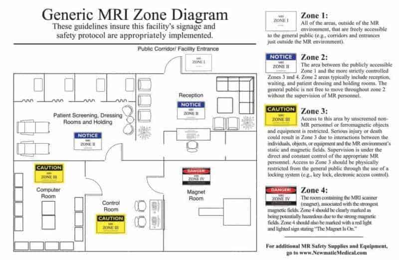
- Go through the spotters’ collection in your department.
- Read the latest ACR Contrast Manual: ACR contrast Manual. You can quote this for contrast related questions that the examiners ask you.
- Do read about the proper use of TLD.
- The Final FRCR: Complete Revision Notes .
- Chapman & Nakielny's Guide to Radiological Procedures
- Chapman & Nakielny's Aids to Radiological Differential Diagnosis
- Radiological Procedures - A Guideline by Bhushan Lakhkar
- Handbook of Interventional Radiologic Procedures – Kandrappa
- Another good book for commonly asked practical questions is Known Unknowns Of Everyday Radiology Practice.
- Cases at RadioGyan.com
- Spotters at RadioGyan.com
- Facebook: Radiology Imaging Cloud .
- Facebook: Radiology Golden Points .
- RadioGyan radiology Telegram Channel , Telegram Group and WhatsApp groups
- Telegram: Dr. Sanjay P sir’s radiology residents group (More than 4000 residents have joined). You can contact him on Telegram. His id is @Sanjaypyadav
- Dr.Raj Ganeshan’s WhatsApp group.
- Radiologist Eye
- Indian Radiologist
- MSKRadioloy4u MSK cases
- Case of the week Barnard Institute of Radiology
- Spotters: Department of Radiodiagnosis JSS Medical College and Hospital, Mysore – Expired!
- Cases: Department of Radiodiagnosis JSS Medical College and Hospital, Mysore- Expired !
- Case of the week: Learning Radiology
- Cases: Aunt Minnie.com
- Spotters: RadioLoksabha
- Cases: Headneckbrainspine.com
- ACR Case of the week
- Radiologymasterclass Quiz
- Radiopedia Playlists
- RadGray.com
- RadCases : Great book for radiology cases.
- Aunt Minnie's Atlas and Imaging-Specific Diagnosis
- Attend conferences which emulate the practical exam pattern like RARE.
- Rapid review of radiology
- Radcases series
- Duke Radiology Case Review: Imaging, Differential Diagnosis, and Discussion
- FRCR 2B Viva: A Case-based Approach
- Radiopaedia playlist
- Stanley radiology conference
- Radiologist eye
- Radiology Spotters Cases Radiogyan
- Oral recall series
- See as many spotters as possible.
- Hardly less than 1 min will be available for spotters.
- For DNB you will be provided with a full set of CT & MRI images instead of f single image. So residents should be able to diagnose the condition in a limited time. Most of the spotter ppts contain only single images. There lies the importance of seeing a full set of images while practicing.
- Be confident & go to the exam with a fresh mind. All the best.
- Stanley viva book
- Try memorizing Chapman & Nakielny's Aids to Radiological Differential Diagnosis as much as possible especially the differentials for chest and bone cases.
- Use mnemonics but do not blurt them in front of examiners as that can be very embarrassing. For eg. the mnemonic for vertebra plana is FETISH, you DON’T want to say that in front of your examiner. That is a sure-shot way to flunk in the exams!
- Try presenting as many cases to the department faculty and amongst yourself. Present cases as you would do in the exam.
- For instruments, viva and interventional radiology go through the following YouTube playlist:
Here is a great video covering common practical questions:
- Revise theory related to radiation protection well. It is commonly asked in exams.
- Get proper sleep the previous night.
- Reach at least half an hour early to the examination center.
- Carry all essentials to the center including hall ticket and a copy of your thesis.
- Read and revise your thesis well. You will be asked questions on that.
- During spotters write COMPLETE answers. For eg. “Sessile osteochondroma involving the distal end of the radius.” rather than just sessile osteochondroma.
- Follow instructions given at the examination center. Do not get in the bad books of the examiners.
- Respect the examiner’s opinion and try not to argue with them.
- Handle films carefully. Examiners have collected cases over their entire career and hence they value them a lot which is obvious. They would not like you handling films in a shabby manner. You can use this pointer to point findings in the films: Extendable Fescue Ball Pen with LED Flashlight and Laser Pointer .

- Most important: Do not cheat!
Your colleague is as clueless as you about that case as you are!
Best of luck and hopefully all of you do well in the exam!
Also, check out this guide for the radiology exam by Dr. Yashwanth:
Tips and Tricks for DNB/MD Radiology Practical Exam
More Radiology resources
- Radiology resources – RadioGyan.com
- Normal Imaging Anatomy modules.: Learn and revise imaging anatomy. Links are divided by anatomy and modality.
- Cases : Radiology cases with important diagnostic points.
- Videos : Radiology videos from other websites with important teaching points highlighted.
- Spotters: Sets of 10 radiology spotters, one minute for each spotter.
- Articles : Descriptive articles on common imaging topics, including Journal club.
- Guide for first-year radiology residents.
- Guide for final year radiology residents.
- Guide to the FRCR exam
- Software: Links to common radiology software.
- Recommended Books for Radiology Residents.
- Radiology Conferences : Updates about upcoming radiology conferences and CMEs.
Best of luck with your exams!
Let us know your exam related tips and tricks in the comments section! Feedback and suggestions can be posted here .
About The Author
Dr. amar udare, md, related posts ↓.

DNB Radiology OSCE – Tips and Tricks

10 thoughts on “Guide for the MD/DMRD/DNB radiology exam!”
beautiful. Thanks for the tips
Glad that you found it useful!
Glad you found it useful, Anstin!
Outstanding guide for preparation
Glad you liked it 🙂
so helpful sir….. thank you sir
Thank you, Yoshita!
I just wanted to ask what are the advantages or role of clearing dnb Radiodiagnosis exam after having done MD Radiodiagnosis
There are no specific advantages as such, especially if you are practicing in India. Some international countries accept DNB as a degree and have higher weightage.
If you are an MD graduate and intend to practice in India, no need to give the exam. I had given it as a practice to revise my concepts during my one year service (Bond) period.
Hope that helps :), Amar.
Leave a Comment Cancel Reply
Your email address will not be published. Required fields are marked *
Get Radiology Updates to Your Inbox!
This site is for use by medical professionals. To continue, you must accept our use of cookies and the site's Terms of Use. Learn more Accept!
Wish to be a BETTER Radiologist? Join 14000 Radiology Colleagues !
Enter your email address below to access HIGH YIELD radiology content, updates, and resources.
No spam, only VALUE! Unsubscribe anytime with a single click.
RSNA 2020 Trending Topics
All virtual rsna 2020 promises attendees a robust program full of human insight/visionary medicine.
While RSNA 2020's all-virtual annual meeting may feel different than previous meetings, attendees can count on RSNA to deliver a meaningful, interactive meeting experience and world-class programming to virtual attendees across the globe.
RSNA 2020, Human Insight/Visionary Medicine , will provide a seamless experience offering interactive science and education programming and an extensive on-demand catalog with more available CME opportunities than ever before. The program, which expands on RSNA’s already successful virtual meeting, offers live Q&A for many sessions that will connect attendees with peers and colleagues of every subspecialty across the world.
To help attendees plan their schedules for this unique meeting, RSNA’s Scientific Program, Refresher Course and Education Exhibits committees chairs and subcommittee chairs offer a preview of the trends, hot topics and trailblazing research in each of the subspecialties available at RSNA 2020.
“While the format may be different, this year’s virtual meeting offers great flexibility for attendees to access a wide range of programming and learning resources,” said Zhen Jane Wang, MD, chair of the RSNA Scientific Program Committee. “Attendees can take advantage of the featured sessions, which offer opportunities to interact with abstract presenters and the chance to access a large number of on-demand sessions at their convenience throughout the week and, with Premium access, after the annual meeting.”
Innovative research continues to push the boundaries in all subspecialties, according to Dr. Wang. In particular, she noted that COVID-19 related topics are prevalent in a number of subspecialties in sessions, exhibits and research. In particular, five Hot Topic sessions update COVID-19 subjects, including non-pulmonary manifestations, neurological and neuroimaging and chest findings, as well as two sessions on radiology department readiness and workforce resilience.
In addition, attendees should look for A Comprehensive Imaging Review of COVID-19 Pneumonia with Focus on RSNA Expert Consensus, Fleischner Society Statement and ACR Recommendations: Challenges, Opportunities and Future Direction .
“Artificial intelligence (AI) remains a hot topic and there is a lot of interesting science on how AI can improve disease-specific diagnoses and how it can be integrated into clinical workflow,” Dr. Wang said. “Also of interest and great relevance to clinical practice is work presented on the validation of various classification systems and reporting systems used by many radiologists.”
Watch the video below for additional insights from Dr. Wang into RSNA 2020 scientific program content:
The education exhibits also highlight subspecialty classification and reporting systems and continue to showcase a wide breadth of innovative research that highlights major trends across all subspecialties, according to Christine O. (Cooky) Menias, MD, chair of the RSNA Education Exhibits Committee.
“In addition to research presented on various reporting systems such as LIRADS and PIRADS, many subspecialties offer exhibits on COVID-19, the unique science around e-cigarette and vaping lung damage, PET/CT and PET/MRI, AI and machine learning (ML) and 3D printing and models,” Dr. Menias said. “The exhibits this year showcase the wide variety of challenges and opportunities facing radiology, including diversity, inclusion and just culture. There are also exhibits about patient care, quality improvement, mentoring and social justice.”
Educational courses remain among the most popular and contain comprehensive content on a wide array of topics vital to clinical practice.
“Artificial intelligence remains a hot and impactful topic across the entire specialty,” said Laura Bancroft, MD, chair of the Refresher Course Committee. “Some notable practice-based courses include Impact of COVID-19 on Workforce Resilience , Mass Casualty Imaging and Workflow , Strategies to Suppress Errors and Biases in Diagnostic Radiology , and Taking Action to Promote Gender Inclusion in Radiology .”
Watch the video below from Dr. Bancroft for insights into the refresher courses at RSNA 2020:
For an overview of this past year’s most impactful research, look for the Special Interest Session, Review of 2020: New Research that Should Impact Your Practice . The two-part Friday Imaging Symposium offers a review of imaging cases in many subspecialties, including neuroradiology, breast, gastrointestinal and cardiothoracic radiology.
RSNA committees are sponsoring a number of sessions. The RSNA Committee on Diversity, Equity & Inclusion presents the Special Interest Session, Exposing Our Blindside and Overcoming Unconscious Bias . The RSNA Public Information Committee presents the Special Interest Session Improving Patient Experience through Human Design Thinking . In addition, the RSNA Professionalism Committee presents Taking Action to Promote Gender Inclusion in Radiology: A Roadmap for Progress . The RSNA Committee on Diversity, Equity & Inclusion also sponsors the Gender-based Harassment and Microaggressions session.
Click on the subspecialties below to preview the trends, hot topics and research available at RSNA 2020.
- Breast Imaging
- Cardiac Radiology
- Chest Radiology
- Emergency Radiology
- Gastrointestinal Radiology
- Genitourinary Radiology/Uroradiology
- Health Service Policy and Research/Policy and Practice
- Informatics
- Molecular Imaging
- Multisystem/Special Interest
- Musculoskeletal Radiology
- Neuroradiology
- Nuclear Medicine
- Obstetric/Gynecologic Radiology
- Pediatric Radiology
- Radiation Oncology and Radiobiology
- Vascular/Interventional
Look for these additional program highlights in each subspecialty.
For More Information
Register for the meeting at RSNA.org/Register .
Read RSNA News stories about RSNA 2020:
RSNA 2020: Program Highlights
Innovations Abound at RSNA 2020 Virtual Exhibition
Embracing Diversity, Equity and Inclusion in Radiology
RSNA Subcommittee Chairs
Scientific Program Subcommittees
Ronald S. Arellano, MD, Vascular and Interventional
Ferco H. Berger, MD, Emergency Radiology
Robert D. Boutin, MD, Musculoskeletal
Ciprian Catana, MD, PhD, Molecular Imaging
Lynn A. Fordham, MD, Pediatric Radiology
Fiona J. Gilbert, MD, Breast
Phillip J. Koo, MD, Nuclear Medicine
Jerome Z. Liang, PhD, Physics
John P. Lichtenberger III, MD, Chest
Desiree E. Morgan, MD, Gastrointestinal
Karen G. Ordovas, MD, Cardiac
Andrew B. Rosenkrantz, MD, Genitourinary
Nabile M. Safdar, MD, Radiology Informatics
Cynthia S. Santillan, MD, Health Services Policy and Research
Anna Shapiro, MD, Radiology Oncology and Radiobiology
Max Wintermark, MD, Neuroradiology
Education Exhibits Subcommittees
Samuel E. Almodovar-Reteguis, MD, Nuclear Medicine
Edson Amaro Jr, MD, PhD, Neuroradiology
Barbaros S. Erdal, PhD, Radiology Informatics
William J. Grande, MD, Vascular/Interventional
Ambrose J. Huang, MD, Musculoskeletal
Kirti M. Kulkarni, MD, Breast
Brent P. Little, MD, Chest
Courtney C. Moreno, MD, Gastrointestinal
Mariam Moshiri, MD, Obstetrics/Gynecology
Nadeem Parkar, MD, Cardiac
Gary R. Schooler, MD, Pediatrics
William F. Sensakovic, PhD, Physics
Anna Shapiro, MD, Radiation Oncology & Radiobiology
Anup S. Shetty, MD, Multisystem/Special Interest
Scott D. Steenburg, MD, Emergency Radiology
Ashish P. Wasnik, MD, Uroradiology
Jeffrey R. Wesolowski, MD, Policy and Practice
Refresher Course Committee
Margarita L. Zuley, MD, Track Chair/Breast Imaging
Maxine S. Jochelson, MD, Track Vice Chair/Breast Imaging
Eric E. Williamson, MD, Track Chair/Cardiac
Jeremy J. Erasmus, MD, Track Chair/Chest
Ioannis Vlahos, MRCP, FRCR, Track Vice Chair/Chest
Petra J. Lewis, MBBS, Track Chair/Education
Douglas S. Katz, MD, Track Chair/Emergency Radiology
Manickam Kumaravel, MD, FRCR, Track Vice Chair/Emergency Radiology
Rathan M. Subramaniam, MD, PhD, Track Chair/Emerging Technology
Diane C. Strollo, MD, Track Chair/Essentials
Judy Yee, MD, Track Chair/GI
Matthew S. Davenport, MD, Track Chair/GU
Stephen C. O'Connor, MD, Track Chair/Hands On
Tabassum A. Kennedy, MD, Track Chair/Head & Neck
Howard P. Forman, MD, Track Chair/Health Policy & Practice
Charles E. Ray Jr, MD, PhD, Track Chair/Interventional
Ajay Gupta, MD, Track Chair/Introduction to Research
Katherine E. Maturen, MD, Track Chair/Introduction to Research
Yoshimi Anzai, MD, Track Chair/Leadership & Management
Bachir Taouli, MD, Track Chair/MR
Leon Lenchik, MD, Track Chair/Musculoskeletal
Linda Probyn, MD, Track Vice Chair/Musculoskeletal
Christopher P. Hess, MD, PhD, Track Chair/Neuro
Ajay Gupta, MD, Track Vice Chair/Neuro
Katherine A. Zukotynski, MD, PhD, Track Chair/Nuclear Medicine
Evis Sala, MD, PhD, Track Chair/Oncologic Imaging
Geetika Khanna, MD, MS, Track Chair/Pediatrics
Adina L. Alazraki, MD, Track Vice Chair/Pediatrics
Lifeng Yu, PhD, Track Chair/Physics (Diagnostic Radiology)
Jon J. Kruse, PhD, Track Chair/Physics (Radiation Oncology)
Tessa S. Cook, MD, PhD, Track Chair/Radiology Informatics
Luciano M. Prevedello, MD, MPH, Track Chair/Radiology Informatics
Christopher J. Roth, MD, Track Chair/Radiology Informatics
Mitchell E. Tublin, MD, Track Chair/Ultrasound
Vincent B. Ho, MD, MBA, Track Chair/Vascular
James M. Kofler Jr, PhD, AAPM Liaison
Ricardo Restrepo, MD, Case-based Chairman
Jorge A. Soto, MD, Case-based Director/Abdomen
Jiyon Lee, MD, Case-based Director/Breast
Diana Litmanovich, MD, Case-based Director/Cardiac
Edward Y. Lee, MD, Case-based Director/CT
Alexander R. Guimaraes, MD, PhD, Case-based Director/MR
Stacy E. Smith, MD, Case-based Director/Musculoskeletal
Amy F. Juliano, MD, Case-based Director/Neuroradiology
Katherine A. Zukotynski, MD, PhD, Case-based Director/Nuclear Medicine/PET-CT
Abbey Winant, MD, Case-based Director/Pediatric Radiology
Andetta R. Hunsaker, MD, Case-based Director/Thoracic
Deborah J. Rubens, MD, Case-based Director/Ultrasound
Sabala Mandava, MD, Associated Sciences Consortium Chairman
Hon’ble Chancellor Vice Chancellor Registrar Principal Vice Principal Medical Superintendent
Anaesthesiology Anatomy Biochemistry Cardiac Anaesthesia Cardiology Cardio vascular & Thoracic Surgery Community Medicine Dermatology (Skin & VD) Emergency Medicine Endocrinology ENT, Head & Neck Surgery Forensic Medicine & Toxicology Gastroenterology
General Medicine General Surgery Interventional Radiology Medical Oncology Microbiology Nephrology Neonatology Neurology Neurosurgery Nuclear Medicine Obstetrics & Gynecology Ophthalmology Orthopedics Pathology
Public Health Research Unit Medical Education MCI Nodal Center Hospital Administration
Pediatrics Pediatric Neurology Pediatric Surgery Pharmacology Physical Medicine & Rehabilitation Physiology Plastic & Reconstructive surgery Psychiatry Radiation Oncology Radiology/Radio-diagnosis Respiratory/Pulmonary Medicine Surgical Oncology Urology
Under Graduate
Post graduate.
Post Doctoral Diploma Courses Public Health Ph.D Hospital Administration Fellowship Courses Allied Courses
Feedback Analysis 2015-20 Feedback Analysis 2021
Syllabus of Courses offered
Under Graduate Post Graduate Post Doctoral Fellowship Courses Ph.D Allied Courses
- KLE Advanced Simulation Centre & Clinical Skills Lab
- Cadaveric Skill Lab
- NIRF Prescribed Format
- JNMC NIRF 2018
- JNMC NIRF 2019
- JNMC NIRF 2020
- JNMC NIRF 2021
- JNMC NIRF 2022
- JNMC NIRF 2023
- JNMC NIRF 2024
- KLE Dr. PBK Hospital & MRC
- KLE Dr.PBK Hospital
- JNMC Women’s & Children’s Health Research Unit
- KLE Society
- Sports Complex
- Convention Center
- Musical Garden
- Shivalaya Temple
- Ganesh Temple
- Attendance Dashboard
- Scientific Society
- Student Association
- Kannada Balaga
- Institutional Ethics Committee
- Internal Complaint Committee
- Anti-Ragging
- Para Medical Course
- Capability Enhancement
- Faculty Login
- Student/Parent Login
- Dissertations
- M.B.B.S Phase III – Part-II
Radio Diagnosis
M.D Dissertation – Ongoing
| Dr.Abhishek Reghunadhan | Dr. Pradeepgoud H. Patil | Comparison between magnetic resonance imaging of knee joint and knee arthroscopy in the dignosis of anterior cruciate ligament tears – A one year hospital based cross sectional study. | 2023-2025 | ||
| Dr.Amruta Ravindra Patil | Dr.Santosh D. Patil | Role of ultrasound elastography in characterization of breast lesion as benign and malignant – A one year hospital based cross | 2023-2025 | ||
| Dr. Chordiya Paawan Sanjay | Dr.Ashwin S. Patil | Hepatic colour doppler study in liver cirrhosis patients and correlation of hepatic hemodynamic with clinical child pugh score – A hospital based cross | 2023-2025 | ||
| Dr. Hanushanth G. | Dr.Rajendra V. Mali | Role of 3 tesla magnetic resonance imaging in the preoperative evaluation of perianal fistulas – A one year hospital based cross | 2023-2025 | ||
| Dr. Harshendra S.G. | Dr. Shreeyansh Rayappa Nandeshwar | Shear wave elastography in evaluation of renal parenchymal stiffness in patients with chronic kidney disease – A one year hospital based cross | 2023-2025 | ||
| Dr.Kavya J Pai | Dr. Pradeepgoud H. Patil | Role of magnetic resonance cholengiopancreatography in patients with pancreatico – biliary pathology – A one year hospital based cross | 2023-2025 | ||
| Dr.M.Vignatha Reddy | Dr. Shreeyansh Rayappa Nandeshwar | 2D Shear wave elastography in to evaluate focal liver lesions – A one year hospital based cross | 2023-2025 | ||
| Dr. Nivya Gudivada | Dr. Shreeyansh Rayappa Nandeshwar | An analogy between clot burden and write ventricular dysfunction in acute pulmonary thromboembolism on computed tomography pulmonary angiography using modified millers score | 2023-2025 | ||
| Dr. Pasurla Purushotham Reddy | Dr.Ashwin S. Patil | Assesment of thyroid nodules using strain ultrasound elstography and its comparison with fine needle aspiration cytology – A one year hospital based cross | 2023-2025 | ||
| Dr. Phalke Rucha Deepak | Dr.Ashwin S. Patil | Assesment grade of hepatic fibrosis in non alcoholic fatty liver disease using shear wave elastography – A one year hospital based cross | 2023-2025 | ||
| Dr. Polireddy Prameela Satya Shivani | Dr.Santosh D. Patil | Prevelance of lumbar intervertebral disc herniation in asymptomatic individuals on magnetic resonance imaging – A one year hospital based cross | 2023-2025 | ||
| Dr.Sahithy Kakkireni | Dr. Pradeepgoud H. Patil | Role of high resolution computed tomography thorax in assessment of various imaging patterns in chronic obstructive pulmonary disease – A one year cross sectional study in a tertiary care hospital | 2023-2025 | ||
| Dr. Vangala Ashwin Kumar | Dr.Santosh D. Patil | Magnetic resonance imaging in evaluation of hypoxic ischemic encephalopathy in children under 5 years of age – A one year hospital based cross | 2023-2025 | ||
| Dr. Sidhant Gupta | Dr. Rajendra V. Mali | Role of magnetic resonance imaging classification system (MRICS) in evaluvation of cerebral palsy in children under five years of age: A one year hospital based cross sectional study | 2023-2025 | ||
| Dr. Anubhav Chandhan | Dr. Rajendra V. Mali | Correlation of High resolution ultrasonography findings with the severity of carpal tunnel syndrome and hand anthoropmetry – A one year hospital based cross sectional study | 2023-2025 | ||
| Dr. Dontula Ujwala | Dr. Santosh D. Patil | Carotid Doppler evaluation of tunica intima media thickness and atherosclerotic plaque characteristics among diabetics and non-diabetics – A one-year hospital based cross sectional study | 2024-2026 | ||
| Dr. Harshita | Dr. Santosh D. Patil | Efficacy of ultrasound in diagnosis of occult spinal dysraphism as compared to MRI: A one year hospital based cross sectional study | 2024-2026 | ||
| Dr. Lakshmi Samhita Vupputuri | Dr. Pradeepgoud H. Patil | Role of contrast enhanced computed tomography in assessment of spleen and splenic vascular involvement in acute pancreatitis and its correlation with modified computed tomography severity index – A one-year hospital based cross sectional study | 2024-2026 | ||
| Dr. Marri Sai Sree | Dr. Pradeepgoud H. Patil | Renal resistive index as a renal function marker in diabetic and non-diabetic chronic kidney disease patients: a cross sectional study in a tertiary care center, Karnataka, India | 2024-2026 | ||
| Dr. Nelluri Krishna Sai | Dr. Rajendra V. Mali | Derivation of syntax scoring in coronary artery disease using computed tomography angiography – A one year hospital based study | 2024-2026 | ||
| Dr. P. Sai Amarthya | Dr. Pradeepgoud H. Patil | Estimation of spleen stiffness using 2D shear wave elastography in patients with liver cirrhosis : A one year hospital based observational study | 2024-2026 | ||
| Dr. Rinjhin Jain | Dr. Santosh D. Patil | Role of MRI parameters in assessing the prevalence of trochlear dysplasia : A cross sectional study | 2024-2026 | ||
| Dr. Sagar Guwalani | Dr. Ashwin S. Patil | Efficacy of contrast enhanced 3D stir flair magnetic resonance imaging in comparison with contrast enhanced T1 weighted magnetic resonance imaging for evaluation of pituitary lessions – A one year hospital based cross sectional study | 2024-2026 | ||
| Dr. Sakshi Arora | Dr. Ashwin S. Patil | Efficacy of acoustic radiation force impulse (ARFI) imaging to differentiate between benign and malignant cervical lymph node in comparison with fine needle aspiration cytology or biopsy – A one-year hospital based cross-sectional study. | 2024-2026 | ||
| Dr. Siva Soorya M. | Dr. Ashwin S. Patil | Efficacy of shear wave elastography in detection of liver stiffness in patients with chronic liver disease and correlation with child turcotte – pugh (CP) score – A one year hospital based cross sectional study | 2024-2026 | ||
| Dr. Analisa Eugenia Ferreira Alvares | Dr. S. R. Nandeshwar | Dr. Manjunath Hukkeri | Diagnostic accuracy of O-RADS ultrasound in classifying adnexal lesions among reproductive age women: A cross sectional study in a teritary care centre in Karnataka, India | 2024-2026 | |
| Dr. Mailare Anuja Angadrao | Dr. Rajendra V. Mali | MRI insights into postpartum neurological complications a one – year cross sectional study at KLE Dr. Prabhakar Kore Hospital, Belagavi | 2024-2026 | ||
| Dr. Shriya Ravichandran | Dr. S. R. Nandeshwar | Dr. K. Ravishankar Naik | Role of arterial spin labeling magnetic resonance perfusion in acute ischemic stroke in a tertiary care centre | 2024-2026 | |
| Dr. Lipi Agarwal | Dr. Rajendra V. Mali | Role of magnetic resonance imaging for correlation of acromial morphology in patient with shoulder pain: A one year hospital based cross sectional study | 2024-2026 | ||
| Dr. Yash Pallod | Dr. Pradeep S. Goudar | Dr. K. Ravishankar Naik | Role of median nerve shear wave ultrasound elastography in diagnosis and grading of severity in carpal tunnel syndrome: A one year tertiary care hospital based cross sectional study | 2024-2026 |
M.D Dissertation – Completed
| 1 | Dr.Shyam.S. | Dr.A.C.Shetti | REAL TIME ULTRASONOGRAPHY IN EVALUATION OF FOCAL LIVER LESIONS. | ||
| 2 | Dr. Praveen kumar | Dr.D.B.Udoshi | STUDY OF EVALUATION OF ROLE OF CT IN PATIENTS WITH CLINICALLY SUSPECTED CARCINOMA LUNG. | ||
| 3 | Dr.Vikrant K. | Dr.R.V.Mali | ROLE OF MR IMAGING IN INTERNAL DERANGEMENTS OF KNEE JOINT. | ||
| 4 | Dr.Gopinath R. | Dr.D.B.Udoshi | MAGNETIC RESONANCE IMAGING OF SUPRA TENTORIAL TUMOURS -A ONE YEAR HOSPITAL BASED CROSS SECTIONAL STUDY | ||
| 5 | Dr.Amarinder Singh | Dr.A.C.Shetti | TRIPHASIC STUDIES IN PATIENTS WITH SUSPECTED HEPATIC TUMOURS USING MULTIDETECTOR CT- A HOSPITAL BASED CROSS SECTIONAL STUDY. | ||
| 6 | Dr.Alla Dinesh | Dr.R.V.Mali | ROLE OF MDCT IN DIAGNOSIS OF CORONARY ARTERY STENOSIS IN PATIENTS WITH LOW PRETEXT LIKELHOOD OF SIGNIFICANT DISEASES. A HOSPITAL BASED CROSS SECTIONAL STUDY. | ||
| 7 | Dr.Priyanka Singh | Dr.R.V.Mali | CHARACTERIZING VASCULAR INVOLVEMENT IN PATIENTS WITH LOWER LIMB ISCHEMIC ARTERIAL DISEASES USING MULTIDETECTOR CT ANGIOGRAPHY- AN OBSERVATIONAL STUDY. | ||
| 8 | Dr.Priyaanka NPR | Dr.A.C.Shetti | MRI EVALUATION OF CLINICALLY SUSPECTED UTERINE & ADNEXAL MASSES – A HOSPITAL BASED CROSS SECTIONAL STUDY. | ||
| 9 | Dr.Dev Thakkar | Dr.D.B.Udoshi | CHARACTERISATION OF WHITE MATTER LESIONS IN CHILDREN & ADOLESCENTS ON MR AN OBSERVATIONAL STUDY. | ||
| 10 | Dr. Tejas B. Gosalia | Dr.Rajendra V.Mali | “CHARACTERIZATION OF INTERSTITIAL LUNG DISEASE USING HRCT IN CLINICALLY SUSPECTED CASES: A HOSPITAL BASED ONE YEAR OBSERVATIONAL STUDY”. | ||
| 11 | Dr.Kiran S.Desai | Dr.Pradeepgoud H.Patil | “MAGNETIC RESONANCE IMAGING IN CHARACTERIZATION OF CONGENITAL AND DEVELOPMENTAL DISORDERS OF SPINE- ONE YEAR HOSPITAL BASED OBSERVATIONAL STUDY” | ||
| 12 | Dr.Vinaykumar C. Udasi | Dr.Ashwin S.Patil | “ROLE OF MULTIDETECTOR-CT IN PATIENTS WITH CLINICALLY SUSPECTED RENAL LESIONS-A HOSPITAL BASED CROSS SECTIONAL STUDY” | ||
| 13 | Dr.Manish Gulabani | Dr.Rajendra V.Mali | “ROLE OF MULTI DETECTOR COMPUTED TOMOGRAPHY IN DETERMINING CONSERVATIVE VERSUS OPERATIVE MANAGEMENT IN CASES OF BLUNT ABDOMINAL TRAUMA-A ONE YEAR CROSS—SECTIONAL STUDY”. | ||
| 14 | Dr.Gauri Gupta | Dr.Ashwin S.Patil | “ROLE OF HIGH RESOLUTION ULTRASOUND IN ASSESSMENT OF CHARACTERISTICS AND EXTENT OF ROTATOR AND NON-ROTATOR CUFF DISORDERS”-A ONE YEAR HOSPITAL BASED CROSS-SECTIONAL STUDY. | ||
| 15 | Dr.Shuchi Agarwal | Dr.Pradeepgoud H.Patil | “EFFICACY OF B-SCAN ULTRASONOGRAPHY IN OCULAR ABNORMALITIES-A ONE YEAR CROSS-SECTIONAL STUDY”. | ||
| 16 | Dr.Aseem Katyal | Dr.R.V.Mali | “CHARACTERIZATION OF IN-VIVO PROTON MAGNETIC RESONANCE SPECTROSCOPIC PATTERNS OF INTRACRANIAL SPACE OCCUPYING LESIONS- A ONE YEAR OBSERVATIONAL STUDY” | ||
| 17 | Dr.Jeevanjot Matharoo | Dr.A.C.Shetti | “ROLE OF MULTIDETECTOR COMPUTED TOMOGRAPHY IN EVALUATION OF ACUTE PANCREATITIS BY MODIFIED COMPUTED TOMOGRAPHY SEVERITY INDEX AND IT’S CORRELATION WITH PATIENT OUTCOME-ONE YEAR HOSPITAL BASED CROSS SECTIONAL STUDY”. | ||
| 18 | Dr.Vishal Deepak Desai. | – Dr.Ashwin S.Patil | Dr. Mahesh Kamte. | “ROLE OF MAGNETIC RESONANCE IMAGING IN DETERMINING MORPHOLOGICAL ABNORMALITIES OF BRAIN IN CHILDREN WITH SEVERE MICROCEPHALY- A ONE YEAR CROSS – SECTIONAL STUDY” | |
| 19 | Dr.Divesh M. Tulsi | Dr.Pradeepgoud H.Patil | “ROLE OF UNENHANCED MULTIDETECTOR COMPUTERIZED TOMOGRAPHY IN DETECTION OF URINARY SYSTEM CALCULI IN COMPARISION WITH ULTRASOUND FINDINGS” – ONE YEAR CROSS SECTIONAL STUDY. | ||
| 20 | Dr.Rahul Nijhara | Dr.R.V.Mali | “MAGNETIC RESONANCE IMAGING STUDY OF TYPES AND INCIDENCE OF INJURIES IN TRAUMATIC KNEE JOINT-A ONE YEAR CROSS – SECTIONAL STUDY”. | ||
| 21 | Dr.Harpreet Singh G. | Dr.A.C.Shetti | “ROLE OF COLOR DOPPLER ULTRASONOLOGY IN EVALUATION OF PORTAL HYPERTENSION – A ONE YEAR CROSS SECTIONAL STUDY”. | ||
| 22 | Dr.Nihaal K Reddy | Dr.Pradeepgoud H.Patil | “ROLE OF MULTI-DETECTOR COMPUTED TOMOGRAPHY IN EVALUATION OF NECK MASSES – A ONE YEAR CROSS SECTIONAL STUDY”. | ||
| 23 | Dr.Rajat Sachdeva | Dr.Ashwin S.Patil | “CHARACTERISATION OF SMALL BOWEL PATHOLOGIES ON MULTI-DETECTOR COMPUTERIZED TOMOGRAPHY ENTEROGRAPHY” – ONE YEAR CROSS SECTIONAL STUDY. | ||
| 24 | Dr. Basavaraj S. | Dr.R.V.Mali | “ULTRASONOGRAPHIC AND COLOR DOPPLER EVALUATION OF CAROTID ARTERY INTIMA MEDIA THICKNESS AND RESISTIVE INDEX IN HYPERTENSIVE PATIENTS COMPARED WITH NORMOTENSIVE, AN OBSERVATIONAL STUDY” | ||
| 25 | Dr.Prakruti | Dr.A.C.Shetti | “ROLE OF MAGNETIC RESONANCE IMAGING IN INTERNAL DERANGEMENT OF THE SHOULDER JOINT: A HOSPITAL BASED OBSERVATIONAL SUDTY” | ||
| 26 | Dr.Chintankumar | Dr.A.S.Patil | ROLE OF MAGNETIC RESONANCE IMAGING IN HIP PATHOLOGIES – A HOSPITAL BASED CASE SERIES STUDY | ||
| 27 | Dr.R.Hemantkumar | Dr.Pradeepgoud H.Patil | “A CROSS SECTIONAL STUDY OF COMPUTED TOMOGRAPHY PULMONANRY ANGIOGRAPHY IN THE SUSPECTED CASES OF PULMONARY EMBOLISM” | ||
| 28 | Dr.Muthu Mahesh. | Dr.V.V.Hattiholi | “CHARACTERIZATION OF THE MAGNETIC RESONANCE IMAGING PATTERNS OF THE CEREBRAL PALSY AND THEIR ASSOCIATION WITH CLINICAL FINDINGS IN CHILDREN: A HOSPITAL BASED OBSERVATIONAL STUDY” | ||
| 29 | Dr. Pranav Mallya | Dr. S.D.Patil | “BONE MINERAL DENSITOMETRY USING DUAL ENERGY X-RAY ABSORPTIOMETRY (DEXA):- A HOSPITAL BASED OBSERVATIONAL STUDY TO LOOK FOR DISCORDANCE IN HIP AND SPINE BONE MINERAL DENSITY” | ||
| 30 | Dr.Rohan M. Bhanushali | Dr.P.H.Patil | ROLE OF MAGNETIC RESONANCE IMAGING IN EVALUATION OF BRAIN LESIONS IN HIV INFECTED PATIENTS- ONE YEAR HOSPITAL BASED CROSS SECTIONAL STUDY. | ||
| 31 | Dr.Predeep S. Goudar | Dr.A.S.Patil | COMPUTED TOMOGRAPHY (CT) GUIDED BIOPSY OF THORACIC LESIONS AT KLES DR.PRABHAKAR KORE HOSPITAL AND MEDICAL RESEARCH CENTRE, BELGAUM A ONE YEAR OBSERVATIONAL STUDY | ||
| 32 | Dr.Amit Garhwal | Dr.A.S.Patil | ROLE OF MAGNETIC RESONANCE IMAGING IN BIOMETRIC EVALUATION OF CORPUS CALLOSUM IN HYPOXIC ISCHAEMIC ENCEPHALOPATHY PATIENTS ONE YEAR HOSPITAL BASED CROSS SECTIONAL STUDY. | ||
| 33 | Dr.Reshma | Dr.S.D.Patil | ROLE OF MR SPECTRESCOPY IN EVALUATION OF BRAIN METABOLITES IN PATIENTS WITH DIABETES MELLITUS II | ||
| 34 | Dr.Sandhya Yadav A. | Dr.V.V.Hattiholi | ROLE OF COMPUTED TOMOGRAPHY IN IMAGING AND CLASSIFICATION OF ACUTE PANCREATITIS BASED ON REVISED ATLANTA CLASSIFICATION – ONE YEAR HOSPITAL BASED CROSS SECTIONAL STUDY. | ||
| 35 | Dr.Aakaar Kapoor | Dr.R.V.Mali | CHARACTERIZATION OF FOCAL LIVER LESIONS USING DIFFUSION WEIGHTED MAGNETIC RESONANCE IMAGING TECHNIQUE IN CORRELATION WITH CONTRAST ENHANCED MULTI SLICE COMPUTED TOMOGRAPHY SCAN – ONE YEAR HOSPITAL BASED CROSS SECTIONAL STUDY. | ||
| 36 | Dr.Amitava Paul | Dr.D.B.Udoshi | COMPUTED TOMOGRAPHIC EVALUATION OF TUMOURS IN RETROPERITONEAL SPACE -A HOSPITAL BASED CROSS SECTIONAL STUDY | ||
| 37 | Dr.Parimala Maguluri | Dr.P.H.Patil | DIFFUSION WEIGHTED MAGNETIC RESONANCE IMAGING AND MR – SPECTROSCOPY IN DIFFERENTIATING POSTERIOR FOSSA TUMORS – HOSPITAL BASED CROSS SECTIONAL STUDY | ||
| 38 | Dr.Advaith Shivapur | Dr.A.S.Patil | ADRENAL INCIDENTALOMAS ON COMPUTED TOMOGRAPHY AND THEIR CHARACTERIZATION INTO BENIGN AND MALIGNANT – A HOSPITAL BASED CROSS SECTIONAL STUDY. | ||
| 39 | Dr.Rithesh A.R. | Dr.S.D.Patil | ULTRASONOGRAPHIC EVALUATION OF GALLBLADDER IN TYPE 2 DIABETES MELLITUS – HOSPITAL BASED CROSS SECTIONAL STUDY. | ||
| 40 | Dr.Pushpendra Shrivastav | Dr.R.V.Mali | ROLE OF 64 SLICE MDCT IN EVALUATION PULMONARY ARTERY ANATOMY IN CHIDREN WITH CYANOTIC CONGENITAL HEART DISEASE AND COMPARISON with 2D ECHOCARDIOGRAPHY | ||
| 41 | Dr.Pallav Gupta | Dr.V.V.Hattiholi | Dr.Jyothi V.Hattiholi | ROLE OF MULTIDETECTOR COMPUTED TOMOGRAPHY IN EVALUATION OF MEDIASTINAL MASS LESIONS BASED ON NEW CLASSIFICATION – HOSPITAL BASED CROSS – SECTIONAL STUDY | |
| 42 | Dr.Janaki Raj | Dr.R.V.Mali | COMPARATIVE STUDY OF MAMMOGRAPHY AND SONOMAMMOGRAPHY WITH FNAC / HISTOPATHOLOGY IN EVALUATING PALPABLE BREAST MASSES- A ONE YEAR HOSPITAL BASED STUDY | ||
| 43 | Dr.Pooja M Kavatagimath | Dr.D.B.Udoshi | EVALUATION OF PERIANAL FISTULA BY MAGNETIC RESONANCE IMAGING –A ONE YEAR CROSS SECTIONAL HOSPITAL BASED STUDY | ||
| 44 | Dr.Gautam Satya Munnangi | Dr.A.S.Patil | ROLE OF MAGNETIC RESONANCE IMAGING IN EVALUATION OF LUMBAR FACET ARTHROPATHY IN LOW BACKACHE- ONE YEAR CROSS SECTIONAL STUDY | ||
| 45 | Dr.Ashwini Hule | Dr.P.H.Patil | MAGNETIC RESONANCE IMAGING EVALUATION OF SPINAL TUBERCULOSIS – A ONE YEAR CROSS SECTIONAL HOSPITAL BASED STUDY. | ||
| 46 | Dr.Chaitra S. | Dr.V.V.Hattiholi | Dr.Jyothi V.Hattiholi | DIAGNOSTIC ACCURACY OF BEDSIDE LUNG ULTRASOUND IN EMERGENCY PROTOCOL FOR THE DIAGNOSIS OF ACUTE RESPIRATORY FAILURE – A ONE YEAR HOSPITAL BASED CROSS SECTIONAL STUDY | |
| 47 | Dr.Vihag Raman | Dr.S.D.Patil | ROLE OF MAGNETIC RESONANCE IMAGING IN ASSESSMENT OF EXTENSOR COMPARTMENT ABNORMALITIES OF KNEE JOINT – A ONE YEAR CROSS SECTIONAL STUDY | ||
| 48 | Dr.Rakesh S. | Dr.A.S.Patil | Dr.Navin Mulimani | EVALUATION OF ENDOVENOUS LASER ABLATION AS A TREATMENT OF VARICOSE VEINS – A ONE YEAR HOSPITAL BASED OBSERVATIONAL STUDY | |
| 49 | Dr.Nagashree S. | Dr.S.D.Patil | VENOUS PHASE DIAMETER OF INFERIOR VENACAVA ON COMPUTED TOMOGRAPHY – A PREDICTOR OF HYPOTENSION IN BLUNT ABDOMINAL TRAUMA – A ONE YEAR HOSPITAL BASED OBSERVATIONAL STUDY | ||
| 50 | Dr.Rahul G Ambi | Dr.R.V.Mali | Dr.Mahesh Kamate | YIELD OF MAGNETIC RESONANCE IMAGING WITH SPECTROSCOPY IN CHILDREN WITH NEW ONSET AFEBRILE SEIZURE BETWEEN ONE MONTH TO TWENTY-FOUR MONTHS OF AGE | |
| 51 | Dr.Bibi Ayesha Pathan | Dr.D.B.Udoshi | Dr.S.I. Neeli. | ROLE OF SONOURETHROGRAPHY IN THE EVALUATION OF ANTERIOR URETHRAL STRICTURE – A ONE YEAR HOSPITAL BASED OBSERVATIONAL STUDY | |
| 52 | Dr.Jyoti Mullur | Dr.P.H.Patil | MULTIDETECTOR COMPUTED TOMOGRAPHIC ANGIOGRAPHY OF CEREBRAL VESSELS IN THE EVALUATION OF SUSPECTED NON-TRAUMATIC SUBARACHNOID HAEMORRHAGE – HOSPITAL BASED OBSERVATIONAL STUDY | ||
| 53 | Dr.Manish Mishra | Dr.V.V.Hattiholi | Dr. Pournima S. Patil | ROLE OF ULTRASONOGRAPHY IN EVALUATION OF ANKLE JOINT PATHOLOGIES IN PATIENTS WITH RHEUMATOID ARTHRITIS – A ONE YEAR HOSPITAL BASED CROSS SECTIONAL STUDY | |
| Dr. Chinmayi C.S. | Dr.Santosh .D.Patil | STUDY OF PREVALENCE OF NON ALCOHOLIC FATTY LIVER DISEASE IN PATIENTS OF TYPE 2 DIABETES MELLITUS AT A TERTIARY REFERRAL HOSPITAL IN NORTH KARNATAKA USING ABDOMINAL ULTRASONOGRAPHY – A ONE YEAR CROSS SECTIONAL STUDY. | |||
| Dr. Manjith N. | Dr.Ashwin .S. Patil | NORMAL RANGE OF VALUES OF LIVER AND SPLEEN SIZE BY ULTRASONOGRAPHY IN CHILDREN IN BELAGAVI (NORTH KARNATAKA)- A ONE YEAR HOSPITAL BASED CROSS SECTIONAL STUDY. | |||
| Dr. Onkar Shivakumar Patil | Dr.Pradeepgoud .H. Patil | STUDY OF PREVALENCE AND SEVERITY OF ATHEROSCLEROTIC DISEASE IN EXTRA CRANIAL CAROTID ARTERIES AMONG TYPE 2 DIABETES MELLITUS PATIENTS USING COLOR DOPPLER SONOGRAPHY – A ONE YEAR HOSPITAL BASED CROSS SECTIONAL STUDY. | |||
| Dr. Nikita N Shet Kanekar | Dr.Ashwin .S. Patil | Dr. Kumar Vinchurkar | COMPARISON OF DIAGNOSTIC ROLE OF DIFFUSION WEIGHTED IMAGING IN THE DIFFERENTIATION OF BENIGN & MALIGNANT CERVICAL GROUP OF LYMPH NODES WITH PATHOLOGICAL CORRELATION – A ONE YEAR OBSERVATIONAL STUDY AT KLES DR PRABHAKAR KORE HOSPITAL AND MRC, BELAGAVI. | ||
| Dr.Tejaswi Ramu | Dr.Virupaxi .V. Hattiholi | EVALUATION OF T2 HYPERINTENSITIES IN SPINAL CORD LESIONS USING MAGNETIC RESONANCE PROTOCOL – A ONE YEAR OBSERVATIONAL STUDY AT TERTIARY CARE HOSPITAL IN NORTH KARNATAKA. | |||
| Dr.Vinay N. Kurki | Dr.Rajendra .V. Mali | ROLE OF THYROID IMAGING, REPORTING AND DATA SYSTEM IN DIFFERENTIATION OF BENIGN AND MALIGNANT THYROID LESIONS AND CYTOPATHOLOGICAL CORRELATION AT TERTIARY CARE CENTRE, BELAGAVI – A ONE YEAR HOSPITAL BASED STUDY. | |||
| Dr. Preetham D S | Dr.Ashwin S. Patil | Dr.Navin Mulimani Dr.Prakash Mahantashetti | COMPARISON OF ARTERIAL SPIN LABELING TECHNIQUE WITH DYNAMIC SUSCEPTIBILITY CONTRAST TECHNIQUE OF PERFUSION MR IMAGING IN ENHANCING BRAIN TUMORS | ||
| Dr.Sandeep M.R. | Dr.Rajendra V. Mali | Dr.Mahesh Kamate | ROLE OF MAGNETIC RESONANCE IMAGING IN EVALUATION OF ACUTE ENCEPHALITIS SYNDROME IN A TERTIARY CARE HOSPITAL – A CROSS SECTIONAL STUDY | ||
| Dr. Kendre Lav Ramrao | Dr.Pradeepgoud.H. Patil | ROLE OF MAGNETIC RESONANCE CHOLANGIO- PANCREATOGRAPHY (MRCP) IN THE EVALUATION OF NORMAL ANATOMICAL VARIATIONS AND ANOMALIES IN BRANCHING PATTERN OF BILIARY SYSTEM – HOSPITAL BASED ONE YEAR OBSERVATIONAL STUDY. | |||
| Dr.Yaramala Satyanarayana Reddy | Dr.Virupaxi V. Hattiholi | ROLE OF COLOR DOPPLER ULTRASONOGRAPHY IN DIFFERENTIATING BENIGN AND MALIGNANT CERVICAL LYMPHADENOPATHY – ONE YEAR HOSPITAL BASED CROSS SECTIONAL STUDY | |||
| Dr.Nitish Makhija | Dr.Santosh D. Patil | EFFECT ON KIDNEY FUNCTION OF INTRAVENOUS CONTRAST ENHANCED CT USING ISOOSMOLAR AND LOW-OSMOLAR IODINATED CONTRAST MEDIUM. | |||
| Dr.Dhananjay Patil | Dr.D.B.Udoshi | ROLE OF MAGNETIC RESONANCE SPECTROSCOPY IN CENTRAL NERVOUS SYSTEM INFECTIONS – A ONE YEAR HOSPITAL BASED OBSERVATIONAL STUDY. | |||
| Dr. Somashekhar Shivappa Sogi | Dr .S.R. Nandeshwar | Dr Ravi Sarvi | RELATIONSHIP BETWEEN FEMORAL ARTERY INTIMA-MEDIA THICKNESS AND ATHEROSCLEROSIS IN PATIENTS WITH CHRONIC KIDNEY DISEASE, A ONE YEAR CROSS SECTIONAL STUDY | ||
| Dr. Assvath Chand O.C. | Dr. Ashwin S.Patil | ACCURACY OF DETECTING PULMONARY HYPERTENSION IN COMPUTED TOMOGRAPHY ON COMPARISON WITH ECHOCARDIOGRAPHY – ONE YEAR CROSS SECTIONAL STUDY. | |||
| Dr.Kyati Sharma | Dr.Rajendra V. Mali | Dr. Mahesh Kamate | ROLE OF MAGNETIC RESONANCE IMAGING OF BRAIN IN EVALUATION OF CHILDREN WITH GLOBAL DEVELOPMENTAL DELAY – ONE YEAR HOSPITAL BASED CROSS- SECTIONAL STUDY. | ||
| Dr.Gouri J. Kavatagimath | Dr.Pradeepgoud.H. Patil | DIAGNOSTIC ACCURACY OF SUSCEPTIBILITY WEIGHTED IMAGING (SWI) TO DIAGNOSE THE CASES OF CEREBRAL VENOUS THROMBOSIS (CVT) COMPARED TO MAGNETIC RESONANCE VENOGRAPHY (MRV) – ONE YEAR HOSPITAL BASED CROSS SECTIONAL STUDY | |||
| Dr.Mahantesh S Magadum | Dr.Virupaxi V. Hattiholi | Dr.R.B.Nerli | DIAGNOSTIC ACCURACY OF MULTI-PARAMETRIC MAGNETIC RESONANCE IMAGING (MPMRI) OF PROSTATE BASED ON PROSTATE IMAGING REPORTING AND DATA SYSTEM (PI-RADS) VERSION 2 IN COMPARISON WITH HISTOPATHOLOGY – ONE YEAR HOSPITAL BASED CROSS-SECTIONAL STUDY | ||
| Dr.Niroop P Kurian | Dr.Santosh D. Patil | MULTIDETECTOR COMPUTED TOMOGRAPHIC ANGIOGRAPHY OF ABDOMEN, IN EVALUATING HEPATIC ARTERY VARIANTS – ONE YEAR HOSPITAL BASED CROSS SECTIONAL STUDY | |||
| Dr. Aakanksha Nayyar | Dr Ashwin S Patil | Dr N S Mahantashetti | ROLE OF TRANS-ABDOMINAL ULTRA-SONOGRAPHIC EVALUATION OF ADRENAL GLAND SIZE IN NEONATES AND COMPARISON WITH CLINICAL DATA-A ONE YEAR HOSPITAL BASED COMPARATIVE STUDY | ||
| Dr. Guthireddy Akhila | Dr Ashwin S Patil | Dr Pravin S Jain | ROLE OF MUSCULOSKELETAL ULTRASOUND IN THE EVALUATION OF JOINT PATHOLOGIES IN PATIENTS WITH HYPERURICEMIA AND GOUT –A ONE YEAR HOSPITAL BASED CROSS-SECTIONAL STUDY | ||
| Dr. Harish Kumar | Dr.Virupaxi V. Hattiholi | 128 MULTI SLICE CT EVALUATION OF NON-TRAUMATIC BOWEL EMERGENCIES-A ONE YEAR HOSPITAL BASED CROSS-SECTIONAL STUDY | |||
| Dr. Kaku Deekshitha Reddy | Dr.Virupaxi V. Hattiholi | SUSCEPTIBILITY WEIGHTED IMAGING IN DAILY PRACTICE: TECHNICAL ASPECTS AND CLINICAL APPLICATIONS- A HOSPITAL BASED OBSERVATIONAL STUDY | |||
| Dr. Kondabattula Rakesh | Dr Rajendra V Mali | Dr Mahesh Kamate | MINERALIZING ANGIOPATHY IN YOUNG CHILDREN- AN OBSERVATIONAL STUDY | ||
| Dr. Kotbagi Chinmayi Mahesh | Dr.Virupaxi V. Hattiholi | EVALUATION OF RING ENHANCING LESIONS IN BRAIN IN CORRELATION WITH ARTERIAL SPIN LABELLING AND MR SPECTROSCOPY | |||
| Dr. Marri Meghana Reddy | Dr.Pradeepgoud .H. Patil | UTILITY OF T2 WEIGHTED AND DIFFUSION WEIGHTED SEQUENCES IN MAGNETIC RESONANCE IMAGING IN DIFFERENTIATION OF BENIGN AND MALIGNANT LIVER LESIONS: BENEFITS OF SINGLE VERSUS COMBINED SEQUENCES A ONE YEAR HOSPITAL BASED OBSERVATIONAL STUDY | |||
| Dr. Nishitha Ashok | Dr Santosh Patil | Dr Bhavana Koppad | EFFECTIVENESS OF LUNG ULTRASONOGRAPHY IN EVALUATION OF NEONATAL PULMONARY DISEASES- A ONE YEAR HOSPITAL BASED OBSERVATIONAL STUDY | ||
| Dr. Nithya Abraham | Dr Rajendra V Mali | ROLE OF MRI IN PAEDIATRIC DEMYELINATING DISORDERS – A ONE YEAR HOSPITAL BASED CROSS-SECTIONAL STUDY | |||
| Dr. Rahul Singh | Dr.Pradeepgoud .H. Patil | ROLE OF HIGH RESOLUTION COMPUTED TOMOGRAPHY THORAX IMAGING IN FOLLOW UP OF COVID-19 PATIENTS- ONE YEAR HOSPITAL BASED CROSS-SECTIONAL STUDY AT KLES DR. PRABHAKAR KORE HOSPITAL & MEDICAL RESEARCH CENTER, BELAGAVI. | |||
| Dr. Raveena Vipin | Dr Ashwin S Patil | Dr N S Mahantashetti | TRANS CRANIAL DOPPLER EVALUATION OF CEREBRAL HEMO DYNAMIC ALTERATION IN PRETERM NEONATES IN EARLY ONSET SEPSIS-A ONE YEAR HOSPITAL BASED OBSERVATIONAL STUDY AT KLE DR. PRABHAKAR KORE HOSPITAL | ||
| Dr. Shivendu Pandey | Dr Santosh Patil | ASSESSMENT OF PERCEPTION EFFECTS OF RADIATION AMONG POST GRADUATES AND CONSULTANTS OF KLE HOSPITAL BELAGAVI. | |||
| Dr. Soham Sen | Dr Rajendra V Mali | ROLE OF COLOR DOPPLER IMAGING IN DIFFERENTIATING BENIGN AND MALIGNANT BREAST MASS A ONE YEAR PROSPECTIVE OBSERVATIONAL STUDY. | |||
| Dr. Suman Agarwal | Dr Santosh Patil | ROLE OF ULTRASOUND IMAGING IN ASSESSMENT OF GROWTH PLATE AND ITS CLINICAL APPLICATIONS-A ONE YEAR HOSPITAL BASED OBSERVATIONAL STUDY | |||
| Dr. Versha Priyadarshini | Dr.Pradeepgoud .H. Patil | ROLE OF COLOR DOPPLER ULTRASOUND IN ASSESSMENT OF COMPLICATIONS OF ARTERIO-VENOUS HAEMODIALYSIS FISTULA ACCESS-A ONE YEAR HOSPITAL BASED OBSERVATIONAL STUDY | |||
| Dr. Nikhil Mahesh | Dr. P. H. Patil | Magnetic Resonance spectroscopy and Diffusion weighted imaging characterization of supratentorial brain tumors – A one-year Hospital Based cross sectional study. | |||
| Dr Ishita Thukral | Dr. R. V. Mali | Role of Imaging (CT & MRI) in Staging of Oral cavity Malignancies with clinico-pathological correlation. | |||
| Dr Divyanshi Jain | Dr. S. R. Nandeshwar | Real time Ultrasonography in evaluation of plantar fascia thickness and Achilles tendon thickness in diabetes mellitus type II – a one-year Hospital based case control study. | |||
| Dr Sankeerthi N.C | Dr. S. R. Nandeshwar | Role of MRI in characterization and categorization of the site and type of spinal dysraphism-A one-year Hospital Based observational study. | |||
| Dr. Rachana Vootukuru | Dr. R. V. Mali | Ultrasonographic evaluation of tenosynovitis of wrist, MCP joints in Rheumatoid Arthritis with clinical and serological correlation – A one-year Hospital based observational study. | |||
| Dr Sourav Mukherjee | Dr. S. R. Nandeshwar | Objective Evaluation Of Cerebral Venous Sinus Attenuation On Non-Contrast Computerized Tomography Of Brain And Correlation With Laboratory Hematocrit And Hemoglobin Indices in KLES Dr Prabhakar Kore Hospital, Belgaum – A cross-sectional study. | |||
| Dr Rhea Badyal | Dr. A. S. Patil | Role of computed tomography in identifying the anatomical variants of nose and paranasal sinuses and to assess their relation in patients having symptoms of sinusitis : A cross sectional study. | |||
| Dr C Rakesh Ruban | Dr. P. H. Patil | Role of Diffusion – weighted magnetic Resonance imaging in evaluation of Renal Masses: one-year Hospital Based Observational Study. | |||
| Dr Prajval U | Dr. R. V. Mali | Prospective and observational study on role of MRI in Acute febrile encephalopathy in children. | |||
| Dr Prachi Gupta | Dr. A. S. Patil | Role of computed Tomography in estimating the prevalence of Renal Artery Disease in Patients of non-Alcoholic fatty Liver Disease – a one-year Hospital Based Prospective comparative study. | |||
| Dr Pooja Gupta | Dr. P. H. Patil | Modified computed Tomography severity index and its correlation with clinical outcome in acute pancreatitis – A one-year Hospital Based cross sectional study. | |||
| Dr Sushmita M Kavatagimath | Dr. Santosh Patil | Collaborative use of susceptibility weighted imaging and with Diffusion weighted imaging and magnetic resonance Angiography in the Assessment of Acute Arterial Stroke using 3T-MRI- a one year Hospital Based Cross sectional study. | |||
| Dr Pritika | Dr. A. S. Patil | Role of 128 slice multi-detector computed tomography in Evaluation of congenital cyanotic heart Diseases. | |||
| Dr Adarsha C | Dr. Santosh Patil | Role of Ultrasonography in Diagnosing Vesico–ureteral reflux in paediatric patients with urinary tract infection – a one-year Hospital based cross-sectional study. | |||
| Dr Sai Sukesh A | Dr. Santosh Patil | Radiomics based approach to quantification of pulmonary nodules on HRCT thorax : A one year hospital based observational study. |
Request a Quote
- Hon’ble Chancellor
- Vice Chancellor
- Vice Principal
- Medical Superintendent
- Biochemistry
- Forensic Medicine
- Microbiology
- Pharmacology
- Community Medicine
- Ophthalmology
- Obst & Gynecology
- Orthopaedics
- Skin & VD
- Pulmnonary Medicine
- Anaesthesiology
- Post Doctoral
- Endocrinology
- Department Public Health
- Research Unit
- Medical Education
- MCI Nodal Center
- Diploma Courses
- Department of Public Health
- Hospital Administration
- Govt-Approval
- KLE Dr. PBK Hospital & MRC
- KLE Dr.PBK Charitable Hospital
- PHC & UHC
- Meenakshi Radiology
- Residents Column

Dr.Sharmela Dhevi.J

MMHRC is good place to learn with support from consultants, seniors, juniors and all other departments. It has case load as like a government hospital and standards like a corporate hospital, gives free hands to pgs and fellows, trains us all to survive and serve others, and gives initiation for rest of the carrier.
Dr.Aitha Saikumar

Dr.Divya pabbisetti

Dr.Priyadarshini

Dr.Sathish Annamalai

Dr.Chetan Sarvakar

Dr.Shanmuga Jayanthan

Dr.Moorthy.N

Dr.Rupasri.T
Get the Reddit app
- Open access
- Published: 19 August 2024
Radiology education for medical students: a qualitative exploration of educational topics, teaching methods and future strategies
- Frederike S. Harthoorn 1 , 2 ,
- Sascha W. J. Scharenborg 1 , 2 ,
- Monique Brink 2 ,
- Liesbeth Peters-Bax 2 &
- Dylan Henssen 2
BMC Medical Education volume 24 , Article number: 891 ( 2024 ) Cite this article
104 Accesses
Metrics details
Imaging techniques play a central role in modern medicine and therefore it would be beneficial for all medical students to incorporate radiology education in medical school curricula. However, a formal undergraduate radiology curriculum with well-defined learning objectives remains lacking in The Netherlands. This study aims to qualitatively ascertain opinions from clinicians (radiologists and non-radiologists) with regard to radiology education in the medical school curricula, including topics, teaching methods and strategies.
A qualitative study with in-depth semi-structured interviews was conducted. Inclusion was carried out until saturation was achieved, after which 2 additional interviews were held. Interviews were conducted using open-ended questions, following a predefined topic list. The constant comparative method was applied in order to include new questions when unexpected topics arose during the interviews. All interviews were transcribed verbatim and coded using a thematic analysis approach. Codes were organized into categories and themes by discussion between the researchers.
Forty-four clinicians were interviewed (8 radiologists, 36 non-radiologists). The three main themes that were derived from the interviews were: (1) expectations of indispensable knowledge and skills on radiology, (2) organization of radiology education within the medical curriculum and (3) promising educational innovations for the radiology curriculum. The qualitative study design provides more in-depth knowledge on clinicians’ views on educational topics.
Conclusions
The themes and statements of this study provided new insights into educational methods, timing of radiology education and new topics to teach. More research is needed to gain consensus on these subjects and inclusion of the opinion of medical students with regard to radiology education is needed.
• Radiology education in medical curricula was considered important by all interviewees
• Integrating radiology and anatomy in Longitudinal learning communities (LLC’s) could be a promising educational strategy
• Integration of ultrasound education in medical curricula should be investigated further
Peer Review reports
Imaging technologies play a central role in the practice of modern medicine. Therefore, it is not surprising that previous studies suggest that all medical students would benefit from (basic) knowledge concerning medical imaging technologies and radiology [ 24 , 37 , 63 ]. However, radiology education is not well integrated in the medical curricula [ 21 , 25 , 29 , 37 ] and students’ lacking knowledge can be potentially dangerous [ 19 , 63 ]. In turn, medical students (including interns) and residents reported a lack of confidence when interpreting radiology examinations, including (chest) radiographs [ 19 , 48 ]. Moreover, lacking radiological knowledge was found to be correlated with an overutilization of medical imaging services [ 27 ], leading to increased societal healthcare-related expenses. Consequently, a need for radiology education in medical schools is recognized among teachers, medical students and curriculum designers [ 1 , 29 , 37 , 42 , 44 , 47 , 48 , 52 , 61 ]. Albeit, the learning objectives of such a radiology curriculum remains a topic of debate [ 61 ]. Therefore, proper identification of useful learning objectives for radiology education in medical curricula should be carried out [ 23 , 54 , 60 , 61 ]. The first step of defining learning objectives is to determine which educational topics are important to teach [ 26 , 34 , 60 , 5 ].
When defining these, it is important to identify the opinions of both clinicians (radiologists and non-radiologists) and medical students since both groups influence which topics are considered important to teach during medical school [ 39 ]. Opinions on this topic diverse, due to the fast technological developments in this broad field, which covers nearly every medical discipline for diagnostic and therapeutic purposes [ 23 , 34 , 63 ]. Several studies have previously aimed to determine radiology curriculum topics by questioning different groups of physicians (both radiologists and non-radiologists) and educational experts using questionnaires [ 32 , 36 , 48 , 53 , 53 , 54 , 55 , 55 , 61 ]. Overall, these studies provided lists of interpretative and non-interpretative skills that respondents agreed on what should be taught in medical school regarding radiology. The most commonly mentioned interpretative skill concerned the systematic approach of reading chest radiographs [ 32 , 36 , 48 , 53 , 53 , 54 , 55 , 55 ]. Suggested non-interpretative skills were more diverse and included (a) the basic physical mechanisms of ionizing radiation, including knowledge on radiation risks [ 32 , 36 , 53 , 53 , 54 , 55 , 55 , 61 ], (b) the principles of justification of procedures (e.g., knowing when to use intravenous contrast agents) [ 32 , 36 , 48 , 53 , 53 , 54 , 55 , 55 , 61 ], and comprehension of the role, indications and limitations of diagnostic imaging (and interventional) techniques [ 32 , 36 , 48 , 53 , 53 , 54 , 55 , 55 , 61 ].
Nevertheless, the aforementioned studies used a survey-based approach in which rather pre-determined information is collected from a large group of participants [ 32 , 36 , 48 , 53 , 53 , 54 , 55 , 55 ]. This study aimed to build on this work by employing an inductive, qualitative approach, allowing for the opportunity to acquire participants’ opinions without any influence of preset questions and to explore these answers to gain more detailed information on a broad range of topics [ 22 , 58 ]. Therefore, it is possible to gain a more accurate insight into the wide diversity of current ideas on education on imaging technology that are continuously changing. Limitations of qualitative research, on the other hand, concern the labor-intensive nature of such studies, which explains why in most fields, qualitative data are lacking. Also, qualitative data are more subjective than quantitative data as the interviewee has more control over the content of the data. Therefore, unnecessary quantification of qualitative data should be avoided as it falsely suggests objective, statistically proven results [ 10 , 38 ].
Consequently, there is a recognized need for enhanced radiology education in medical schools among teachers, students, and curriculum designers. This study aimed to 1) Identify key topics that should be included in a radiology curriculum, 2) Determine effective teaching methods for radiology education and 3) Propose strategies for integrating radiology education into existing medical school curricula. Therefore, we qualitatively investigated the perspectives of clinicians (both radiologists and non-radiologists) on radiology education in medical curricula.
An exploratory inductive qualitative study focusing on the role of radiology education in medical curricula was performed. A pragmatic qualitative approach was used with the aim to identify topics in radiology education that clinicians considered important to embed in the medical curriculum. A sample of clinicians involved in medical education in the Netherlands was asked to provide their insights using in-depth semi-structured interviews. Interviews were performed following an inductive iterative process using the constant comparative method [ 31 ]. This implies that if new topics arose during interviews, it was possible to explore these topics and thereby allowing new topics to be added to the interview guide during the experiment. The interview guide is provided in Table 1 . After interviewing, a thematic approach was used to analyze the data.
Relevant scientific literature was reviewed on learning objectives and teaching methods in radiology education in medical school. After reviewing the available literature, two researchers (F.H. en D.H.) constructed a topic list. An inductive iterative interviewing process was carried out using the constant comparative method [ 51 ]. Therefore, new topics could be added to the topic list during the interviews.
Participants
A list of eligible clinicians was constructed by reviewing hospitals and general practitioners within the training region of the university medical center in the east of The Netherlands (OpleidingsRegio Oost-Nederland). The contact person of each practice or department that provided a mandatory internship within the medical curriculum or an elective internship in radiology was contacted by e-mail in order to recruit eligible clinicians. Only clinical specialties embedding radiological imaging in their daily clinical practice were deemed eligible. Therefore, clinicians of the department of psychiatry, dermatology and ophthalmology were excluded from this study. The remaining clinicians were eligible if they participated in any medical curriculum in the Netherlands, regardless of being involved in the Bachelor’s or Master’s phase. Additionally, clinicians needed to be board-certified and actively working medical specialists, general practitioners or residents in radiology. Moreover, board-certified radiologists of the same training region who were involved in (any) medical curriculum in the Netherlands were included to provide more insight into what these “imaging experts” considered important to teach. Eligible clinicians were contacted by use of e-mail. After no initial response, the eligible clinicians were contacted again two weeks later. A third reminder was sent after a longer period of time, which varied from two weeks to three months. If no response was received, the participant was excluded from further inclusion.
Ethical statement
This study was approved by the ethics committee of the Netherlands Association of Medical Education (NVMO, case number 2023.2.9). Before being interviewed, clinicians confirmed to participate in the study. Informed consent was obtained from all clinicians prior to the interview in which the clinicians consented to have the interview audio-recorded for further analyses. Moreover, all methods were carried out in accordance with relevant guidelines and regulations. All recorded data was stored on a secured disc, to which only one researcher (F.H.) had access. Transcribed data was stored and analyzed anonymously.
Individual semi-structured interviews were conducted by one of the researchers (F.H.). Clinicians decided in which way the interviews were held: in person, via electronic telecommunication software (i.e. Skype version 8.65.0.78; Skype Technologies, Luxembourg City, Luxembourg Palo Alto, CA, United States) or by telephone. In addition, four clinicians provided extensive answers to interview questions via e-mail. These data were also used in the data analysis. The interviews started with a short introduction of the research content followed by an open question on the participant’s thoughts on this matter. During the interviews, the interviewer used open-ended questions and encouraged the clinicians to speak openly and express their opinions, thoughts and considerations. The interviewer explained that there were no relations with the board of examiners, the university medical center educational board or the educational management team. In order to ensure reliable data, all interviews were audio-recorded and transcribed verbatim afterwards. Each transcript was thereafter analyzed using direct content analysis [ 30 ]. Starting after the first interview had taken place; transcriptions were coded line-by-line, through which a code list was created. Coding was continued after each interview. Inclusion of new participants was halted when no new topics and codes arose from this process, indicating that data saturation occurred. To confirm data saturation, two additional interviews were held. When confirmed, inclusion of new participants was stopped.
Data analysis
The interview transcripts were analyzed qualitatively. The first four transcripts were independently analyzed by two researchers (F.H. and B.v.W.). Coding results were compared and discrepancies were resolved by discussion. If necessary, a third more experienced investigator (D.H.) could be asked to help resolve issues. Since there were no major discrepancies, further coding and analysis were carried out independently by one of the researchers (F.H.), who met periodically with one of the other researchers (D.H.) to discuss codes and themes until consensus was reached. The coding process was performed using Atlas.ti software, version 8.2.29.0 (ATLAS.ti Scientific Software Development GmbH, Berlin, Germany). The constructed codebook was organized into categories and themes which arose after discussion of all the different codes between two of the researchers (F.H. and D.H.). Categories were used to group codes, which were then grouped into several themes. The categories and themes were shared with the other researchers in order to assess their validity.
A total of 97 eligible clinicians (radiologists; 10.3% and non-radiologists; 89.7%) were contacted by one of the researchers (F.H.) via e-mail between July and October 2020 (Table 2 ). Non-responders were excluded after a period of six months after the first e-mail was sent ( n = 44). Clinicians were also excluded if they expressed to have no active involvement in medical curricula ( n = 3) (Fig. 1 ). Of the included clinicians, four reactions were received via e-mail, while the other forty respondents provided their input by participating in an interview. The interviews lasted between 17 and 59 min. Participant characteristics are displayed in Table 2 .

Selection of the clinicians. 1 Six clinicians were not available due to lack of time . 2 Three contacted clinicians were excluded since they no longer worked for the specific training region
Ten categories of items were distilled from the transcribed codes, which were arranged in the following three themes (Fig. 2 ).

An overview of the subcategorized themes. Three themes accompanied by ten categories were derived from the interviews during the analysis after qualitative exploration of the opinions of clinicians and general practicioners on imaging technologies in medical school curricula
Theme 1: Expectations with regard to educational topics in radiology education
Anatomical knowledge.
Interviewees advocated that students need to be able to identify important anatomical landmarks and gross anatomical structures on the different radiologic imaging techniques. Knowledge of anatomy was believed to be the foundation of understanding a radiologic image by both radiologists and non-radiologists.
“It starts with that [knowledge of the human anatomy], as this forms the foundation of radiology. Then, you can also start interpreting medical images” – General Practitioner
“… but I sense that there is little attention for forming an idea on the anatomical relations. And in the end, that is the essence…” – Surgeon
Conversely, Computed Tomography scans (CT-scans) and Ultrasound (US) were suggested as ideal tools to teach anatomy in medical school. This was believed to benefit both anatomy education and radiology education. This combination provides clinical significance to anatomical structures as well as a three-dimensional insight into the anatomy. Furthermore, it would lead to early exposure to medical imaging in the curriculum. Magnetic Resonance Imaging scans (MRI-scans) were both suggested and dissuaded as a teaching tool because of their complexity.
Skills in interpretation
Interviews with both radiologists and non-radiologists revealed that the ability to interpret a wide range of radiological studies should not be included as a learning objective in medical school. Learning to interpret specific radiological studies (e.g., CT study of the thorax, brain MRI) should be incorporated in post-academic education for residents in training, as there is a greater exposure to these specific radiological studies during this period. Nevertheless, interviewees stated four things a medical student should be able to do concerning radiographs: (1) distinguish abnormal from normal (recognizing gross abnormalities), (2) identify some very common pathologies (e.g., pulmonary infiltrates, common bone fractures, joint luxation, pulmonary edema, hemorrhage, ischemia and malignancies), (3) identify acute diagnoses (e.g., vertebral fractures and pneumothorax on radiographs) and (4) acquire a systematic approach when reading radiographs (both chest radiographies and musculoskeletal radiographies). The extent to which these skills should be mastered under supervision was scarcely discussed and varied greatly.
Basic technological knowledge
Knowledge on the techniques of the four major different imaging modalities (radiography, CT, MRI and ultrasound) was regarded important as this provides knowledge on (contra-)indications and strengths and weaknesses of each imaging modality. It can also help a student interpret medical images as it helps to understand which structures are visible and why they are displayed in the way they are (e.g., the differences in size while comparing an AP- and PA-radiograph). It could also help students to understand the content of a radiological report (e.g., helping to understand why radiologists discuss patient positioning in their reports).
“You have to know the basics. You can order radiographs, a CT-scan, or an ultrasound or an MRI-scan. And the reason why you would choose one option or another is always different, but you always visualize something with it. I consider it important to know what a specific imaging technique shows you.” – Emergency doctor.
More specifically, it was considered important to have knowledge on the basics of ionization radiation, including its hazardous effects. For MR imaging, knowledge of the basic differences between T1-weighted-, T2-weighted-, and fluid-attenuation inversion recovery (FLAIR) sequences were disclosed as important subjects to master for medical students.
In addition, the impact that a radiological examination has on a patient (both mentally as well as physically) should also be embedded in the medical curriculum. This would also help future healthcare professionals to inform their patients properly in order to achieve well-informed consent.
“...I think that it is good to know because we receive a lot of questions from patients about radiologic studies” – General Practitioner
Comprehension of the role, indications and limitations of diagnostic imaging techniques
The most common (contra-)indications and limitations of the most frequently used modalities are seen as imperative knowledge which a student should acquire in medical school. This includes insights in accuracy rates of different radiological imaging methods and how these rates are influenced by other factors, as well as the costs of the different modalities. It should be noted that some clinicians mentioned that keeping up with the quickly changing indications could be a challenge and another participant did not find knowledge in indication important. All believed that you should always consult a radiologist when in doubt.
The benefits and drawbacks of the use of contrast agents, especially in CT imaging, and its (contra-)indications are worth emphasizing, for it has been mentioned multiple times in the interviews and is apart from one explicit modality.
“I believe that it is very important that you know which radiological examinations are available and what you can use each one of them for. I also believe that it is very important that students are aware of the costs of the different imaging modalities and that they also take this into account when making a decision. And that they realize which study is useful for a specific question” – General Practitioner.
Implications of radiology use in clinical practice
As each medical specialty has some level of experience with certain radiological imaging methods, it is important that students learn which techniques are used in various settings.. This was reported as a learning goal which should be achieved through experience-based learning (i.e., during internships). Also, clinicians expressed that it was paramount that students learn to write a concise though complete request for radiological imaging. In addition, students need to learn to look critically and should learn how to implement the radiologist's conclusion in the clinical setting for further medical management and/or follow-up.
Finally, students should also learn to consult the radiologist when questions arise regarding the most optimal imaging method or the radiological conclusion and how to interpret it.
“Radiological findings are subjected to interpretation: someone sees an abnormality and expects it to be something. And those expectations are supported or undermined by the clinical presentation and you have to either provide this knowledge to the radiologist or have to take this into account yourself”—ENT-specialist.
“I noticed that they [students and junior doctors] have no comprehension of contrast agents and therefore just follow guidelines which state to ‘Check renal function’. They have no idea why and whether they have to order for contrast agents” – Radiologist.
Theme 2: Teaching strategies with regard to radiology education
Timing and emphasizing responsibilities.
Most interviewees were convinced that during the Bachelor’s phase (i.e., the first three years of the university curriculum), imaging technology education needs to focus on the differences between modalities from a technical point of view. During those three years, radiological images should be used to help students understand the technical basis of imaging and recognize anatomical structures. This should gradually evolve into using radiological images to recognize simple pathology at the end of the Bachelor’s phase (e.g., bone fractures, pneumonia, pneumothorax). During the Master’s phase (last three years of university curriculum), the interviewees considered applied radiology as an important learning goal. This education could then be combined with recapitulating the anatomy.
“I think that it should definitely be addressed in the Bachelor’s phase, but that the subjects in radiology that are embedded in an internship should be addressed in more detailed and specific way before that internship. I am actually getting thrilled by that idea”—General practitioner.
It was believed that students will get more familiar with radiology when learning about imaging technologies is combined with anatomy and repeated over the years. Doing this while emphasizing different aspects of radiology during different learning phases of students, was also believed to result in a greater feeling of competence for medical students, especially with regard to chest radiographs and musculoskeletal radiographs. Therefore, radiology education during the Master’s phase of medicine should also focus on basic, structured interpretation of chest radiographs.
Assessment during internships of other disciplines
Interviewees suggested incorporating Entrusted Professional Abilities (EPAs) for radiology in the internships, so that radiology knowledge can be reviewed and improved continuously. Therefore, the knowledge of radiology can be monitored during the internships in the same way the discipline of radiology is integrated through all the different specialisms in medicine.
Theme 3: Promising educational strategies in radiology education
Longitudinal subject planning.
The idea of Longitudinal Learning Communities (LLCs) in radiology was discussed during all interviews. LLCs were defined as a community-based approach to learning during a time period of more than 1 year, encouraging meaningful student interaction and small-group learning as well as peer-group evaluation. LLCs were believed to help students to develop a collaborative approach to clinical practice, particularly in radiology. Clinicians believed that a timely repetition of anatomical and radiological knowledge before an internship would result in an improved learning experience.
Three clinicians, all non-radiologists, did not support more radiology education in already overcrowded medical curricula. One participant explicitly expressed that an LLC in radiology would take up too much time. Other interviewees (both radiologists and non-radiologists), however, considered radiology to be important enough to devote attention to, for example by use of LLCs. One participant also suggested saving time by combining the LLC with anatomy and physiology education throughout the medical curriculum. The learning materials used in such LLCs on radiology education were discussed as well. Suggested teaching methods included e-learnings and interactive workgroups. Additionally, the use of clinical cases during education as a form of applied radiology was expressed by many. Nevertheless, discrepancies remained with regard to the different teaching forms. Proposed forms were interactive teaching forms, clinical cases, lectures, computer orientated education, e-learnings, workshops, self-study, seminars, learning during the internships themselves (via specific educational moments, multidisciplinary meetings, during consulting hours at the outpatient department, radiology meetings, before surgery or via assignments). Clinicians expressed that they found it difficult to decide which educational methods would create the best learning environment for students.
Some additions to the described LLC were mentioned during the interviews. Several clinicians, both radiologists and non-radiologists, suggested adding practical ultrasound education to the LLC’s. One participant highlighted the importance of recapitulation shortly before practical education, also called in-time learning. This person believed that students would benefit more from good references, so they would know where to look when they need it and have clear learning objectives for radiology during their internships.
‘…I strongly believe that just in time learning would be a valuable option. If you simply teach students in-time where to find specific knowledge on radiology, they will use it when they need it the most. Then, all they need to do is practice their knowledge” – Geriatrician.
‘If you learn about radiological examinations relevant in the clinical practice that you are about to embark in, you will learn the basics just prior to your internship and the clinical context will help you to complete the picture. Together, I would consider this a rich learning experience for students” – Radiologist.
Internship in radiology
Due to a lack of time in the medical curriculum, most of the interviewees would not opt for the incorporation of a mandatory internship in radiology. Nevertheless, it was considered an important elective internship. Only one participant believed it was important to create time for a mandatory internship.
On the other hand, interviewees expressed that some practical experience in radiology for all medical students would be beneficial to: (1) gain insight into the tasks of a radiologist, (2) become aware of one’s own strengths and limitations regarding reading radiological examinations and (3) learn how to establish an optimal collaboration between radiologist and clinician. It was mentioned that such “intern days” could be integrated into the proposed LLCs in radiology or in various internships such as emergency and internal medicine or surgery.
“I consider it a good idea to offer it as an internship for choice, apart from the LLC”- Internal medicine doctor
Intracurricular primary radiologic skills
There was some discussion with regard to learning the skill of interpretation of a chest radiograph and the skill to perform a point-of-care ultrasound (POCUS). Chest radiography in itself takes a prominent place in radiology education and was believed to deserve a specific view on learning goals. There is an emerging use of POCUS in health care and the opinions on what should be taught on this subject diverse widely. Some interviewees thought that integrating POCUS as an intra-curricular learning goal would take up too much time to really let students master this skill. On the other hand, others were eager to implement teaching POCUS in the medical curriculum as it could serve as an extension on the physical examination with immediate results, low costs and high mobility with hand-held devices. It was mentioned that since so little is taught on ultrasound, there is so much to gain out of a bit more education.
“If you ask me, we will all throw out our stethoscope and let everyone have an ultrasound machine and I do believe that time will come. I just do not know how soon” – Emergency doctor
This study elucidated the views of both radiologists and non-radiologists and grouped these views in three themes: 1) Expectations with regard to educational topics in radiology education; 2) Teaching strategies with regard to radiology education; and 3) Promising educational strategies in radiology education.
These findings are largely corroborated by others. For example, Subramaniam et al. [ 53 , 55 , 55 ] also showed that radiology education should include the teaching of (contra-)indications for different imaging techniques, skills to systematically review chest and musculoskeletal radiographs, skills to identify gross abnormalities on radiographs and teaching students how to fit important findings in the clinical setting. However, contradictory to the studies of Subramaniam et al., interviewees did not express the reading of abdominal radiographs as an educational topic, which can be explained by the ongoing development of radiology in the clinical setting [ 2 , 57 ]. At the time of the publication of the papers of Subramaniam et al., abdominal radiographs had a more prominent clinical role than today.
Integration of radiology and anatomy education
Interviewees in this study stated that basic anatomical knowledge is needed to fully comprehend imaging studies. However, as less time is being assigned to anatomy education in medical curricula [ 18 , 35 , 4 ], learning about radiological examinations could become more complicated for students. Also, as Kourdioukova et al. [ 33 ] mention in their paper, Problem Based Curricula create a building block approach in which radiology and radiologic anatomy is relatively underrepresented in examinations. Integration of applied anatomy and applied radiology has been commonly suggested to optimize quality of anatomy and radiology education in modern medical curricula, [ 4 , 14 , 28 , 36 ]. This was also objectified as radiology small group teaching significantly improved anatomy scores [ 8 , 9 ] and radiology skills [ 40 ]. Additionally, combining radiology and anatomy education has been described to be easily implementable in existing preclinical curricula, because it requires few additional resources [ 62 ]. Integration of radiology education with other disciplines has also been suggested [ 42 ]. Interestingly, in the current study, MRI sequence which were considered basic knowledge comprised T1-weighted images, T2-weighted images and FLAIR images, whereas other sequences were not mentioned. Fat suppression techniques were not discussed, although several advantages are well-known in for example neuroradiology [ 56 ] and imaging of the musculoskeletal system [ 16 ]. Also, the use of diffusion-weighted imaging was not mentioned as part of the basic knowledge that a medical student must obtain. Possibly, clinicians omitted these sequence as the physical concepts are somewhat more complex to explain to students during rotations. However, the exact motives remain elusive. Together with the positive feedback to the LLC in the interviews of this study, a balanced integration of radiology education in various subjects of teaching could be a promising next step for radiology educators.
Other innovative teaching methods which might play a role in the future of anatomy and radiology education, such as augmented reality, virtual reality and combined use of these techniques with radiological data were not mentioned during the interviews. Nevertheless, several publications point out the possible advantages of each individual technique [ 6 , 3 , 12 , 13 , 41 ].
Radiology education topics: reading chest radiographs and practical teaching of ultrasound skills
Although in this study chest radiographs were considered an important educational topic in medical school, there was a wide diversity in opinion to what extent a student should master this subcategory of imaging technologies. Even though this study was not able to provide results to what extent of supervision level or entrusted professional activity a medical student should master this skill, this study was the first that objectified this wide diversity in opinions. We believe this should be investigated more profoundly to be able to create a properly adjusted learning objective on this topic. Especially since Eisen and colleagues found that only 15% of their study population, consisting of medical students, interns, residents and fellows, felt capable to interpret chest radiographs independently in an academic medical center setting [ 19 ]. This lack of confidence has been found by others as well [ 7 , 11 ].
Lastly, teaching ultrasound was a topic of debate in our study, which was widely discussed among the interviewees. This observed discrepancy is in line with literature on this topic [ 36 , 50 ]. Although ultrasound has been described as an educational tool to improve anatomy knowledge, physical examination skills, increase diagnostic accuracy and advance patient safety, the evidence regarding the effects of ultrasound education on these outcomes is very limited [ 20 ]. Nevertheless, various studies reported that medical students consider ultrasound education as valuable [ 15 , 17 , 46 , 49 , 59 ]. Despite this increased demand of ultrasound education in medical school, studies showed that hands-on education of ultrasound is taught at a minority of universities in Europe and the United States [ 43 , 45 ]. More research is needed to either create insight into the learning objectives of ultrasound in medical curricula or to chart the potential benefits of teaching ultrasound in medical school. Additionally, the effects of using ultrasound for educational goals on learning outcomes should be studied as well.
Strengths and limitations
The major strength of this study was the qualitative study design as a recent review highlighted that quality research is needed to investigate when and how radiology should be included in medical education [ 8 , 9 ]. A second strength concerns the exploration of the thoughts and opinions of a wide variety of clinicians included in this study. The sparse availability of recent scientific literature on the teaching of a dynamic subject like medical imaging illustrates that this is a relatively understudied domain and, simultaneously, shows the importance of the present work. This work, however, is not without its limitations. One limitation of this study was formed by the strictly defined inclusion criteria which only allowed clinicians from one region within The Netherlands to participate. The ideas on this topic within this region can differ from others since every training region has its own personal and cultural view on certain subjects and specific spearheads. This limitation regarding generalizability of the reported themes might also exist for the clinicians’ views on radiology education in countries other than The Netherlands. Secondly, this study population cannot be considered as a generalizable population of clinicians which are involved in medical education. For example, the number of radiologists participating in this study was larger as compared to the number of general practitioners (Table 2 ). Therefore, radiologists were overrepresented in the study population. In addition, some medical disciplines, such as psychiatry, ophthalmology and dermatology were excluded from this interview study due to the fact that these clinicians do not frequently encounter radiology. However, the risk of potential bias is limited as the nature of this study and research question did not warrant the inclusion of these clinicians. Furthermore, a limitation of the qualitative study design concerns the relative subjectivity of the results as participants hold control over the content of the data. This prevents quantification of the results and warrants future studies to investigate the statistical significance of the here described findings [ 10 , 38 ]. Additionally, it must be noted that clinicians are no education experts. Future implementation of these results should be carried out in close collaboration with education experts.
This qualitative study provided more in-depth knowledge on well-known topics with regard to radiology education in medical curricula. More knowledge with regard to educational methods, timing of radiology education was distilled and several new topics arose. This includes thoughts on educating ultrasound skills to undergraduates and the views on a longitudinal learning community in radiology in order to integrate imaging technologies in a problem based medical curriculum. It was recommended that radiology education should be more embedded in the medical curriculum and various educational strategies and topics to achieve this were suggested. Nevertheless, to which extent these educational topics should be mastered, what resulting learning objectives will need to entail and how to evaluate them need further research.
Availability of data and materials
The dataset generated from the interviews and analyzed during the current study are not publicly available since individual privacy could potentially be compromised but are available from the corresponding author on reasonable request.
Abbreviations
Longitudinal learning community/curriculum
Afaq A, McCall J. Improving Undergraduate Education in Radiology. Acad Radiol. 2002;9(2):221–3.
Article Google Scholar
Artigas Martin JM, Marti de Gracia M, Rodriguez Torres C, Marquina Martinez D, Parrilla Herranz P. Routine abdominal X-rays in the emergency department: a thing of the past? Radiologia. 2015;57(5):380–90.
Google Scholar
Belfi LM, Dean KE, Jordan SG. I.C.A.R.U.S. in Flight: A Radiology Simulator Teaches Imaging Appropriateness, Anatomy, and Image Interpretation Skills. Acad Radiol. 2022;29(Suppl 5):S94–102.
Bergman EM, van der Vleuten CP, Scherpbier AJ. Why don’t they know enough about anatomy? A narrative review. Med Teach. 2011;33(5):403–9.
Centra, N. N. F. v. U. M. (2020). "Medical Training Framework 2020."
Chen Y, Zheng K, Ye S, Wang J, Xu L, Li Z, Meng Q, Yang J, Feng ST. Constructing an experiential education model in undergraduate radiology education by the utilization of the picture archiving and communication system (PACS). BMC Med Educ. 2019;19(1):383.
Cheung T, Harianto H, Spanger M, Young A, Wadhwa V. Low accuracy and confidence in chest radiograph interpretation amongst junior doctors and medical students. Intern Med J. 2018;48(7):864–8.
Chew C, Cannon P, O’Dwyer PJ. Radiology for medical students (1925–2018): an overview. BJR Open. 2020;2(1):20190050.
Chew C, O’Dwyer PJ, Young D, Gracie JA. Radiology teaching improves Anatomy scores for medical students. Br J Radiol. 2020;93(1114):20200463.
Chivanga SY. Qualitative research methodology and numbers. Journal of Social Sciences. 2016;47(2):119–22.
Christiansen JM, Gerke O, Karstoft J, Andersen PE. Poor interpretation of chest X-rays by junior doctors. Dan Med J. 2014;61(7):A4875.
Chytas D, Salmas M, Demesticha T, Noussios G, Paraskevas G, Chrysanthou C, Asouhidou I, Katsourakis A, Fiska A. A Review of the Use of Virtual Reality for Teaching Radiology in Conjunction With Anatomy. Cureus. 2021;13(12):e20174. https://doi.org/10.7759/cureus.20174 .
Creagh S, Pigg N, Gordillo C, Banks J. Virtual medical student radiology clerkships during the COVID-19 pandemic: Distancing is not a barrier. Clin Imaging. 2021;80:420–3.
Darras KE, Spouge RJ, de Bruin ABH, Sedlic A, Hague C, Forster BB. Undergraduate Radiology Education During the COVID-19 Pandemic: A Review of Teaching and Learning Strategies [Formula: see text]. Can Assoc Radiol J. 2021;72(2):194–200.
Davis JJ, Wessner CE, Potts J, Au AK, Pohl CA, Fields JM. Ultrasonography in Undergraduate Medical Education: A Systematic Review. J Ultrasound Med. 2018;37(11):2667–79.
Del Grande F, Santini F, Herzka DA, Aro MR, Dean CW, Gold GE, Carrino JA. Fat-suppression techniques for 3-T MR imaging of the musculoskeletal system. Radiographics. 2014;34(1):217–33.
Dietrich CF, Goudie A, Chiorean L, Cui XW, Gilja OH, Dong Y, Abramowicz JS, Vinayak S, Westerway SC, Nolsoe CP, Chou YH, Blaivas M. Point of Care Ultrasound: A WFUMB Position Paper. Ultrasound Med Biol. 2017;43(1):49–58.
Drake RL, McBride JM, Lachman N, Pawlina W. Medical education in the anatomical sciences: the winds of change continue to blow. Anat Sci Educ. 2009;2(6):253–9.
Eisen LA, Berger JS, Hegde A, Schneider RF. Competency in chest radiography. A comparison of medical students, residents, and fellows. J Gen Intern Med. 2006;21(5):460–5.
Feilchenfeld Z, Dornan T, Whitehead C, Kuper A. Ultrasound in undergraduate medical education: a systematic and critical review. Med Educ. 2017;51(4):366–78.
Glenn-Cox S, Hird K, Sweetman G, Furness E. Radiology teaching for interns: Experiences, current practice and suggestions for improvement. J Med Imaging Radiat Oncol. 2019;63(4):454–60.
Grossoehme DH. Overview of qualitative research. J Health Care Chaplain. 2014;20(3):109–22.
Gunderman RB. Ingredients of successful medical student teaching. AJR Am J Roentgenol. 2004;182(5):1115–7.
Gunderman RB. Medical students are our future. J Am Coll Radiol. 2005;2(9):795–7.
Gunderman RB, Siddiqui AR, Heitkamp DE, Kipfer HD. The vital role of radiology in the medical school curriculum. AJR Am J Roentgenol. 2003;180(5):1239–42.
Gunderman RB, Stephens CD. Teaching medical students about imaging techniques. AJR Am J Roentgenol. 2009;192(4):859–61.
Hendee WR, Becker GJ, Borgstede JP, Bosma J, Casarella WJ, Erickson BA, Maynard CD, Thrall JH, Wallner PE. Addressing overutilization in medical imaging. Radiology. 2010;257(1):240–5.
Heptonstall NB, Ali T, Mankad K. Integrating Radiology and Anatomy Teaching in Medical Education in the UK–The Evidence, Current Trends, and Future Scope. Acad Radiol. 2016;23(4):521–6.
Holt NF. Medical students need more radiology education. Acad Med. 2001;76(1):1.
Hsieh HF, Shannon SE. Three approaches to qualitative content analysis. Qual Health Res. 2005;15(9):1277–88.
Kolb S. Grounded theory and the constant comparative method: Valid research strategies for educators. Journal of Emerging Trends in Educational Research and Policy Studies. 2012;4:83–6.
Kondo KL, Swerdlow M. Medical student radiology curriculum: what skills do residency program directors believe are essential for medical students to attain? Acad Radiol. 2013;20(3):263–71.
Kourdioukova EV, Valcke M, Derese A, Verstraete KL. Analysis of radiology education in undergraduate medical doctors training in Europe. Eur J Radiol. 2011;78(3):309–18.
Lewis PJ, Shaffer K. Developing a national medical student curriculum in radiology. J Am Coll Radiol. 2005;2(1):8–11.
Louw G, Eizenberg N, Carmichael SW. The place of anatomy in medical education: AMEE Guide no 41. Med Teach. 2009;31(5):373–86.
Mirsadraee S, Mankad K, McCoubrie P, Roberts T, Kessel D. Radiology curriculum for undergraduate medical studies–a consensus survey. Clin Radiol. 2012;67(12):1155–61.
Moloney BM, McCarthy CE, Byrne D, McVeigh TP, Kerin MJ, McCarthy PA. Teaching Radiology to Medical Students—There Is a Need for Change to Better Prepare Students for Clinical Practice. Acad Radiol. 2017;24(4):506–13.
Monrouxe LV, Rees CE. When I say... quantification in qualitative research. Med Educ. 2020;54(3):186–7.
Mullins ME, Lieberman G, Shaffer K, Novelline RA. Factors to consider in the revision of educational objectives for medical students in radiology clerkships. J Am Coll Radiol. 2005;2(1):55–60.
Murakami T, Tajika Y, Ueno H, Awata S, Hirasawa S, Sugimoto M, Kominato Y, Tsushima Y, Endo K, Yorifuji H. An integrated teaching method of gross anatomy and computed tomography radiology. Anat Sci Educ. 2014;7(6):438–49.
Olivares-Perez ME, Graglia S, Harmon DJ, Klein BA. Virtual anatomy and point-of-care ultrasonography integration pilot for medical students. Anat Sci Educ. 2022;15(3):464–75.
Pascual TN, Chhem R, Wang SC, Vujnovic S. Undergraduate radiology education in the era of dynamism in medical curriculum: an educational perspective. Eur J Radiol. 2011;78(3):319–25.
Phelps A, Wan J, Straus C, Naeger DM, Webb EM. Incorporation of Ultrasound Education Into Medical School Curricula: Survey of Directors of Medical Student Education in Radiology. Acad Radiol. 2016;23(7):830–5.
Prezzia C, Vorona G, Greenspan R. Fourth-year medical student opinions and basic knowledge regarding the field of radiology. Acad Radiol. 2013;20(3):272–83.
Prosch H, Radzina M, Dietrich CF, Nielsen MB, Baumann S, Ewertsen C, Jenssen C, Kabaalioglu A, Kosiak W, Kratzer W, Lim A, Popescu A, Mitkov V, Schiavone C, Wohlin M, Wustner M, Cantisani V. Ultrasound Curricula of Student Education in Europe: Summary of the Experience. Ultrasound Int Open. 2020;6(1):E25–33.
Recker F, Barth G, Lo H, Haverkamp N, Nurnberg D, Kravchenko D, Raupach T, Schafer VS. Students’ Perspectives on Curricular Ultrasound Education at German Medical Schools. Front Med (Lausanne). 2021;8: 758255.
Robinson AE, Voci S. On Teaching Radiology to Medical Students. Acad Radiol. 2002;9(2):224–5.
Saha A, Roland RA, Hartman MS, Daffner RH. Radiology medical student education: an outcome-based survey of PGY-1 residents. Acad Radiol. 2013;20(3):284–9.
Smith JP, Kendall JL, Royer DF. Improved medical student perception of ultrasound using a paired anatomy teaching assistant and clinician teaching model. Anat Sci Educ. 2018;11(2):175–84.
So S, Patel RM, Orebaugh SL. Ultrasound imaging in medical student education: Impact on learning anatomy and physical diagnosis. Anat Sci Educ. 2017;10(2):176–89.
Srivastava P, Hopwood N. A Practical Iterative Framework for Qualitative Data Analysis. Int J Qual Methods. 2009;8(1):76–84.
Straus CM, Webb EM, Kondo KL, Phillips AW, Naeger DM, Carrico CW, Herring W, Neutze JA, Haines GR, Dodd GD 3rd. Medical student radiology education: summary and recommendations from a national survey of medical school and radiology department leadership. J Am Coll Radiol. 2014;11(6):606–10.
Subramaniam RM, Beckley V, Chan M, Chou T, Scally P. Radiology curriculum topics for medical students: students’ perspectives. Acad Radiol. 2006;13(7):880–4.
Subramaniam RM, Kim C, Scally P, Tress B. Medical Student Radiology Training. Acad Radiol. 2003;10(3):295–300.
Subramaniam RM, Sherriff J, Holmes K, Chan MC, Shadbolt B. Radiology curriculum for medical students: clinicians’ perspectives. Australas Radiol. 2006;50(5):442–6.
Sundermann B, Billebaut B, Bauer J, Iacoban CG, Alykova O, Schülke C, Gerdes M, Kugel H, Neduvakkattu S, Bösenberg H, Mathys C. Practical Aspects of novel MRI Techniques in Neuroradiology: Part 1–3D Acquisitions, Dixon Techniques and Artefact Reduction. Rofo. 2022;194(10):1100–8.
Tam W. Current abdominal X-rays practice in accident and emergency. J Med Imaging Radiat Sci. 2024;55(2):297–306. https://doi.org/10.1016/j.jmir.2023.07.018 .
Tenny S, Brannan JM, Brannan GD. Qualitative Study. In: StatPearls [Internet]. Treasure Island: StatPearls Publishing; 2024.
Webb EM, Cotton JB, Kane K, Straus CM, Topp KS, Naeger DM. Teaching point of care ultrasound skills in medical school: keeping radiology in the driver’s seat. Acad Radiol. 2014;21(7):893–901.
Webb EM, Naeger DM, Fulton TB, Straus CM. Learning objectives in radiology education: why you need them and how to write them. Acad Radiol. 2013;20(3):358–63.
Webb EM, Naeger DM, McNulty NJ, Straus CM. Needs Assessment for Standardized Medical Student Imaging Education: Review of the Literature and a Survey of Deans and Chairs. Acad Radiol. 2015;22(10):1214–20.
Wilson JS, Alvarez J, Davis BC, Duerinckx AJ. Cost-effective teaching of radiology with preclinical anatomy. Anat Sci Educ. 2018;11(2):196–206.
Zwaan L, Kok EM, van der Gijp A. Radiology education: a radiology curriculum for all medical students? Diagnosis (Berl). 2017;4(3):185–9.
Download references
Acknowledgements
The authors would like to acknowledge Beau van Woudenberg, MSc, for his help with coding the interviews and with his valuable insights into qualitative research methods. No potential conflict of interest was reported.
Authors’ information
Not applicable.
No funding was received for conducting this study.
Author information
Authors and affiliations.
Radboud University Nijmegen, Nijmegen, The Netherlands
Frederike S. Harthoorn & Sascha W. J. Scharenborg
Department of Medical Imaging, Radboud University Medical Center, Geert Grooteplein Zuid 22, 6525 GA, Nijmegen, The Netherlands
Frederike S. Harthoorn, Sascha W. J. Scharenborg, Monique Brink, Liesbeth Peters-Bax & Dylan Henssen
You can also search for this author in PubMed Google Scholar
Contributions
FH contributed to the design of the study, the acquisition, analysis and interpretation of data and drafted the work. SS and MB have substantively revised the work. LPB designed the study and substantively revised the work. DH supervised the whole project and consequently contributed to the design of the study and to revisions of the work. All authors approve the submitted version of this article and have agreed to both to be personally accountable for the author’s own contributions and to ensure that questions related to the accuracy or integrity of any part of the work are appropriately investigated, resolved and the resolution is documented in the literature.
Corresponding author
Correspondence to Dylan Henssen .
Ethics declarations
Ethics approval and consent to participate.
This study was approved by the ethics committee of the Netherlands Association of Medical Education (NVMO, case number 2023.2.9). Before being interviewed, clinicians confirmed to participate in the study. Informed consent was obtained from all clinicians prior to the interview in which the clinicians consented to have the interview audio-recorded for further analyses. Moreover, all methods were carried out in accordance with relevant guidelines and regulations.
Consent for publication
Not applicable since data presented in this paper are anonymous.
Competing interests
The authors declare no competing interests.
Additional information
Publisher’s note.
Springer Nature remains neutral with regard to jurisdictional claims in published maps and institutional affiliations.
Rights and permissions
Open Access This article is licensed under a Creative Commons Attribution 4.0 International License, which permits use, sharing, adaptation, distribution and reproduction in any medium or format, as long as you give appropriate credit to the original author(s) and the source, provide a link to the Creative Commons licence, and indicate if changes were made. The images or other third party material in this article are included in the article's Creative Commons licence, unless indicated otherwise in a credit line to the material. If material is not included in the article's Creative Commons licence and your intended use is not permitted by statutory regulation or exceeds the permitted use, you will need to obtain permission directly from the copyright holder. To view a copy of this licence, visit http://creativecommons.org/licenses/by/4.0/ . The Creative Commons Public Domain Dedication waiver ( http://creativecommons.org/publicdomain/zero/1.0/ ) applies to the data made available in this article, unless otherwise stated in a credit line to the data.
Reprints and permissions
About this article
Cite this article.
Harthoorn, F.S., Scharenborg, S.W.J., Brink, M. et al. Radiology education for medical students: a qualitative exploration of educational topics, teaching methods and future strategies. BMC Med Educ 24 , 891 (2024). https://doi.org/10.1186/s12909-024-05879-0
Download citation
Received : 03 April 2024
Accepted : 08 August 2024
Published : 19 August 2024
DOI : https://doi.org/10.1186/s12909-024-05879-0
Share this article
Anyone you share the following link with will be able to read this content:
Sorry, a shareable link is not currently available for this article.
Provided by the Springer Nature SharedIt content-sharing initiative
- Radiology education
- Medical education
- Medical school curriculum
- Qualitative research
BMC Medical Education
ISSN: 1472-6920
- General enquiries: [email protected]
COMMENTS
Guide for First-Year Radiology Residents; FRCR Exam: THE Most Comprehensive Guide (2022)! ... If anyone has an issue with any radiology thesis topics displayed here, you can message us here, and we can delete them. These are only sample guidelines. Thesis guidelines differ from institution to institution. Image source: Thesis complete! (2018).
A dissertation is an essential part of the radiology curriculum for an MD, DNB, or DMRD degree programme. Dissertations in radiology can be very tricky and challenging due to the complexity of the subject. Students must conduct thorough research to develop a first-class dissertation that makes a valuable contribution to the file of radiology.
This page aims to provide students studying health sciences with a comprehensive collection of radiology research paper topics to inspire and guide their research endeavors. By delving into various categories and exploring ten thought-provoking topics within each, students can gain insights into the diverse research possibilities in radiology.
Follow. 4 min read. ·. Feb 9, 2023. Radiology is a crucial medical specialty involving imaging techniques such as X-rays, MRI, CT, and ultrasound to diagnose and treat various medical conditions ...
Topics for a Radiology dissertation. Multislice CT scan, barium swallow and their role in the estimation of the length of oesophageal tumors. Malignant Lesions-A Prospective Study. Ultrasonography is an important tool for the diagnosis of acute abdominal disease in children.
101 Radiology Thesis Topics. These topics cover a broad spectrum of radiology, from various imaging modalities to applications in diverse medical conditions, diagnostics, and advancements in the field. If you are stuck with your Radiology Thesis writing, send your requirement to [email protected] and get instant Radiology Thesis ...
Focal Spot/Area/Zone is a term used in radiology to refer to the area of the body that is being imaged. It is the area where the X-ray beam is focused and is usually the size of a pinhead. Popular examples include mammograms, which focus on the breast tissue, and CT scans, which focus on the head or chest.
Here is a detailed guide on Radiology Thesis topics with more than 300 topics for reference: Radiology Thesis - More than 400 Research Topics (2022)! Get enrolled in radiology organizations and societies as first-year radiology residents.
Board of Radiology CORE exam. There is a need for improved learning resources on the topic, but current literature does not provide much information about what types of study materials residents value or creating effective media for teaching MRI physics. This thesis project explores the MRI physics resource needs of radiology residents,
Yale offers incredible research opportunities. Resident research spans topics in clinical radiology, outcomes research, image processing, artificial intelligence, healthcare policy, and many other fields. We collaborate with numerous clinical departments, basic and translational research centers at the hospital, as well as with faculty across ...
April 25, 2023. Research is critical to the future growth of radiology. The specialty has a rich history in innovation and today's investigators ensure a bright future for radiology by uncovering new discoveries and advancing radiologic research. Innovations in radiology have led to better patient outcomes through improved screening ...
In addition to my top five resources, of course, there are a ton more. Some of the other resources that I have used include Felsons Roentgenology , E-Anatomy (application), headneckbrainspine.com, and Lieberman's eRadiology. Although I poked fun at it above, I still need to mention the radiology requisites series in a better light.
August 22, 2019. Academic 2018-2019 year was exciting for Yale Radiology Medical Student Thesis Program. Four medical student candidates successfully completed their thesis requirements this year; the quality of the scientific work led to multiple journal publications. There was an emphasis on artificial intelligence (AI) methodology.
His Ph.D. thesis aimed Achilles tendon pathology (2010). He is an active member of the Board of the Swedish Society of Musculoskeletal Radiology. Besides, he is responsible for the education program in musculoskeletal radiology for residents in radiology and medical students.
Residents need to cover the main chapters of these textbooks as they go through the respective rotation. You can see more specific reading recommendations (chapters, papers, etc.) and additional textbook recommendations from each division (via the residency website intranet) and discuss reading topics with attendings on service.
Fostering radiology research among medical students can enhance a student's interest and understanding of radiology and research. It increases the academic productivity of the mentor and the department. Radiology faculty and departments should actively seek to recruit and engage students in research. Once involved, students benefit greatly from being given clear responsibility, close ...
The following publications in Radiology were the most-viewed articles of 2021, in order: Six-month Follow-up Chest CT Findings after Severe COVID-19 Pneumonia ( 1) Lymphadenopathy in COVID-19 Vaccine Recipients: Diagnostic Dilemma in Oncologic Patients ( 2) Parsonage-Turner Syndrome Following COVID-19 Vaccination: MR Neurography ( 3)
Radiology Thesis Topics. Computed tomographic (CT) evaluation of laryngeal pathologies. Role of elastography as an adjuvant imaging modality to x-ray mammography and sonomammography in evaluating breast lesions. Comparative study of modified ct severity index to ct severity index in evaluation of acute pancreatitis with its clinical outcome.
Diagnostic Radiology Berry series is a good book that covers most of the topics for the radiology exam. Here is the complete list of recommended books: Recommended Books for Radiology Residents . One day prior to the exam you should have a single book to revise so keep one book for each system and whatever you feel is missing add on to that book.
Click on the subspecialties below to preview the trends, hot topics and research available at RSNA 2020. Breast Imaging. Cardiac Radiology. Chest Radiology. Emergency Radiology. Gastrointestinal Radiology. Genitourinary Radiology/Uroradiology. Health Service Policy and Research/Policy and Practice. Informatics.
Title of the Dissertation. Duration (From-To) 1. Dr. Nikhil Mahesh. Dr. P. H. Patil. Magnetic Resonance spectroscopy and Diffusion weighted imaging characterization of supratentorial brain tumors - A one-year Hospital Based cross sectional study. 2022-2024. 2. Dr Ishita Thukral.
Residents Column. MMHRC is a very good postgraduate teaching institute with higher level of tertiary care. Thesis topic: Role of diffusion tensor imaging and tractography in preoperative evaluation and characterisation of brain tumours. MMHRC is good place to learn with support from consultants, seniors, juniors and all other departments.
Go to Residency r/Residency • by cmmrs PGY1 View community ranking In the Top 1% of largest communities on Reddit. Need some easy to do thesis topic ideas in radiology . Any modality with easy methodology. Perferably usg. comment sorted by Best ...
Imaging techniques play a central role in modern medicine and therefore it would be beneficial for all medical students to incorporate radiology education in medical school curricula. However, a formal undergraduate radiology curriculum with well-defined learning objectives remains lacking in The Netherlands. This study aims to qualitatively ascertain opinions from clinicians (radiologists and ...